MEG Detection of Attention and Memory Processes in Individuals with Dyslexia S.M. Bowyer1,2,3, L. Pawluk4, A. Olszewski4, M.L. Gallaway4, A. Mansour4, D. Jacobson4, L. Erdodi4, P. Lewandowski-Powley 4, J. Moran1, N. Tepley1, and R. Lajiness-O'Neill1,4 1
3
Henry Ford Hospital, Detroit, MI, USA, 2 Wayne State University, Detroit, MI, USA, Oakland University, Rochester, MI USA, 4 Eastern Michigan University, Ypsilanti, MI, USA
Abstract— Atypical right and bilateral hemispheric activations in temporoparietal regions have been reported in individuals with dyslexia during nonword reading. We hypothesized similar aberrant activity would be evident during verbal and spatial working memory (WM) in individuals with dyslexia compared to controls as measured by MEG, suggesting inefficiencies in fundamental attentional processes necessary for reading. Nine controls and seven individuals with dyslexia underwent MEG to detect cortical activity during verbal and spatial working memory (VWM, SWM). Differential patterns of activation in acquired (AD) compared to developmental dyslexia (DD) were also examined in three 14-year-old males. Subject’s MEG field responses were measured to visual presentations of a series of upper case letters (verbal) or squares presented in one of twelve locations around an imaginary circle (spatial). Brain activity was analyzed with MR-FOCUSS [1]. Results revealed that VWM and SWM activated earlier in dyslexia compared to controls, particularly in superior frontal (SF) regions during VWM and temporal regions during SWM. Individuals with dyslexia demonstrated higher amplitudes of activation in left parahippocampal regions during VWM. Additionally, lower mean amplitudes were observed in dyslexia in right superior temporal regions and the right precentral gyrus during VWM. During SWM, controls demonstrated later activation in precentral and middle and inferior temporal regions. During SWM, higher normalized mean amplitudes were noted in right superior temporal regions in controls relative to dyslexia. Results of WM performance in DD compared to AD suggest more effortful processing in left temporal regions relative to right in individuals with DD possibly suggestive of neural reorganization in AD. Overall findings suggest that children and young adults with dyslexia display aberrant working memory systems which may result in fewer resources available for the maintenance of information during reading and other verbally mediated tasks. Keywords— MEG, Dyslexia, Verbal Working Memory, Spatial Working Memory.
I. INTRODUCTION Working memory (WM) has been shown to be closely associated with reading ability and literacy in typically developing children [2]. Atypical right and bilateral
hemispheric brain activation in temporoparietal regions is reported during nonword reading in individuals with dyslexia during functional neuroimaging [3], although less is known about the relative contribution of WM for reading. While there have been important links found between WM, language, and reading in children and adults with dyslexia [1, 4] recent reports have not found WM to be predictive of reading ability in dyslexia [5]. This study sought to examine differences in WM during MEG in controls compared to those with dyslexia and examine differential effects of verbal compared to spatial working memory.
II. METHODS A. Participants Nine controls (Males = 8; Mean Age = 26) (Mean (SD) Full Scale IQ (FSIQ) = 115 (14)) and seven individuals with dyslexia (Males = 5; Mean Age = 24) (Mean (SD) FSIQ = 112 (18)) underwent MEG to detect cortical activity during verbal and spatial working memory (VWM, SWM) [6]. Dyslexia was diagnosed based on a test of phonological decoding or reading if performance was below the 25th percentile. Additionally, three right-handed, 14-year-old adolescent males were also compared on WM tasks to examine developmental trends. One boy was a neurotypical control (NC), one boy had developmental dyslexia (DD), and one boy had acquired dyslexia resulting from an early brain trauma (AD). B. Measures Subjects’ MEG field responses were measured to visual presentations of a series of upper case letters (verbal) or squares presented in one of twelve locations around an imaginary circle (spatial). Each stimulus was presented for two seconds in three-second intervals. The paradigm was a 2 n-back task. 148 channel whole head MEG [4D
S. Supek and A. Sušac (Eds.): Advances in Biomagnetism – BIOMAG2010, IFMBE Proceedings 28, pp. 346–349, 2010. www.springerlink.com
MEG Detection of Attention and Memory Processes in Individuals with Dyslexia
Neuroimaging, Magnes WH2500)] collected cortical activity. All data processing was performed with MEGTools (Moran 2005) using MATLAB. Data were forward and backward filtered 1-50Hz. Independent Component analysis (ICA) was used to remove heart artifact from the raw MEG data. Then singular valued decomposition (SVD) of MEG data was used if needed to eliminate and other noise components, such as dental artifact, not removed by the ICA and frequency filtering. Evoked magnetic field data from -0.2 - 0.65 seconds after the stimulus onset was analyzed with MR-FOCUSS [1], a current distribution technique. An average normalized amplitude per active sources in 54 brain regions based on MNI coordinates was calculated. The left and right hemisphere regions of interest (ROI) were summed and averaged based on total active sources for that ROI to generate a normalized mean amplitude for the ROI over the 650 ms epoch to compare groups. The amplitude normalization procedure allows us to compare across brains by collapsing 4134 brain regions into 54 areas of interest defined by MNI/Talairach coordinates. Group differences were examined with a t-statistic. The pvalues were not corrected for multiple comparisons given the small N and to decrease the probability of a Type II error.
347
A
B
C
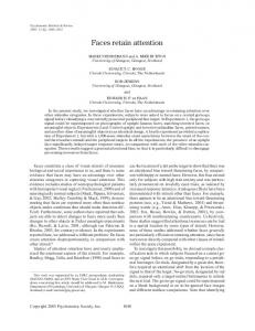
III. RESULTS Verbal Working Memory. In individuals with dyslexia, significantly earlier activations were primarily noted in superior frontal (SF) regions. On average, individuals with dyslexia displayed a mean onset in SF regions at ≈210 ms, while controls demonstrate activation at ≈325 ms, t(15) = 2.02, p = .05. See Figure 1 (A and B). Subjects with dyslexia also demonstrated significantly higher amplitudes in the left parahippocampal regions, t(15) = 2.18, p = .048. See Figure 1 (C and D). Normalized mean amplitudes were significantly lower in the right precentral region, t(15) = 2.45, p = .029 and right superior temporal region, t(15) = 3.341, p = .005 in dyslexia. Figure 2 displays a comparison of control vs. subjects with dyslexia ROI amplitudes, note increased amplitude for control subjects in the superior temporal region.
D
Fig. 1 Verbal Working Memory. Note pronounced left superior frontal activation in (A) control subject at about ≈300 ms compared to subject with (B) dyslexia who displays alternate, right inferior frontal activation at this time revealing possible reversed hemispheric processing during VWM. Note pronounced activation in left parahippocampal region in dyslexia (C) relative to control subject (D) at about ≈ 350ms possibly suggesting compensatory recruitment of inferior hippocampal memory regions rather than a frontoparietal working memory network when attempting working memory tasks
IFMBE Proceedings Vol. 28
348
S.M. Bowyer et al.
temporal and frontal while predominately right temporoparietal activations were noted in AD. In general, during WM, the NC showed pronounced activation in the superior frontal parietal cortices. The clinical subjects displayed a higher number of significant amplitudes of activation and more variability in neural activity throughout the 650 ms epoch.
Verbal Working Memory 1.600
Normalized Mean Amplitudes
1.400
1.200 1.000 0.800 0.600
0.400 0.200 0.000 Control
Dyslexia
L. Parahippocampal
Control
Dyslexia
R. Precentral
Control
Dyslexia
R. Superior Temporal
Fig.
2 Comparison of control subjects vs. subjects with dyslexia for 3 normalized ROI amplitudes during verbal working memory
Spatial Working Memory. During spatial working memory, significantly earlier activations occurred in bilateral precentral, t(15) = 3.59, p = .008, and middle and inferior temporal regions, t(15) = 2.86, p = .02, in dyslexia compared to controls. The onset of temporal activation was noted at ≈150 ms in subjects with dyslexia ≈150 ms while similar regions activated in controls at ≈350 ms. On average, over the 650 ms, more robust amplitudes were noted in right superior temporal regions in controls compared to those with dyslexia t(15) = 2.579, p = .023. See Figure 3. This increase was similar to that found in the VWM. See Figure 2
Fig.
4 Butterfly plot showing all 148 MEG channel waveforms during Verbal Working Memory task in Neurotypical Control (NC) at 240 ms arising from left parietal activation
Fig.
5 Butterfly plot showing all 148 MEG channel waveforms during Verbal Working Memory task in Developmental Dyslexia (DD) at 244ms arising from left frontal activation
Spatial Working Memory 1.800
Normalized Mean Amplitudes
1.600 1.400 1.200 1.000 0.800 0.600 0.400 0.200 0.000
Control
Dyslexia R. Superior Temporal
Fig. 3 Normalized amplitudes during spatial working memory reveal higher amplitudes in right superior temporal ROI during spatial working memory in controls compared to dyslexia
Fig.
Developmental Trends. During onset of the VWM task, left parietal activation was observed in NC around ≈240 ms that was not observed in the DD or AD subjects. See Figure 4. The most robust activations in DD were bilateral
During spatial WM, clear right hemisphere dominance was observed in NC. The opposite pattern characterized the DD. The AD produced symmetric bilateral activation throughout the task.
6 Butterfly plot showing waveform from the 148 sensor array of MEG during Verbal Working Memory task in Acquired Dyslexia at 240 ms arising from right temporoparietal activation
IFMBE Proceedings Vol. 28
MEG Detection of Attention and Memory Processes in Individuals with Dyslexia
IV. DISCUSSION Individuals with dyslexia demonstrated a pattern of activation during both VWM and SWM suggestive of frontotemporal inefficiencies and or impairment, particularly in superior frontal and temporal regions. In general, individuals with dyslexia tended to demonstrate an earlier pattern of activation during WM tasks. Consistent with prior investigations of orthographic recall, structures in basal temporal regions were more heavily recruited during VWM tasks [7]. That is, individuals with dyslexia may be relying more heavily on ventral or basal temporal language areas and orthographic recall during performance of the VWM task rather than dorsal frontotemporal or frontoparietal regions and the phonological loop. A similar pattern of early bilateral frontal activation in dyslexia differentiated controls from those with dyslexia during SWM. In addition, middle and inferior temporal regions were found to be active significantly earlier in the dyslexic population. Interestingly, individuals with dyslexia also displayed decreased activation in right superior temporal regions during SWM; a finding not previously reported. Increased amplitudes in right superior temporal regions during both the WM tasks in controls may be primarily due to the visual nature of the verbal working memory task. In examining developmental differences in acquired compared to developmental dyslexia, we report a gradient of deviation from NC during VWM, with the AD being a closer approximation of the normative brain activity than the DD. This may suggest a time-related reorganization of neural activity post trauma versus a relatively persistent deviation associated with developmental dyslexia. In spatial WM, the aberrant lateralization observed in dyslexics also depicts the AD (bilateral) as an intermediate condition between NC (right dominance) and DD (left dominance). This pattern could be interpreted as evidence for plasticity in acquired neurological disorders.
V. CONCLUSION While verbal memory inefficiencies have been a consistently reported finding in dyslexia, the literature is equivocal with respect to the relative importance of verbal
349
working memory processes necessary for reading efficiency in dyslexia. In addition, limited studies have examined spatial working memory in dyslexia despite the visual nature of reading and common visual processing problems such as transpositions with orthographs. The current MEG study reveals differences in latency in frontal regions and amplitudes in bilateral superior temporal regions in controls compared to dyslexics. This suggests inefficiency in dorsal frontoparietal and superior temporal regions in dyslexia during working memory for which compensatory reversed hemispheric or bilateral ventral temporal activation may occur.
ACKNOWLEDGEMENT NIH/NINDS Grant R01-NS30914 to N. Tepley.
REFERENCES 1. Moran J, Bowyer S, Tepley N. (2005) Multi-Resolution FOCUSS: source imaging technique applied to MEG data. Brain Topo 18:1-17. 2. Bayliss DM, Jarrold C, Gunn DM, Baddeley AD (2003). The complexities of complex span: explaining individual differences in working memory in children and adults. J Exp Psychol Gen. Mar;132(1):71-92. 3. Simos PG, Fletcher JM, Foorman BR, Francis DJ, Castillo EM, Davis RN, Fitzgerald M, Mathes PG, Denton C, Papanicolaou AC. (2002). Brain activation profiles during the early stages of reading acquisition. J Child Neurol. Mar;17(3):159-63. 4. McNamara JK, Wong B. (2003). Memory for everyday information in students with learning disabilities. J Learn Disabil. 2003 SepOct;36(5):394-406. 5. Willcutt EG, Pennington BF, Olson RK, Chhabildas N, Hulslander (2005). Neuropsychological analyses of comorbidity between reading disability and attention deficit hyperactivity disorder: in search of the common deficit. J. Dev Neuropsychol. ;27(1):35-78. 6. Functional MRI studies of spatial and nonspatial working memory. (1998). D'Esposito M, Aguirre GK, Zarahn E, Ballard D, Shin RK, Lease J. Brain Res Cogn Brain Res. Jul;7(1):1-13. 7. Cohen L, Cohen S, Dehaene L, Naccache et al. (2000) The visual word form area: Spatial and temporal characterization of an initial stage of reading in normal subjects and posterior split brain patients. Brain 123:291-307. Author: Institute: Street: City: Country: Email:
Susan Bowyer, Ph.D. Henry Ford Hospital, Neuromagnetism Lab CFP 78/79, 2799 West Grand Blvd. Detroit USA
[email protected] or
[email protected]
IFMBE Proceedings Vol. 28