Uncorrected Version. Published on July 3, 2007 as DOI:10.1189/jlb.0307140
Mesenchymal stem cells fail to trigger effector functions of cytotoxic T lymphocytes Ida Rasmusson,*,1,2 Michael Uhlin,†,1 Katarina Le Blanc,* and Victor Levitsky† *Division of Clinical Immunology and †Microbiology and Tumor Biology Center, Karolinska Institutet, Stockholm, Sweden
Abstract: Mesenchymal stem cells (MSCs), isolated from adult human bone marrow, have immunomodulatory properties. The functional outcomes of MSCs–CTL interactions remain poorly characterized. In this study, we demonstrate that MSCs remain resistant to CTL lysis, even after pulsing with the specific synthetic peptide at high concentrations, in spite of surface expression of the relevant MHC class I allele. MSCs were also much less sensitive to lysis by an allo-specific CTL clone as compared with HLA-matched lymphoblastoid cell lines. MSCs induced CD25 up-regulation, albeit at relatively low levels, and were unable to induce CD3 or CD8 down-regulation at the surface of CTLs. MSCs also failed to induce IFN-␥ and TNF-␣ production by the CTLs. Furthermore, peptide-pulsed MSCs were inefficient in stimulating tyrosine phosphorylation in specific CTLs. Our results demonstrate that MSCs induce only an abortive activation program in fully differentiated, effector CTLs, which does not involve activation of major CTL effector functions. These data may have important implications for the development of therapeutic strategies based on administration of in vitro-expanded MSCs. J. Leukoc. Biol. 82: 000 – 000; 2007. Key Words: marrow stromal cells 䡠 effector CD8 T cells 䡠 activation 䡠 EBV 䡠 alloantigen
INTRODUCTION Human mesenchymal stem cells (MSCs) isolated from adult bone marrow can inhibit mitogen- and alloantigen-induced proliferation of PBMCs; however, the mechanisms of this phenomenon are still unknown. MSCs can inhibit naı¨ve as well as memory T cell responses [1] and affect CD4⫹ and CD8⫹ T cell subsets [2– 4]. In some experimental systems, the inhibitory effect could be reproduced using soluble factors released by MSCs [2, 4, 5], such as PGE2 [6]. However, we have shown that PGE2 was important only in mitogen-stimulated cultures and did not play a role in the suppression induced by MSCs in alloantigen-stimulated PBMCs [7], suggesting the existence of other soluble, inhibitory factors released by MSCs. We previously found that MSCs are capable of inhibiting the induction of CTL responses in vitro [5], and these findings were 0741-5400/07/0082-0001 © Society for Leukocyte Biology
further confirmed by others [8]. It is still not clear, however, whether MSCs also exert an inhibitory effect on activated effector CTLs, which represent a cellular subset mediating graft-versus-host disease (GVHD) in transplant recipients and are used for adoptive cancer immunotherapy. Specific activation of CTLs leads to phosphorylation of a number of signaling molecules, increased production of cytokines, and up-regulation of the IL-2R ␣-chain (CD25). Activation of CTLs also induces release of cytolytic granules, the process termed degranulation, and up-regulates expression of ligands for death receptors, such as FasL and TRAIL (reviewed in ref. [9]). In the present study, we demonstrate that MSCs used as APCs fail to induce IFN-␥ and TNF-␣ production in human CD8⫹ CTL clones and trigger weaker up-regulation of CD25 and down-regulation of the TCR complex as compared with the effects exerted on these molecules upon CTL stimulation with a lymphoblastoid cell line (LCL). This correlated with reduced, overall phosphorylation of cellular proteins detected in CTLs after MSC-induced stimulation. The potential biological implications of this phenomenon are discussed.
MATERIALS AND METHODS Antibodies and reagents The phosphotyrosine-specific antibody (4G10) was purchased from Upstate Biotechnology (Waltham, MA, USA) and actin-specific mAb from Sigma (Gillingham, Dorset, UK; AC-74). PE-labeled antibodies against CD25 (M-A251), CD3 (UCHT1), and CD8 (RPA-T8) were purchased from Becton Dickinson (San Jose, CA, USA). Antibodies recognizing all classical and nonclassical MHC molecules (w6/32) and A2/B17 or A2/Aw69 HLA molecules, were produced as supernatants of HB95, HB54, and HB117 hybridoma, respectively.
Generation and maintenance of MSCs For MSC isolation, bone marrow aspirates were obtained from the iliac crest of healthy adult volunteers, and mononuclear cells were separated by Percoll gradient centrifugation as described previously [7]. Briefly, bone marrow mononuclear cells were plated at 160,000 cells/cm2, and suspension cells were washed away after 48 h. When adherent colonies had formed, the cells were trypsinizied and replated at 4000 cells/cm2. The cells were classified as MSCs based on their ability to differentiate into bone, fat, and cartilage [10] and by
1
These authors contributed equally to the work. Correspondence: Division of Clinical Immunology, F79, Karolinska University Hospital, SE-141 86 Stockholm, Sweden. E-mail:
[email protected] Received March 8, 2007; revised May 10, 2007; accepted May 21, 2007. doi: 10.1189/jlb.0307140 2
Journal of Leukocyte Biology Volume 82, October 2007
Copyright 2007 by The Society for Leukocyte Biology.
1
flow cytometric analysis (positive for CD29, -44, -73, -105, and -166 and HLA class I but negative for CD14, -34, and -45). MSCs of Passages 3– 8 have been used in this study.
Cell lines, CTL cultures, and clones The HLA A11-transfected subline of the HLA class I-negative mutant cell line C1R [11] was generated by transfection with a pHEBO vector-based HLA A11 expression vector [12] and maintained in RPMI-1640 medium supplemented with 100 g/ml streptomycin, 100 UI/ml penicillin, and 10% FCS (complete medium) with 200 g/ml hygromycin B. EBV-specific CTL cultures and clones specific to the EBV nuclear antigen 4-derived epitope IVTDFSVIK (IVT) were obtained from HLA A11-positive, healthy, Caucasian EBV carriers by stimulation of their PBMCs with a B95.8 EBV strain-transformed, autologous LCL, as described previously [13], to induce activation and proliferation of EBV-specific T cells. After three consecutive restimulations, the cultures were expanded in RPMI with 10% FCS supplemented with 10 U/ml recombinant IL-2 and 30% (vol/vol) culture supernatant from the gibbon lymphoma line MLA144 [14]. Single-cell cloning was done by limiting dilution in 96-well plates with the above medium and 100,000 irradiated (3000 rad), allogeneic, PHA-activated PBLs as feeder [15]. Growing cultures were transferred into 48-well plates and fed twice a week by replacing half of the medium. The EBV specificity and HLA class I restriction of the CTL preparations were investigated by testing their cytotoxic activity against a panel of EBV-positive and -negative targets including autologous LCLs, allogeneic LCLs matched through single HLA class I alleles, at least two cell lines for each allele, PHA-activated blasts, HLA-mismatched LCLs, and the prototype NK-sensitive target K562. Alloreactive CTL clones were expanded in a similar manner. HLA A2-restricted CTLs specific to the BMLF1derived peptide epitope GLCTLVAML (GLC) [16] were derived from the peripheral blood of a healthy EBV carrier using FACS sorting of lymphocytes, which were stained specifically with GLC-containing HLA A2 tetramer purchased from ProImmune Ltd. (Oxford, UK). Sorted lymphocytes were subcloned and expanded as described above.
Synthetic peptides Synthetic peptides were synthesized by the Merrifield solid-phase method at Alta Bioscience (University of Birmingham, School of Biochemistry, Birmingham, UK) and purified by HPLC on SuperPac Pep-S 5 mm reverse-phase columns (Pharmacia, Uppsala, Sweden). The purified peptides were dried using SpeedVac (Pharmacia) and diluted in DMSO at a concentration of 1 ⫻ 10⫺2 M, as determined by Biuret assays.
T cell activation assays Target cells were pulsed with the indicated peptide for 1 h at 37°C, washed, and mixed with CTLs at an E:T ratio of 3:1 and incubated for 20 h in 5 ml tubes (1 million T cells/ml). The cells were washed in PBS and stained with PE-labeled CD3-, CD8-, or CD25-specific antibodies and analyzed on a FACS analyzer using CellQuest software (Becton Dickinson Labware, Franklin Lakes, NJ, USA). IFN-␥ and TNF-␣ were measured by intracellular fluorescent staining after activation with peptide-pulsed targets. CTLs were treated with golgistop (Becton Dickinson) for 1 h to sequester newly synthesized proteins in the Golgi complex. After incubation with targets, the cells were fixed using cytofix/cytoperm solution and stained with PE- or allophycocyanin-conjugated antibodies for 30 min on ice.
Cytotoxicity assay Standard, 4 h 51Cr-release assays were performed as described previously [15]. Target cells were preincubated with the relevant peptide at a range of peptide concentrations and labeled with 0.1 Ci/106 cells Na51CrO4 at 37°C for 1 h (Amersham Pharmacia Biotech UK Ltd., Buckinghamshire, UK). After extensive washing, target cells were incubated with HLA-A11 or A2-restricted, EBV-specific CTLs at an E:T ratio of 10:1 or 3:1 in triplicates for 4 h at 37°C. 51 Cr release in the supernatants was measured by a ␥-counter (Wallac Sverige AB, Stockholm, Sweden).
Analysis of protein tyrosine phosphorylation by immunoblotting CTLs incubated with the target cells, unpulsed or pulsed with the indicated synthetic peptides for 1, 10, and 20 min in 5 ml tubes (1 million T cells/ml),
2
Journal of Leukocyte Biology Volume 82, October 2007
were lysed in electrophoresis sample buffer (2⫻105 cells in 10 l) consisting of 50 mM Tris, pH 6.8, 1 mM EDTA, 150 mM NaCl, 10% glycerol, 5% ME, 1% bromophenol blue, and 1% Triton X-100. Cell lysates were separated on a SDS-PAGE. The gels were blotted onto nitrocellulose filters, which were then incubated with the phosphotyrosine-specific antibody diluted in PBS containing 5% skimmed milk. After incubation with anti-mouse or -rabbit HRPconjugated antibodies, the blots were visualized by SuperECL (Amersham Pharmacia Biotech UK Ltd.). Images were acquired on a FujiFilm phosphoimager using Las-100Pro ImageReader Version 2.1 software and analyzed using ScienceLab’98 Image Gauge Version 3.4X image analysis software.
Statistical analysis The Student’s t-test was used for statistical calculations. P values below 0.05 were considered statistically significant.
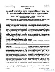
RESULTS Peptide-pulsed MSCs are resistant to lysis by peptide-specific MHC class I-restricted CTLs To analyze how the efficiency of MSC lysis by CTLs is affected by the amount of antigen presented at the cell surface, we pulsed HLA A2 or HLA A11-positive MSCs with the GLC or IVT peptide, respectively, using a range of peptide concentrations. The HLA A2-restricted GLC peptide is derived from the EBV lytic cycle protein encoded by the BMLF1 open-reading frame of the EBV genome, and the HLA A11-restricted CTL peptide epitope IVT is derived from the EBV nuclear antigen 4 expressed in EBV-transformed, lymphoblastoid cells. CTLmediated lysis of peptide-pulsed targets was assessed using standard 51Cr-release assays with CTL clones of the relevant specificity as effectors and compared with lysis of peptidepulsed C1R/A2 or C1R/A11 cells. GLC-specific effectors efficiently lysed C1R/A2 cells pulsed with the peptide at a concentration of 10⫺6 M, and HLA A2-positive MSCs remained virtually resistant to CTL lysis after pulsing with GLC peptide at all concentrations used in our assays (Fig. 1a). C1R/A11 cells were killed efficiently after pulsing with the IVT peptide at a concentration of 10⫺9 M, and the half-maximal lysis of these targets was observed at a peptide concentration as low as 10⫺10 M. Specific CTL lysis of IVT-pulsed, A11-positive MSCs never exceeded the level of half-maximal lysis of C1R/A11 cells, and this efficiency of killing was observed only at a 1000 times higher concentration of the IVT peptide (10⫺7 M; Fig. 1b). Only a slight increase in killing of IVT peptide-pulsed MSCs was observed following treatment of the target cells with exogenously added IFN-␥. Lysis by HLA A11-specific, allogeneic CTL clones was assayed against LCLs and MSCs. As shown in Figure 1c, lysis of A11-positive MSCs was comparable with the A11-negative control.
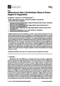
MSCs express intermediate-to-high levels of classical MHC class I alleles The level of MHC class I expression at the cell surface is one of the key parameters determining the efficiency of CTL lysis. Therefore, we analyzed expression of total MHC class I and the relevant individual class I alleles, HLA A2 and A11, on the surface of MSCs. Expression of surface MHC class I by MSCs was comparable with that detected on C1R/A2 or C1R/A11 cells (Fig. 2), and treatment of MSCs with IFN-␥ increased http://www.jleukbio.org
Fig. 1. CTL-mediated lysis of peptide-pulsed target cells. CTL lysis of C1R/A2 or C1R/A11 cells and A2 or A11 expressing MSCs were assessed using A2 (a)- or A11 (b)-restricted CTLs specific for the GLC or IVT peptide, respectively. Lysis was monitored by 4 h standard chromium release assay and is presented as percentage of specific lysis. The target cells were pulsed with the relevant synthetic peptide at the indicated concentrations. The graphs show the means and SEM for three experiments. (c) Lysis of the indicated targets by a HLA A11-specific, allogeneic CTL clone. MSCs were tested mock-treated or preincubated in the presence of IFN-␥.
MHC class I expression only slightly (data not shown). These results rule out the possibility that resistance of MSCs to CTL lysis is a result of low levels of MHC class I expression at the cell surface. Moreover, the variations in HLA expression observed between different MSCs could not be correlated to differences in lysis. We also analyzed the surface expression of nonclassical MHC class I molecules HLA-E and -G, which are known to inhibit activation of T cells [17, 18] and NK cells [19, 20] and therefore, could potentially suppress activation of CTLs. However, expression of these molecules at the surface of MSCs was not detected (data not shown).
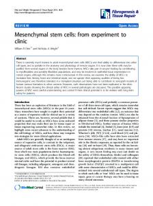
MSCs do not induce IFN-␥ or TNF-␣ production in CTLs Upon activation, CTLs produce cytokines important for their effector functions. We assessed production of IFN-␥ and TNF-␣ in CTLs activated with C1R/A2 or A2-positive MSCs, pulsed or unpulsed with the GLC peptide. Triggering with peptide-pulsed C1R/A2 cells increased the percentage of IFN-␥- and TNF-␣-producing CTLs significantly, as was determined by intracellular staining with lymphokine-specific antibodies (Fig. 3). In contrast, incubation with peptidepulsed MSCs did not lead to any detectable induction of IFN-␥ and TNF-␣ secretion by specific CTLs.
Modulation of TCR, CD8 coreceptor, and IL-2R ␣-chain expression by MSC-mediated, CTL triggering Specific CTL triggering leads to a change in the expression of a number of surface molecules. Up-regulation of CD25 (IL-2R ␣-chain) enables T cells to respond to IL-2, which promotes cell proliferation. The efficiency of TCR engagement on acti-
Fig. 2. Expression of total HLA class I and HLA A alleles on target cells. Expression of total MHC class I or HLA A2 molecules was determined by immunostaining and FACS analysis as described in Materials and Methods. The filled graphs show total HLA class I expression on C1R/A2 (a) and MSCs (b). The dark lines show A2 allele expression, and the dashed lines are the negative control, which represents staining with a CD3-specific antibody. The figure shows one representative experiment out of three.
Rasmusson et al. Activation of cytotoxic T cells by mesenchymal stem cells
3
Fig. 3. MSCs fail to induce cytokine production in triggered CTLs. GLC-specific CTLs were preincubated with control or GLC peptide-pulsed C1R/A2 cells or A2positive MSCs. Production of IFN-␥ and TNF-␣ was revealed by intracellular staining with the relevant, specific antibodies and FACS analysis. The dashed lines indicate staining with an isotype control antibody (Ctrl), filled graphs show staining obtained after preincubation of CTLs with nonpulsed target cells (Unstim), and the dark line histograms show staining of CTLs stimulated by the peptide-pulsed targets (Stim). The upper and lower panels show results obtained with C1R/A2 cells or MSCs as target cells. The figure shows one representative experiment out of three.
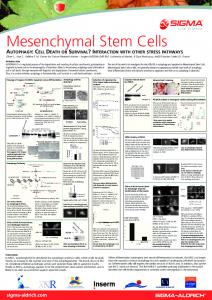
vated CTLs is also reflected in the level of TCR and CD8 down-regulation at the cell surface [21]. CTLs were activated by peptide-pulsed C1R/A2 or A2-positive MSCs, and levels of surface expression of CD25, CD3, and CD8 were determined 20 h after triggering (Fig. 4). Activation with peptide-pulsed C1R/A2 cells resulted in more than tenfold up-regulation of CD25 expression and considerable CD3 and CD8 down-regulation at the surface of CTLs. In contrast, MSC-mediated triggering induced low CD25 up-regulation (approximately threefold, P⬍0.05, compared with C1R) but failed to induce CD3 or CD8 down-regulation in specific CTLs (not significant compared with C1R).
Triggering with MSCs results in reduced tyrosine phosphorylation of proteins in CTLs TCR triggering leads to rapid phosphorylation of signaling molecules downstream of the TCR/CD3 complex. We analyzed the level of phosphorylation in IVT peptide-specific CTLs after their activation with peptide-pulsed C1R/A11 cells or A11positive MSCs for 1, 10, and 20 min. Immunoblotting with a phosphotyrosine-specific antibody revealed reduced levels of tyrosine phosphorylation in total cell lysates of MSC-activated CTLs as compared with CTLs triggered with C1R/A11 cells (Fig. 5). In addition, the response to MSCs was delayed, as the
Fig. 4. Alterations in surface expression of CD3, CD8, and CD25. (a) IVT-specific CTLs were stimulated with peptide-pulsed C1R/A11 cells or A11-positive MSCs. Expression of CD3 and CD8 on the surface of activated CTLs was monitored by immunostaining and FACS analysis as described in Materials and Methods. The data are shown as percentage of CD3 or CD8 down-regulation on activated CTLs as compared with cells preincubated with control APCs without the peptide. (b) The experiments were performed essentially as described in a. Expression of CD25 is presented as percentage of its up-regulation on the surface of CTLs, preincubated with C1R/A2, C1R/A11, or MSCs expressing the relevant class I alleles. The means and SEM of data obtained in three independent experiments performed with different APCs and CTLs are shown in the figure.
4
Journal of Leukocyte Biology Volume 82, October 2007
http://www.jleukbio.org
Fig. 5. Tyrosine phosphorylation in peptide-specific CTLs after C1R/A11 or MSCmediated triggering. Tyrosine phosphorylation was analyzed in IVT-specific CTLs after incubation with peptide-pulsed or unpulsed targets (C1R/A11 or A11-positive MSCs) at an E:T ratio of 3:1. The figure shows three separate time-points of incubation: 1, 10, and 20 min. Position 1, CIR/ A11, peptide-pulsed; 2, C1R/A11, unpulsed; 3, A11-positive MSCs, peptidepulsed; 4, A11-positive, unpulsed MSCs. The figure shows one representative blot out of three performed experiments.
maximal level of phosphorylation in C1R/A11-triggered T cells was observed after 1 min of activation, and MSC-stimulated T cells expressed the highest level of phosphorylated proteins 10 min after triggering. The level of protein phosphorylation decreased after 20 min of activation in CTLs stimulated with peptide-pulsed C1R/A11 cells, as compared with CTLs cocultured with the same APCs but without peptide. This was probably a result of activation of the dephosphorylation process, which quenches signal transduction after CTL activation. In contrast, activation-induced increase of protein phosphorylation was still observed in CTLs stimulated for 20 min with peptide-pulsed MSCs, again consistent with a delayed kinetics of post-TCR-triggering events initiated in CTLs by specific antigen presented on the surface of MSCs.
DISCUSSION In coculture experiments, MSCs fail to induce proliferation of allogeneic lymphocytes [1, 10, 22–24]. For transplantation across MHC barriers, to be successful, allogeneic MSCs must not only fail to induce activation of CD4⫹ and CD8⫹ responses but also escape lysis by CD8⫹ effectors [5, 25]. Indeed, following priming of CTLs by allogeneic lymphocytes, donor lymphocytes but not donor MSCs were lysed by the CTLs in cocultures [5]. In this study, we further explore the finding that MSCs are resistant to lysis by fully differentiated, effector CTLs. We show that MSCs exogenously loaded with the relevant MHC class I peptide epitopes remain resistant to lysis. The activation program induced by MSCs in CTLs appears to be incomplete or abortive and is characterized by inefficient up-regulation of CD25 on the surface of activated cells without concomitant stimulation of TNF-␣ and INF-␥ secretion, which are readily released by CTLs on triggering with other types of APCs. In our experiments, MSCs were lysed by specific CTLs inefficiently, despite intermediate-to-high expression of HLA class I, corroborating previous findings that PBMCs do not proliferate in response to MSCs, even if the latter are exposed to IFN-␥, which induces high levels of HLA classes I and II expression [24]. In addition, MSCs did not induce production of the proinflammatory cytokines IFN-␥ and TNF-␣. This is consistent with studies reported previously, indicating that MSCs do not induce production of IFN-␥ or increased expres-
sion of lymphocyte activation markers CD25, CD38, and CD69, whereas fibroblasts do trigger these changes in T cells [1, 22, 23, 26]. The effect could reflect an inefficient activation or active suppression of effector CTLs by MSCs or both. When MSCs are present in MLC, T, B, and dentritic cell proliferation is suppressed through MHC-independent mechanisms [1, 2, 4, 8, 27]. The factor mediating suppression by MSCs appears to be soluble, as proliferation is still reduced if MSCs and lymphocytes are separated in a transwell system [2– 4, 7, 28]. MSCs suppress the formation of effector CTLs if present during the stimulation phase but do not affect lysis of third-party targets by effectors formed already [5]. This, however, does not exclude the possibility that MSCs actively suppress other T cell activation events, e.g., lymphokine production and proliferation. Triggering of cytotoxic activity usually requires a low level of activating signal, and lytic activity remains intact in CTLs, which are nonresponsive to the specific stimulus in terms of other parameters of activation [21, 29, 30]. In accordance with this possibility, the analysis of protein phosphorylation performed in this study suggests that MSCs affect the amplitude and probably the quality of signal transduction events induced by specific TCR triggering in CTLs. Our data are generally consistent with an inability of MSCs to trigger CTL activation. However, the delayed kinetics of tyrosine phosphorylation in CTLs suggests that MSCs may modulate signal transduction actively. Resistance of MSCs to CTLmediated lysis may be important for their capacity to suppress immune activation across major and minor histocompatibility barriers. Several animal studies have shown that allogeneic MSCs persist when transplanted, without immunosuppression, across MHC barriers [31–34]. Low levels of engraftment were also observed after infusion of haploidentical MSCs to a patient with severe aplastic anemia and in patients treated with third-partyderived, HLA-mismatched MSCs for steroid-resistant GVHD after allogeneic stem cell transplantation [35, 36]. We reported recently about fully mismatched, allogeneic MSCs transplanted into an immunocompetent fetus with osteogenesis imperfecta [37]. Persistence of transplanted cells 9 months post-transplant and the lack of immunoreactivity when patient lymphocytes were re-exposed to the MSC graft in vitro indicate that MSCs can be tolerated when transplanted across MHC barriers in humans.
Rasmusson et al. Activation of cytotoxic T cells by mesenchymal stem cells
5
Studies in experimental animals and patients suffering from severe GVHD suggest that MSCs may be immunosuppressive in vivo [36, 38 – 41]. In contrast, Sudres et al. [42] reported recently that although MSCs suppress alloantigen-induced proliferation in vitro, the cells failed to prevent GVHD in a murine model. Previous reports indicate that MSCs derived from C57BL/6 mice, as used by the authors, evoke an immune response with subsequent graft rejection in BALB/c recipients, although no immunoreactivity was detected in MSC lymphocyte cocultures prior to transplant [43]. Immunoreactivity, not initially detectable in vitro, was also reported between other strains of mice and in xenogenic systems [44, 45]. Clearly, species-specific differences exist as suppression by mouse MSCs is cell– cell contact-dependent and induces a cell-division arrest not observed in human lymphocytes [1, 22, 46 – 48]. Autologous and allogeneic MSCs are evaluated currently in clinical trials as suppressors of pathologic immune responses and treatment of organ damage. Passive or active resistance of MSC to CTLs may be important for transplantation across MHC barriers as well as for the capacity of MSCs to inhibit ongoing GVHD and autoimmune reactions.
14.
15.
16.
17.
18.
19.
20.
REFERENCES 21. 1. Krampera, M., Glennie, S., Dyson, J., Scott, D., Laylor, R., Simpson, E., Dazzi, F. (2003) Bone marrow mesenchymal stem cells inhibit the response of naive and memory antigen-specific T cells to their cognate peptide. Blood 101, 3722–3729. 2. Di Nicola, M., Carlo-Stella, C., Magni, M., Milanesi, M., Longoni, P. D., Matteucci, P., Grisanti, S., Gianni, A. M. (2002) Human bone marrow stromal cells suppress T-lymphocyte proliferation induced by cellular or nonspecific mitogenic stimuli. Blood 99, 3838 –3843. 3. Djouad, F., Plence, P., Bony, C., Tropel, P., Apparailly, F., Sany, J., Noel, D., Jorgensen, C. (2003) Immunosuppressive effect of mesenchymal stem cells favors tumor growth in allogeneic animals. Blood 102, 3837–3844. 4. Tse, W. T., Pendleton, J. D., Beyer, W. M., Egalka, M. C., Guinan, E. C. (2003) Suppression of allogeneic T-cell proliferation by human marrow stromal cells: implications in transplantation. Transplantation 75, 389 – 397. 5. Rasmusson, I., Ringden, O., Sundberg, B., Le Blanc, K. (2003) Mesenchymal stem cells inhibit the formation of cytotoxic T lymphocytes, but not activated cytotoxic T lymphocytes or natural killer cells. Transplantation 76, 1208 –1213. 6. Aggarwal, S., Pittenger, M. F. (2005) Human mesenchymal stem cells modulate allogeneic immune cell responses. Blood 105, 1815–1822. 7. Rasmusson, I., Ringden, O., Sundberg, B., Le Blanc, K. (2005) Mesenchymal stem cells inhibit lymphocyte proliferation by mitogens and alloantigens by different mechanisms. Exp. Cell Res. 305, 33– 41. 8. Potian, J. A., Aviv, H., Ponzio, N. M., Harrison, J. S., Rameshwar, P. (2003) Veto-like activity of mesenchymal stem cells: functional discrimination between cellular responses to alloantigens and recall antigens. J. Immunol. 171, 3426 –3434. 9. Walczak, H., Krammer, P. H. (2000) The CD95 (APO-1/Fas) and the TRAIL (APO-2L) apoptosis systems. Exp. Cell Res. 256, 58 – 66. 10. Le Blanc, K., Tammik, L., Sundberg, B., Haynesworth, S. E., Ringden, O. (2003) Mesenchymal stem cells inhibit and stimulate mixed lymphocyte cultures and mitogenic responses independently of the major histocompatibility complex. Scand. J. Immunol. 57, 11–20. 11. Zemmour, J., Little, A. M., Schendel, D. J., Parham, P. (1992) The HLA-A,B “negative” mutant cell line C1R expresses a novel HLA-B35 allele, which also has a point mutation in the translation initiation codon. J. Immunol. 148, 1941–1948. 12. Zhang, Q. J., Gavioli, R., Klein, G., Masucci, M. G. (1993) An HLA-A11specific motif in nonamer peptides derived from viral and cellular proteins. Proc. Natl. Acad. Sci. USA 90, 2217–2221. 13. de Campos-Lima, P. O., Levitsky, V., Brooks, J., Lee, S. P., Hu, L. F., Rickinson, A. B., Masucci, M. G. (1994) T cell responses and virus evolution: loss of HLA A11-restricted CTL epitopes in Epstein-Barr virus
6
Journal of Leukocyte Biology Volume 82, October 2007
22.
23.
24.
25.
26.
27.
28.
29.
30.
31.
32.
isolates from highly A11-positive populations by selective mutation of anchor residues. J. Exp. Med. 179, 1297–1305. Rabin, H., Hopkins III, R. F., Ruscetti, F. W., Neubauer, R. H., Brown, R. L., Kawakami, T. G. (1981) Spontaneous release of a factor with properties of T cell growth factor from a continuous line of primate tumor T cells. J. Immunol. 127, 1852–1856. Torsteinsdottir, S., Masucci, M. G., Ehlin-Henriksson, B., Brautbar, C., Ben Bassat, H., Klein, G., Klein, E. (1986) Differentiation-dependent sensitivity of human B-cell-derived lines to major histocompatibility complex-restricted T-cell cytotoxicity. Proc. Natl. Acad. Sci. USA 83, 5620 – 5624. Steven, N. M., Annels, N. E., Kumar, A., Leese, A. M., Kurilla, M. G., Rickinson, A. B. (1997) Immediate early and early lytic cycle proteins are frequent targets of the Epstein-Barr virus-induced cytotoxic T cell response. J. Exp. Med. 185, 1605–1617. Le Gal, F. A., Riteau, B., Sedlik, C., Khalil-Daher, I., Menier, C., Dausset, J., Guillet, J. G., Carosella, E. D., Rouas-Freiss, N. (1999) HLA-Gmediated inhibition of antigen-specific cytotoxic T lymphocytes. Int. Immunol. 11, 1351–1356. Malmberg, K. J., Levitsky, V., Norell, H., de Matos, C. T., Carlsten, M., Schedvins, K., Rabbani, H., Moretta, A., Soderstrom, K., Levitskaya, J., Kiessling, R. (2002) IFN-␥ protects short-term ovarian carcinoma cell lines from CTL lysis via a CD94/NKG2A-dependent mechanism. J. Clin. Invest. 110, 1515–1523. Braud, V. M., Allan, D. S., O’Callaghan, C. A., Soderstrom, K., D’Andrea, A., Ogg, G. S., Lazetic, S., Young, N. T., Bell, J. I., Phillips, J. H., Lanier, L. L., McMichael, A. J. (1998) HLA-E binds to natural killer cell receptors CD94/NKG2A, B and C. Nature 391, 795–799. Adrian Cabestre, F., Moreau, P., Riteau, B., Ibrahim, E. C., Le Danff, C., Dausset, J., Rouas-Freiss, N., Carosella, E. D., Paul, P. (1999) HLA-G expression in human melanoma cells: protection from NK cytolysis. J. Reprod. Immunol. 43, 183–193. Valitutti, S., Muller, S., Dessing, M., Lanzavecchia, A. (1996) Different responses are elicited in cytotoxic T lymphocytes by different levels of T cell receptor occupancy. J. Exp. Med. 183, 1917–1921. Maitra, B., Szekely, E., Gjini, K., Laughlin, M. J., Dennis, J., Haynesworth, S. E., Koc, O. N. (2004) Human mesenchymal stem cells support unrelated donor hematopoietic stem cells and suppress T-cell activation. Bone Marrow Transplant. 33, 597– 604. Klyushnenkova, E., Mosca, J. D., Zernetkina, V., Majumdar, M. K., Beggs, K. J., Simonetti, D. W., Deans, R. J., McIntosh, K. R. (2005) T cell responses to allogeneic human mesenchymal stem cells: immunogenicity, tolerance, and suppression. J. Biomed. Sci. 12, 47–57. Le Blanc, K., Tammik, C., Rosendahl, K., Zetterberg, E., Ringden, O. (2003) HLA expression and immunologic properties of differentiated and undifferentiated mesenchymal stem cells. Exp. Hematol. 31, 890 – 896. Angoulvant, D., Clerc, A., Benchalal, S., Galambrun, C., Farre, A., Bertrand, Y., Eljaafari, A. (2004) Human mesenchymal stem cells suppress induction of cytotoxic response to alloantigens. Biorheology 41, 469 – 476. Beyth, S., Borovsky, Z., Mevorach, D., Liebergall, M., Gazit, Z., Aslan, H., Galun, E., Rachmilewitz, J. (2005) Human mesenchymal stem cells alter antigen-presenting cell maturation and induce T-cell unresponsiveness. Blood 105, 2214 –2219. Le Blanc, K., Rasmusson, I., Gotherstrom, C., Seidel, C., Sundberg, B., Sundin, M., Rosendahl, K., Tammik, C., Ringden, O. (2004) Mesenchymal stem cells inhibit the expression of CD25 (interleukin-2 receptor) and CD38 on phytohaemagglutinin-activated lymphocytes. Scand. J. Immunol. 60, 307–315. Groh, M. E., Maitra, B., Szekely, E., Koc, O. N. (2005) Human mesenchymal stem cells require monocyte-mediated activation to suppress alloreactive T cells. Exp. Hematol. 33, 928 –934. Uhlin, M., Sandalova, E., Masucci, M. G., Levitsky, V. (2005) Help signals provided by lymphokines modulate the activation and apoptotic programs induced by partially agonistic peptides in specific cytotoxic T lymphocytes. Eur. J. Immunol. 35, 2929 –2939. Uhlin, M., Masucci, M. G., Levitsky, V. (2005) Regulation of lck degradation and refractory state in CD8⫹ cytotoxic T lymphocytes. Proc. Natl. Acad. Sci. USA 102, 9264 –9269. Devine, S. M., Cobbs, C., Jennings, M., Bartholomew, A., Hoffman, R. (2003) Mesenchymal stem cells distribute to a wide range of tissues following systemic infusion into nonhuman primates. Blood 101, 2999 – 3001. Kraitchman, D. L., Heldman, A. W., Atalar, E., Amado, L. C., Martin, B. J., Pittenger, M. F., Hare, J. M., Bulte, J. W. (2003) In vivo magnetic resonance imaging of mesenchymal stem cells in myocardial infarction. Circulation 107, 2290 –2293.
http://www.jleukbio.org
33. Hill, J. M., Dick, A. J., Raman, V. K., Thompson, R. B., Yu, Z. X., Hinds, K. A., Pessanha, B. S., Guttman, M. A., Varney, T. R., Martin, B. J., Dunbar, C. E., McVeigh, E. R., Lederman, R. J. (2003) Serial cardiac magnetic resonance imaging of injected mesenchymal stem cells. Circulation 108, 1009 –1014. 34. Pochampally, R. R., Neville, B. T., Schwarz, E. J., Li, M. M., Prockop, D. J. (2004) Rat adult stem cells (marrow stromal cells) engraft and differentiate in chick embryos without evidence of cell fusion. Proc. Natl. Acad. Sci. USA 101, 9282–9285. 35. Fouillard, L., Bensidhoum, M., Bories, D., Bonte, H., Lopez, M., Moseley, A. M., Smith, A., Lesage, S., Beaujean, F., Thierry, D., Gourmelon, P., Najman, A., Gorin, N. C. (2003) Engraftment of allogeneic mesenchymal stem cells in the bone marrow of a patient with severe idiopathic aplastic anemia improves stroma. Leukemia 17, 474 – 476. 36. Ringden, O., Uzunel, M., Rasmusson, I., Remberger, M., Sundberg, B., Lonnies, H., Marschall, H. U., Dlugosz, A., Szakos, A., Hassan, Z., Omazic, B., Aschan, J., Barkholt, L., Le Blanc, K. (2006) Mesenchymal stem cells for treatment of therapy-resistant graft-versus-host disease. Transplantation 81, 1390 –1397. 37. Le Blanc, K., Gotherstrom, C., Ringden, O., Hassan, M., McMahon, R., Horwitz, E., Anneren, G., Axelsson, O., Nunn, J., Ewald, U., NordenLindeberg, S., Jansson, M., Dalton, A., Astrom, E., Westgren, M. (2005) Fetal mesenchymal stem-cell engraftment in bone after in utero transplantation in a patient with severe osteogenesis imperfecta. Transplantation 79, 1607–1614. 38. Le Blanc, K., Rasmusson, I., Sundberg, B., Gotherstrom, C., Hassan, M., Uzunel, M., Ringden, O. (2004) Treatment of severe acute graft-versushost disease with third party haploidentical mesenchymal stem cells. Lancet 363, 1439 –1441. 39. Chung, N. G., Jeong, D. C., Park, S. J., Choi, B. O., Cho, B., Kim, H. K., Chun, C. S., Won, J. H., Han, C. W. (2004) Cotransplantation of marrow stromal cells may prevent lethal graft-versus-host disease in major histocompatibility complex mismatched murine hematopoietic stem cell transplantation. Int. J. Hematol. 80, 370 –376. 40. Zappia, E., Casazza, S., Pedemonte, E., Benvenuto, F., Bonanni, I., Gerdoni, E., Giunti, D., Ceravolo, A., Cazzanti, F., Frassoni, F., Mancardi, G., Uccelli, A. (2005) Mesenchymal stem cells ameliorate experimental
41.
42.
43. 44.
45.
46.
47.
48.
autoimmune encephalomyelitis inducing T-cell anergy. Blood 106, 1755–1761. Gerdoni, E., Gallo, B., Casazza, S., Musio, S., Bonanni, I., Pedemonte, E., Mantegazza, R., Frassoni, F., Mancardi, G., Pedotti, R., Uccelli, A. (2007) Mesenchymal stem cells effectively modulate pathogenic immune response in experimental autoimmune encephalomyelitis. Ann. Neurol. 61, 219 –227. Sudres, M., Norol, F., Trenado, A., Gregoire, S., Charlotte, F., Levacher, B., Lataillade, J. J., Bourin, P., Holy, X., Vernant, J. P., Klatzmann, D., Cohen, J. L. (2006) Bone marrow mesenchymal stem cells suppress lymphocyte proliferation in vitro but fail to prevent graft-versus-host disease in mice. J. Immunol. 176, 7761–7767. Eliopoulos, N., Stagg, J., Lejeune, L., Pommey, S., Galipeau, J. (2005) Allogeneic marrow stromal cells are immune rejected by MHC class I- and class II-mismatched recipient mice. Blood 106, 4057– 4065. Grinnemo, K. H., Mansson, A., Dellgren, G., Klingberg, D., Wardell, E., Drvota, V., Tammik, C., Holgersson, J., Ringden, O., Sylven, C., Le Blanc, K. (2004) Xenoreactivity and engraftment of human mesenchymal stem cells transplanted into infarcted rat myocardium. J. Thorac. Cardiovasc. Surg. 127, 1293–1300. Nauta, A. J., Westerhuis, G., Kruisselbrink, A. B., Lurvink, E. G., Willemze, R., Fibbe, W. E. (2006) Donor-derived mesenchymal stem cells are immunogenic in an allogeneic host and stimulate donor graft rejection in a nonmyeloablative setting. Blood 108, 2114 –2120. Bartholomew, A., Sturgeon, C., Siatskas, M., Ferrer, K., McIntosh, K., Patil, S., Hardy, W., Devine, S., Ucker, D., Deans, R., Moseley, A., Hoffman, R. (2002) Mesenchymal stem cells suppress lymphocyte proliferation in vitro and prolong skin graft survival in vivo. Exp. Hematol. 30, 42– 48. Maccario, R., Podesta, M., Moretta, A., Cometa, A., Comoli, P., Montagna, D., Daudt, L., Ibatici, A., Piaggio, G., Pozzi, S., Frassoni, F., Locatelli, F. (2005) Interaction of human mesenchymal stem cells with cells involved in alloantigen-specific immune response favors the differentiation of CD4⫹ T-cell subsets expressing a regulatory/suppressive phenotype. Haematologica 90, 516 –525. Glennie, S., Soeiro, I., Dyson, P. J., Lam, E. W., Dazzi, F. (2005) Bone marrow mesenchymal stem cells induce division arrest anergy of activated T cells. Blood 105, 2821–2827.
Rasmusson et al. Activation of cytotoxic T cells by mesenchymal stem cells
7