MethodsX MethodsX 00 (2015) 1–5 www.elsevier.com/locate/procedia
Development of a computational system for IMRT radiation therapy planning based on MCNPx code with graphical interface for 3D voxel model - SOFT-RT Fonseca, Telma C. F.a , Campos, Tarcisio P. R. deb a Centre
for Development of Nuclear Technology, Brazilian Nuclear Energy Commission (CDTN/CNEN)- MG, Brazil b University of Federal of Minas Gerais - Dep. Of Nuclear Engineering DEN/UFMG - MG, Brazil
Abstract This paper presents a computational software system called SOFT-RT, which allows the simulation of an entire Intensity Modulated Radiation Therapy - IMRT treatment. Starting from: • a three-dimensional voxel model based on computer tomographic (CT) or magnetic resonance (MRI) images of the patient. • the creation of the full treatment planning using combinations of multiple intensity-modulated fields coming from different beam directions producing a sculpted radiation dose that maximizes the tumour dose. • simulation using MCNPx code, to calculated the dose absorbed from each treatment plan. • and the dose results are shown at the SOFT-RT output module.
c 2014 Published by Elsevier Ltd.
Keywords: MCNPX, SOFT-RT, IMRT, VOXEL MODEL
2
/ MethodsX 00 (2015) 1–5
Indroduction Intensity-modulated radiation therapy (IMRT) is an advanced radiotherapy technique able to deliver high doses of radiation to the target volume (tumor), while preserving the surrounding tissues. This is possible by the modulation of radiation beam usually carried out by multileaf collimators that conform the radiation beam during the irradiation. Due to the high complexity of the dose distribution achieved, a detailed study of these treatments is required [1, 2]. For this task we developed the software SOFT-RT which allows the simulation of an entire IMRT treatment protocol. Starting from a three-dimensional voxel model of a patient, it is possible to extract a region of interest (ROI) including only the tumor and part of the surrounding tissues. Thus, a more accurate analysis and additional planning can be performed, taking into account where exactly the radiation beam passes through and which organs/tissues are exposed and how much dose is absorbed by the surrounding normal critical structures. Therefore, dose to the target (tumor) can be maximized, while, at the same time, the surrounding healthy tissues are preserved.
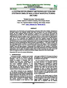
Method Imaging The digitalization of images is processing by converting a set of image (photos, x-rays, texts, etc.) through scanner or digital capture. The SISCODES [3] assembles its three-dimensional voxel model, based on a set of CT (Computed Tomography) or MR (Magnetic Resonance) images of the patient. During the processing of segmentation a set of pixel images (2D) is attached to generate a voxel volume (3D). Each voxel is assigned to its representative tissue which is stored into a database. For each tissue in the database, its density, material composition, colors, among other features are associated or added at the moment of the creation of the 3D voxel model. The SOFT-RT graphic interface generates a three-dimensional visualization of the voxel model, such a view may help the understanding of the anotomy of the patient, in a general aspect. Morover, it provides a better understanding of the treatment planning whereby allowing to release a greater dose in the tumor and preserving surrounding normal tissues. Figure 1 shows two voxel models, teh first is that of an Ear voxel model with some hidden/unhidden structures and the second is that of a Brain voxel model, which depicts its structures, including in details the brain anatomy. SOFT-RT features Different visualization possibilities are offered by the software. It is possible to cut through the tumor such that its width and height can be measured and its precise contours can be defined with respect to the selected beam orientation. Also, it is possible to perform the rotation and translation of the model around the axis of the coordinate system. As well, hide and unhide different organs/tissues to provide a better view of all surrounded organs/tissues nearby to the area requiring treatment. SOFT-RT was developed using the robust and multiplatform C++ programming language with the free open source OpenGL (Open Graphics Library) graphics packages [4] and the GLUI library [5] for the user interface. OpenGL was chosen to represent a matrix of blocks using its library of graphical routines and bi and three-dimensional modelling, since it is open domain, it is portable and quick to run. With this library, one can develop interactive applications and generated images of 3D scenes, with a high degree of realism. Figure 2 shows the three-dimensional visualization of the Brain voxel model, some hidden and unhidden structures and SORF-RT main option’s menu. Also, shown in red is the radiation beam and grey is the tumor modulated by the multileaf collimator. In addition, some other features of the SOFT-RT commands and options available: I Corresponding author at. Present address: CDTN - Centro de Desenvolvimento da Tecnologia Nuclear - Metrologia das Radiações Ionizantes/Radioproteção e Dosimetria - Belo Horizonte/MG - Brazil. Phone (Office) +55 031 3069 3496. Email address:
[email protected] (Fonseca, Telma C. F.)
/ MethodsX 00 (2015) 1–5
3
Figura 1. Three-dimensional voxel models: above the Ear and below the Brain
Figura 2. Main screen of the software SOFT-RT Planning system. Brain voxel model
• Viewing windows: 2D and 3D voxel model • Zoom in and out (both view windows) • Detailed command instructions: dimensions of the voxel model, angle of rotation of the beam, central position of the tumor, irradiation field, etc. • 3D coordenates axis (X, Y and Z) • Controlling of vectors that represent the direction of the beams and its radiation portals (collimators) • Rotation and translation function of the voxel model • Hidden/Unhidden organ/tissues • Create an MCNPx input file • Convert MCNPx output file to a SOFT-RT output file format. • Also, it is possible to use SOFT-RT with the Microsoft Windows or GNU Linux operational systems.
/ MethodsX 00 (2015) 1–5
4
SOFT-RT x MCNPx simulation The SOFT-RT planning system generates input files to be used in the General Purpose Monte Carlo transport code (MCNPx) [6]. All the parameters required to perform the simulation are forwarded and the interactions of the incident beam with the tumor and adjacent healthy tissues are performed. The simulation process is repeated for each selected orientation of the incident beam holding in a specific window (multileaf collimator). After all the simulations have been computed, the results are read in the SOFT-RT output-system. The 3D voxel model visualization is shown in a transparent glass procedure in which the adopted gray scale values at each voxel depends on the mass density of the correlated tissue. The doses are plotted using different colors defined by the isodose scale and are superimposed to the patient voxel model. Figure 3 shows a three-dimensional visualization of the Brain model in gray scale. It shows the plotting of the dose result obtained from the simulations with three different orientated beams and also, only the tumor with its total absorbed dose after removal of some organs/tissues.
Figura 3. SOFT-RT Out-put system. Brain voxel model
Summary and Addition Information The present article described the main features of the software SOFT-RT which is able to create and manage the IMRT protocol. The Brain and Ears voxel models, including in details all their anatomy, were shown. The SOFT-RT enables observation and controlling of vectors that represent the direction of the beams and its radiation portals (collimators). Also, it is possible to remove tissues from the model allowing to observe where the radiation beams are passing through inside the model, nearby of the tumor and healthy organs. This software allows the analysis of results from the MCNPx simulations by the visualization of the three dimensional voxel model in gray scale with the total dose plotted in colors. Some simulations using the Brain voxel model were already performed whose main goal was the validation of the system, but the results were not yet compared with a real case of IMRT treatment. Our early efforts are to focus on the validation of this software but comparing a IMRT simulated protocol versus a real case treatment. As well as, a improvement of the simulation time. In this case, the use of GPUs as generalpurpose processors [7] are going to be tested.
/ MethodsX 00 (2015) 1–5
5
Acknowledgements I like to thank CAPES - Coordination for the Improvement of Higher Education Personnel - for financial support of this research. Referências [1] MACISZEWSKI Wieslaw, Waldemar SCHARF, “Particle Accelerators for Radiotherapy. Present Status and Future,” Warsaw Poland, October 27, 2004. [2] Wuu CS, Xu Y., Three-dimensional dose verification for intensity modulated radiation therapy using optical CT based polymer gel dosimetry. Med Phys. 2006 May;33(5):1412-9. [3] TRINDADE, Bruno Machado and CAMPOS, Tarcisio Passos Ribeiro de. Sistema computacional para dosimetria de nêutrons e fótons baseado em métodos estocásticos aplicado a radioterapia e radiologia. Radiol Bras [online]. 2011, vol.44, n.2, pp. 109-116. ISSN 0100-3984. http://dx.doi.org/10.1590/S0100-39842011000200011. [4] OPENGL. The Industry’s Foundation for High Performance Graphics. Available in:http://www.OpenGL.org. Last accessed: September 2014 [5] GLUI User Interface Library. Available in:http://glui.sourceforge.net. Last accessed: September 2014 [6] Pelowitz D, James MR, McKinney GW, Durkee JW, Fensin ML, Hendricks JS, Mashnik SG, Verbeke JM, Waters LS. MCNPX 2.7.D extensions. In: Los Alamos National Security. LA-UR-10-07031. Los Alamos, NM: Los Alamos National Security; 2010. [7] Bert J1, Perez-Ponce H. Geant4-based Monte Carlo simulations on GPU for medical applications. Phys Med Biol. 2013 Aug 21;58(16):5593-611. doi: 10.1088/0031-9155/58/16/5593. Epub 2013 Jul 29.