Regeneration and transformation methods
Module 2
Regeneration and transformation methods
Hanh Trinh, David Barker and Pascal Ratet
Local organiser : Pascal Ratet
2-1
Regeneration and transformation methods
Regeneration and transformation methods
1. INTRODUCTION..........................................................................................................
3
2. AGROBACTERIUM-BASED IN VITRO TRANSFORMATION OF M. TRUNCATULA …………….……........……………...………………….….....
4
3. AGROBACTERIUM RHIZOGENES-BASED TRANSFORMATION OF M. TRUNCATULA .............…………………………………………………….…..... 10 4. LITERATURE CITED…………………..……………………………………………. 14 5. TIME SCALE …………………..................................................................................
15
Additional protocol…………………...................................................................……….. 16
2-2
Regeneration and transformation methods
1. INTRODUCTION Plant transformation technologies have had a considerable impact on plant biology understanding and on plant biotechnology. Generally the Agrobacterium-mediated T-DNA transfert process is used to deliver the gene of interest to the plant genome. The pathogenic bacteria Agrobacterium has the capacity to transfer part of its DNA (called the T-DNA) into the nuclear genome of plants cells (reviewed in Tinland, 1996). Two types of Agrobacterium strains are used for plant genetic transformation. In the A. tumefaciens strains, the T-DNA genes encode oncogenes that will induce the formation of a tumor on the infected plant tissue. In the A. rhizogenes strains, the T-DNA genes encode oncogenes that will induce the production of adventitious roots called the hairy root tissue. This later is used to produced rapidly chimaeric plants with untransformed aerial part and transgenic roots cotransformed with the Ri T-DNA and the construct of interest (see chapter III). This T-DNA plant transformation process is used to introduce genes in plants but also to inactivate plant genes by insertion mutagenesis. The T-DNA transfer to the plant nucleus depends on the expression of the bacterial vir genes that delimit the extent of the DNA sequence transferred to the nucleus, by recognizing specific sequences called T-DNA right and left borders (RB and LB). In between these borders any DNA construct can be introduced and transferred into the plant genome. This is the base for the construction of transgenic plants. For this, the oncogenes are deleted from the T-DNA and replaced by a gene of interest generally associated to a selectable gene. This T-DNA construct can be placed on another replicon (binary vector) than the vir genes, making the transformation system more versatile. The integration of the T-DNA in the genome probably depends on the plant DNA reparation machinery. Generally one copy of the T-DNA is inserted randomly in the plant genome, and gene fusions studies indicated that these insertions preferably occur in transcribed regions or in their vicinity (Koncz et al., 1989 ; Mathur et al., 1998). An excellent review on binary vectors, Agrobacterium strain and their use was published recently by Hellens et al. (2000). For M. truncatula any binary vector can be used for transformation experiments. Those confering Basta resistance will be the most useful but hygromycin and kanamycin can also be used as selectable marker. The transformation protocols vary from one plant to the other but mostly rely on in vitro regeneration of the transformed tissues. For Arabidopsis however, efficient in planta transformation protocols were developed (Bechtold et al., 1993 ; Clough and Bent, 1998 ; Richardson et al, 1998). These in planta simplified transformation protocols have allowed the large scale production of transgenic plants necessary for T-DNA tagging strategies. In vitro transformation protocols have been described for M. truncatula and will be presented in the course. In addition, a protocol for hairy root production will also be described.
2-3
Regeneration and transformation methods
2. AGROBACTERIUM -BASED IN VITRO TRANSFORMATION OF MEDICAGO TRUNCATULA Contributed by Pascal Ratet and Hanh Trinh 2.1. Introduction Several laboratories have reported the in vitro transformation and regeneration of M. truncatula cv. Jemalong through somatic embryogenesis or direct organogenesis (Nolan et al. 1989 ; Chabeau et al., 1996 ; Trieu and Harrison, 1996). The laboratory in Gif-sur-Yvette has developed a very efficient transformation/regeneration protocol based on somatic embryogenesis, for another M. truncatula ecotype : R108-1 (Hoffmann et al., 1997 ; Trinh et al., 1998 ; Kamaté et al., 2000). With this protocol production of transgenic plants is done routinely in the laboratory using various Agrobacterium tumefaciens strains. Most of the Agrobacterium strains used in the laboratory work. However, strain LBA4404 (Hellens et al., 2000) currently used for tobacco transformation is not efficient at all for Medicago transformation. In the laboratory, we normally use the Agrobacterium tumefaciens strain EHA105 (Hellens et al., 2000). 2.2. Transformation Growth conditions of the plant material before transformation Germinating seedlings are grown in sand for two weeks. Plantlets are then transferred to plastic pots with vermiculite in the green house (sixteen hour day period, 16°C night to 22°C day temperature with additional light : 200 µE/m2/s) or in growth chambers (sixteen hour day period, 16°C night to 22°C day temperature with 200 µE/m2/s light) for three weeks, watering them twice a day alternatively with nutrient solution (Soluplant, N/P/K : 18/6/26, Duclos International, Lunel Viel, France) and water. Five to eight weeks plants can be used for in vitro transformation experiments. Leaf sterilisation - Items required : . healthy 5 to 8 weeks plants . 50 ml falcon tubes . teepol . sterile water . Na-hypochloride solution (6° Cl) - Use leaves from 4 to 6 weeks old plants. These leaves should be round shape, healthy, without too many hairs. Plan five leaflet per plate. Each leaflet can generate at least one transformed plant. - Sterilisation is done in a 50 ml falcon tube. Leaflets are first rinsed in tap water containing two to three drops of teepol. Inverse several times the tube in order to wet all the leaves. Rinse with tap water.
2-4
Regeneration and transformation methods
Replace water with Na-hypochloride solution (6° Cl) and leave the tube for 7 mn. Return the tube (top on the bottom) and leave it for an additional 7 mn. Under sterile conditions, rinse the leaflet three times with sterile water. All the following manipulations are done under sterile conditions !!!! Agrobacterium culture and explant preparation Items required : . overnight 30 ml Agrobacterium culture in YEB medium (OD600nm= 0,6), prepared from a 2 ml overnight preculture . sterile petri dishes . sterile forceps and scalpels . sterile vacuum flask . sterile SH3a liquid medium - Centrifuge a 30 ml Agrobacterium culture at 3000 g for 20 mn and resuspend gently the pellet in 50 ml sterile SH3a liquid medium. - Cut the leaflet in square peaces with a sterile scalpel, and place them in a vacuum flask containing the SH3a solution with Agrobacterium. Mix gently to separate the plant pieces. Infiltration Items required . Agrobacterium culture . vacuum pump . shaking table This infiltration step to introduce bacteria inside the plant tissue results in high frequency of transformation. - Apply vacuum to the leaf explants in the SH3a solution with the Agrobacterium for 20 mn at 650 psi. Release slowly the vacuum and place the closed recipient on a shaking table at room temperature for 1 to 2 h to allow the tissue to recover from the infiltration procedure. 2.3. Regeneration Cocultivation Items required : . sterile forceps . laminar flow . sterile petri dishes . SH3a solid medium . sterile disposable pipets . plastic film . plant growth room
2-5
Regeneration and transformation methods
This step allows the bacteria in contact with the plant cells to transfer the T-DNA to the plant nucleus. This is during this period of time that the transformation occurs. - Under the sterile laminar flow, transfer the explants to a petri dish and remove the bacterial solution with a pipet. This bacterial solution (GMO) should be sterilized with Nahypochloride before discarding it. - Transfer the leaf explants to solid SH3a medium without antibiotics. The lower side of the leaf explant should be in contact with the medium. The plates are sealed with plastic film and incubated for a maximum of 2 days in the dark in the plant growth culture room (24°C). During this cocultivation step care should be taken that the agrobacteria do not over-grow the leaf explants. Callus formation step Items required : . laminar flow . sterile forceps . SH3a solid medium with antibiotics and selection compounds . plant growth chamber This step will allow the transformed tissue to multiply and form calli. In addition, the hormone treatment will induce the embryogenesis process. At the end of the callus formation step, the pre-embryos will be formed. - The leaf explants are transferred from the cocultivation medium to SH3a medium with 800 mg/l augmentin (to select against the agrobacteria) and with 3 mg/l basta to select for the transformed cells. If a vector conferring kanamycin resistance is used, basta is replaced with 40 mg/ml kanamycin. If the vector confers hygromycin resistance 10 mg/l hygromycin B is used. - The material is placed in the dark in the growth chamber (24°C) for 5 to 6 weeks and checked regularly for contaminations. The material (calli) is transferred to new medium every two week. Embryogenesis Items required : . SH9 solid medium . plant growth chamber At this step the calli are transferred to hormone free medium and grown in the light. These two changes will induce the embryogenesis followed by plantlet development. - calli at this stage should be friable. Transfer calli to SH9 medium (with basta) and place them in the light (130 mE ; alternating tubes Mazdafluor Prestiflux-HF Incandia : 4A TF"P"58W/inc and Mazdafluor Blanc Industrie 33 : 6J TF58W/BI ) in the growth chamber (24°C, 12 h photoperiod). If at this stage the bacteria start to grow again, the calli should be transferred to SH9 medium with basta and augmentin. - The pre-embryos from the calli will then develop in to true embryos that will later develop leaflets. These plantlets are then transferred to 1/2 SH9 medium to induce rooting. When
2-6
Regeneration and transformation methods
rooting starts, the plantlets are transferred to 1/2 SH9 square plates (120X120 mm) which are placed vertically in the growth chamber to allow roots to grow along the medium (M. truncatula R108 does not like to have roots growing inside the medium). Transfer of transgenic plants to the green-house Items required : . greenhouse . plastic trays with transparent lids . sterile sand The plant material transferred from the in vitro conditions to the green house is very sensitive to the change in the humidity conditions. Thus to avoid significant loss, plants should be maintained at the beginning of the transfer in water-saturated conditions and then adapted progressively to the normal green house conditions. - Plantlets that have developed few leaves and roots on 1/2 SH9 medium are planted in sand in trays with a transparent lid and watered alternatively with tap water and nutrient solution. A plate is placed under the tray in order to keep it with water. The sand should always be humid. The lid is maintained closed for the first days to maintain the plants in an atmosphere saturated with water. Then the lid is progressively open to reduce slowly the humidity level. At the end of the second week, the lid can be completely removed. The plants are then watered alternatively with nutrient solution and water. When they are big enough they can be transferred to normal pots. 2.4. References - Chabaud, M., Larsonneau, C., Marmouget, C., and Huguet T. (1996) Transformation of barrel medic (Medicago truncatula Gaertn.) by Agrobacterium tumefaciens and regeneration via somatic embryogenesis of transgenic plants with the MtENOD12 nodulin promoter fused to the gus reporter gene. Plant Cell Rep 15 : 305-310. - Hellens, R., Mullineaux, P., Klee, H. (2000) A guide to Agrobacterium binary Ti vectors. Trends in Plant Sci. 5 : 446-451. - Hoffmann, B., Trinh, T.H., Leung, J., Kondorosi, A., and Kondorosi, E. (1997) A new Medicago truncatula line with superior in vitro regeneration, transformation, and symbiotic properties isolated through cell culture selection. Mol Plant-Microbe interact 10 : 307-315. - Kamaté, K., Rodriguez-Llorente, I.D., Scholte, M., Durand, P., Ratet, P., Kondorosi, E., Kondorosi, A., and Trinh, T.H. (2000) Transformation of floral organs with GFP in Medicago truncatula. Plant Cell Rep. 19 : 647-653. - Koncz, C., Martini, N., Mayerhofer, R., Koncz-Kalman, Z., Korber, H., Redei, G.P., and Schell, J. (1989) High-frequency T-DNA-mediated gene tagging in plants. Proc. Natl. Acad. Sci. USA 86 : 8467-8471. - Mathur, J., Szabados, L., Schaefer, S., Grunenberg, B., Lossow, A., Jonas-Straube, E., Schell, J., Koncz, C., and Koncz-Kàlmàn, Z. (1998) Gene identification with sequened T-DNA tags generated by transformation of Arabidopsis cell suspension. Plant J. 13 : 707-716.
2-7
Regeneration and transformation methods
- Nolan, K.E., Rose, R.J., and Gorst, J.R. (1989) Regeneration of Medicago truncatula from tissue culture : increased somatic embryogenesis using explants from regenerated plants. Plant Cell Rep. 8 : 278-281. - Tinland B. (1996) The integration of T-DNA into plant genomes. Trends in Plant Sci. 1 : 178-184. - Trieu, A.T., and Harrison, M.J. (1996) Rapid transformation of Medicago truncatula: regeneration via shoot organogenesis. Plant Cell Rep 16 : 6-11 - Trinh, T.H., Ratet, P., Kondorosi, E., Durand, P., Kamaté, K., Bauer, P., and Kondorosi, A. (1998) Rapid and efficient transformation of diploid Medicago truncatula and Medicago sativa ssp. falcata in vitro lines improved in somatic embryogenesis. Plant Cell Rep 17 : 345355. 2.5. Transformation medium for Medicago truncatula R-108-1 SH3a (SHMab in Trinh et al., 1998) (for 1 L of medium) N6 major
100 ml
SH minor
1 ml
SH vitamins
1 ml
EDFS
20 ml (stock solution)
Myo-inositol
100 mg
Sucrose
30 g
2-4 D
4 mg
BAP
0.5 mg
PH
5.8
H2O
QSP 1 L
Phytagel
3g
SH9 (SHM2 in Trinh et al., 1998) (for 1 L of medium) N6 major
100 ml
SH minor
1 ml
SH vitamins
1 ml
EDFS
20 ml (stock solution)
Myo-inositol
100 mg
Sucrose
20 g
PH
5.8
H2O
QSP 1 l
Kalys agar
7g or 9g when the plates are placed vertically
2-8
Regeneration and transformation methods
1/2 SH9 (SHM2/2 in Trinh et al., 1998) (for 1 L of medium) N6 major
50 ml
SH minor
0.5 ml
SH vitamins
0.5 ml
EDFS
10 ml (stock solution)
Myo-inositol
50 mg
Sucrose
10 g
PH
5.8
H2O
QSP 1 l
Kalys agar
7g
N6 major ( For 1 L) MgSO4.7H2O (dissolve completely)
1.85 g
KNO3
28.30 g
(NH4)2SO4
4.63 g
CaCl2.2H2O
1.66 g
KH2PO4
4.00 g
H2O
QSP 1 l
SH minor ( For 100 ml) MnSO4.H2O
1g
H3BO3
500 mg
ZnSO4.7H2O
100 mg
KI
100 mg
NaMoO4.2H2O
10 mg
CuSO4.5H2O
20 mg (CuSO4=12.8 mg)
CoCl2.6H2O
10 mg
H2O
QSP 100 ml
SH vitamin ( For 100 ml) Nicotinic acid
500 mg
Thiamine.Hcl (vitamin B1)
500 mg
Pyridoxine.Hcl (vitamin B6)
500 mg
H2O
QSP 100 ml
- N6 medium : Chu, C.C., Proc. Sym. Plant tissue Culture. Science Press, Peking : pages 4550 (1978). - SH medium : Schenk, R.U.,and Hildebrandt, A. C., Can. J. Bot. 50 :199-204 (1972).
2-9
Regeneration and transformation methods
- EDFS (ethylenediamine-tetraacetic acid ferric-sodium salt, Sigma : 15708-41-5), stock solution : 7 g/l ; take 20 ml of this stock solution for 1L of culture medium. - Kalys agar HP 696-7470 (Kalys, 39 Avenue Jean Lebas, 59100 Roubaix, France) - Phytagel (Sigma : P-8169) - 2-4 D (2-4-dichlorophenoxyacetic acid, Sigma : D-8407) - BAP (6-benzylaminopurine). YEB Agrobacterium culture medium (for 1liter) - Bacto beef extract : 5 g, Bacto yeast extract : 1 g, Peptone : 5 g, Saccharose : 5 g, magnesium sulfate : 2 ml of a 1M solution. PH : 7.2 Add 15 g/l Bacto agar for solid medium. Selection compounds - augmentin (amoxicilline-Acide clavulanique : 1 g/200 mg ; GlaxoSmithKline, 92731 Nanterre Cedex, France) : 800 mg/l - basta (gluphosinate-amonium, Hoechst schering AgrEvo GmbH, Frankfurt/Main) : 3 mg/l Selection for transformed cells can be done also using kanamycin or hygromycin in M. truncatula. - kanamycin (Sigma : 25389-94-0) : 40 mg/l - hygromycin (Hygromycin B, Boehringer, Mannheim, Germany) : 10 mg/l
3. AGROBACTERIUM RHIZOGENES-BASED TRANSFORMATION OF MEDICAGO TRUNCATULA Developed by : Aurélien Boisson-Dernier, Mireille Chabaud, Charles Rosenberg and David G. Barker (Molecular Plant Microbe Interactions , 2001, Vol. 14, 695-700) Laboratoire de Biologie Moléculaire des Relations Plantes-Microorganismes, CNRS-INRA UMR215, BP27, 31326 Castanet-Tolosan cedex, France Tel : (33)561285046 Fax : (33)561285061 e-mail :
[email protected] 3.1. Introduction Since the adoption of M. truncatula as model legume approximately 10 years ago, efficient transformation/regeneration protocols using Agrobacterium tumefaciens have been described for at least two M. truncatula ecotypes (Chabaud et al., 1996 ; Trinh et al., 1998), as well as a recent transformation protocol making use of in planta infiltration (Trieu et al., 2000). However, in parallel to these often lengthy and labour-intensive approaches it is important to develop more rapid procedures which allow transgene constructs to be stably integrated and expressed in vivo. In the case of root tissues, this can be achieved through the
2-10
Regeneration and transformation methods
use of Agrobacterium rhizogenes, a soil pathogen which generates adventitious, genetically (Ri T-DNA) transformed roots at the site of inoculation in many dicots (Chilton et al., 1982). A. rhizogenes-mediated transformation in legumes was first described for Lotus corniculatus several years ago (Jensen et al., 1986 ; Petit et al., 1987) and it was subsequently shown that such transgenic roots could be directly nodulated (Hansen et al., 1989). Since then, the usefulness of the "composite plant" approach (so-called because transformed roots are formed on a non-transformed plant) as a shortcut for generating transgenic nodules has been extended to other legumes such as clover, vetch, soybean and lotus. The following protocol, adapted and optimised for A. rhizogenes-mediated transformation of M. truncatula allows fast and efficient production of transgenic roots, with the additional possibility of antibiotic (Km) selection for roots expressing the co-transferred transgene of interest (cloned in a binary vector carrying a KmR gene). Composite M. truncatula plants can be efficiently nodulated by the N-fixing symbiotic partner Sinorhizobium meliloti and transgenic roots can be clonally propagated for studies of either symbiotic endomycorrhizal root colonisation or interactions with pathogenic/parasitic organisms such as root nematodes. This technique has been recently published in the journal MPMI (Boisson-Dernier et al., 2001), and additional general information about the transformation protocol and its applications can be found in this article. Protocol for A. rhizogenes transformation of M. truncatula (with direct antibiotic selection for transformed roots) Items required : . Agrobacterium culture on TY plates. . plates with TY solid medium plus the appropriate antibiotics . square petri dishes (12x12 cm) with sterile Fahraeus medium plus Km at 25 mg/l . M. truncatula seeds . Agar plates (15 g/l agar in tap water, sterilized) . sterile bench . plant growth cabinets at 20°C and 25°C (16-h photoperiod) . sterile forceps and scalpels 1. Surface sterilize seeds of M. truncatula and germinate on inverted agar plates at 14°C. 2. Streak A. rhizogenes strain ARqua1 (Quandt et al., 1993) containing the binary vector of interest on an agar plate of TY medium with appropriate antibiotics and grow for approximately 48h. One plate of A. rhizogenes is sufficient for approximately 30 inoculated plants. Note that in order to use direct antibiotic selection (kanamycin) for transgenic roots it is necessary that the binary vector carries a KmR gene (generally nptII) under the control of a constitutive plant promoter. 3. After approximately 30 hr germination, seedlings should have a radicle length of about 1 cm. Under the laminar flow hood, place the seedlings in a glass Petri dish containing water
2-11
Regeneration and transformation methods
to avoid dessication of the radicle. Cut the radicle approximately 3 mm from the root tip with a sterile scalpel holding the tip with the forceps (see below). Hold the tip of the seedling with the forceps
Cut the radicle here with a scalpel
After removing the radicle tip take hold of the seedling by the cotyledons and coat the sectioned surface with A. rhizogenes by lightly scraping on the surface of the plate. The seedling is then placed on a Petri dish (approx. 4-6 cm from the top) containing agar with modified Fahraeus medium and Km at 25 mg/l (see below). To help prevent the seedlings from falling when the dish is placed vertically the agar is poured so as to generate a slant (approx. 5° angle) . 6-7 seedlings can be placed on a single square Petri dish (12x12 cm). Seal the dish with parafilm but make several incisions with a scalpel to allow for gas exchange (very important). Place the Petri dish at an angle of approx. 45° for 2-3 days (again to reduce the risk of the seedlings falling) and then vertically for a further 4-5 days in a 20°C growth room (16-h photoperiod). Co-transformation at 20°C is twice as efficient as at 25°C. Note that it is important to include controls without A. rhizogenes inoculation to confirm that rooting is inhibited in the presence of kanamycin. 4. Approximately 7 days after inoculation transfer the plates to a growth room at 25°C (16-h photoperiod). The first co-transformed roots (i.e. those having integrated both the Ri T-DNA and the binary vector T-DNA) should begin to appear at this stage, with growth clearly initiating from the inoculated radicule section. 50-75 % of inoculated plants should produce transformed roots. Roots which develop in the air should be gently pushed back onto the agar surface to ensure uptake of the antibiotic. 5. 2-3 weeks after inoculation, the transformed roots should be sufficiently well developed for experimental studies (e.g. direct Nod factor treatment or transfer to aeroponics for nodulation or excision to generate clonal transgenic root lines). Under optimal conditions an average of four Km-resistant transformed roots should be obtained per inoculated plant (see BoissonDernier et al., 200 1)
Additional general remarks : 1. Healthy fast-growing transgenic roots are more easily obtained from this radicle sectioning procedure as compared with a classical hypocotyl-wounding approach. 2-12
Regeneration and transformation methods
2. Note that, in general, S. meliloti-elicted nodulation of M. truncatula on agar medium is often delayed and inefficient. For nodulating transgenic roots of composite plants we recommend initial transfer to aeroponic growth chambers. Transfer of plants to solid substrate in pots should also be possible, although this has not yet been tested. 3. Whilst Km-resistance is an efficient means of directly selecting for transgenic roots which have integrated the binary vector T-DNA, preliminary experiments suggest that hygromycin is an unsuitable antibiotic for selection. If it is not possible to use kanamycin, transformation can be performed in the absence of direct selection. Under these conditions non-transformed laterals will appear approx. 3-4 days after sectioning, usually emerging from just above the section. Furthermore, in the absence of selection, only a proportion of the Ri-transformed roots (about 60 %) will also be co-transformed with the binary vector T-DNA. Whilst Kmresistant selection works well on agar medium, it does not seem to be as effective on certain other solid supports such as phytagel. Aerial plant development of such composite plants is unaffected by the presence of Km in the medium.
Modified Fahraeus medium used for M. truncatula transformation Stock
Final
solution
concentration
Calcium chloride CaCl 2
0.9 M
0.9 mM
Magnesium sulfate MgSO4
0.5 M
0.5 mM
Potassium dihydrogen phosphate KH2PO4
0.7 M
0.7 mM
Di-sodium hydrogen phosphate Na2HPO4
0.4 M
0.8 mM
Ferric citrate
20 mM
20 µM
Ammonium nitrate NH4NO3
1M
0.5 mM
Manganese chloride MnCl 2
1 mg / ml
100 µg / L
Copper sulfate CuSO4
1 mg / ml
100 µg / L
Zinc chloride ZnCl2
1 mg / ml
100 µg / L
Boric acid H3BO3
1 mg / ml
100 µg / L
Sodium molybdate Na2MoO4
1 mg / ml
100 µg / L
Macro-elements :
Micro-elements :
Agar
15 g / L
- Adjust to pH 7.5 before autoclaving - For antibiotic selection add 25 mg/l Km after autoclaving
2-13
Regeneration and transformation methods
4. LITERATURE CITED - Boisson-Dernier, A., Chabaud, M., Garcia, F., Bécard, G., Rosenberg, C., and Barker, D.G. (2001) Hairy roots of Medicago truncatula as tools for studying nitrogen-fixing and endomycorrhizal symbioses. Mol. Plant-Microbe Interact. 14 : 693-700. - Chabaud, M., Larsonneau, C., Marmouget, C., and Huguet, T. (1996) Transformation of barrel medic (Medicago truncatula Gaertn.) by Agrobacterium tumefaciens and regeneration via somatic embryogenesis of transgenic plants with the MtENOD12 nodulin promoter fused to the gus reporter gene. Plant Cell Rep. 15 : 305-310. - Chilton, M.D., Tepfer, D.A., Petit, A., David, C., Casse-Delbart, F., and Tempé, J. (1982) Agrobacterium rhizogenes inserts T-DNA into the genome of the host plant root cells. Nature 295 : 432-434. - Hansen, J., Jorgensen, J.E., Stougaard, J., and Marcker, K.A. (1989). Hairy roots - a short cut to transgenic root nodules. Plant Cell Rep. 8 : 12-15. - Jensen, J.S., Marcker, K.A., Otten, L., and Schell, J. (1986) Nodule-specific expression of a chimaeric soybean leghaemoglobin gene in transgenic Lotus corniculatus. Nature 321 : 669674. - Petit, A., Stougaard, J., Kuhle, A., Marcker, K.A., and Tempé, J. (1987) Transformation and regeneration of the legume Lotus corniculatus: a system for molecular studies of symbiotic nitrogen fixation. Mol. Gen. Genet. 207 : 245-250. - Quandt H.J., Pühler A., and Broer I. (1993) Transgenic root nodules of Vicia hirsutea : a fast and efficient system for the study of gene expression in indeterminate-type nodules. Mol. Plant Microbe Interact. 6 : 699-706. - Trieu, A.T., Burleigh, S.H., Kardailsky, I.V., Maldonado-Mendoza, I.E., Versaw, W.K., Blaylock, L.A., Shin, H., Chiou, T.J., Katagi, H., Dewbre, G.R., Weigel, D., and Harrison, M.J. (2000) Technical Advance : Transformation of Medicago truncatula via infiltration of seedlings or flowering plants with Agrobacterium. Plant J. 22 : 531-541. - Trinh, T.H., Ratet, P., Kondorosi, E., Durand, P., Kamaté, K., Bauer, P., and Kondorosi, A. (1998) Rapid and efficient transformation of diploid Medicago truncatula and Medicago sativa ssp. falcata lines improved in somatic embryogenesis. Plant Cell Rep. 17 : 345-355.
2-14
Regeneration and transformation methods
5. TIME SCALE Day 1 (20th November) - Agrobacterium culture innoculation (30 mn) for in vitro transformation and A. rhizogenes transformation. - Seed sterilisation and transfer of the seeds on plates for germination (1h30 mn) - Nod factor treatment of already prepared transgenic hairy roots (30 mn) Day 2 (21st November) - Inoculation of the seedlings with A. rhizogenes (2h) - X-Gluc staining of the Nod factor treated hairy roots (1h) - in vitro transformation/regeneration: preparation of the leaf material, transformation and transfer of the material to regeneration plates (3h) Day 3 (22th November) - Observation of the X-Gluc staining (30 mn) Day 4 (23rd November) - Observation of the in vitro transformation Day 5 (24th November) - Transfer of plant explants to selection medium. Day 7th to 10th (26th to 29th November) - Observation of the A. rhizogenes transformed root seedlings and transformed leaf tissue.
2-15
Regeneration and transformation methods
Additional Protocol
Protocol for transformation via Agrobacterium tumefaciens and regeneration in vitro of Medicago truncatula Jemalong genotype 2HA. Mireille CHABAUD, Laboratoire de Biologie Moléculaire des Relations PlantesMicroorganism, INRA-CNRS, Castanet-Tolosan, France.
This protocol has been optimised for transformation and regeneration of the highly embryogenic genotype 2HA (named 2HA3-9-10-3 by Nolan et al. 1989) derived from Jemalong. With A17 (a typical Jemalong genotype) the regeneration step is less efficient because of the significantly lower frequency of somatic embryogenesis.
Basic Principles : Leaflets from axenic 2HA plantlets grown in vitro are wounded and cocultivated with the hypervirulent A. tumefaciens strain AGL1. Co-cultivation lasts 3 days and is performed under continuous kanamycin selection (our binary vector carries the nptII gene under the control of the nos promoter). Explants are then decontaminated by extensive washing in liquid medium in the presence of antibiotics. Regeneration of transformed cells involves callogenesis followed by somatic embryogenesis.
Particular features of this protocol : - use of the hypervirulent strain AGL1 - co-cultivation of explants and Agrobacterium in the presence of kanamycin - extensive decontamination of the explants in liquid medium following co-cultivation. Under these optimised conditions, 25 % of the 2HA explants give rise to transgenic kanamycin-resistant plants within 4-5 months.
PROTOCOL DETAILS 3 weeks prior to transformation Preparation of explants -Seed sterilization : see accompanying protocol. -Seed germination : seeds are placed in Petri dishes containing solid SHb10 medium. Petri dishes are placed in a dark chamber upside down at 14°C for 2 days. -Plantlet growth : germinated seeds (with radicules 0.5 to 1 cm long) are planted in Magenta boxes (4 plantlets/Magenta box) containing ≅ 60 ml SHb10 medium, and incubated in a growth chamber at 25°C, with a 16h/8h photoperiod for 2 to 3 weeks.
2-16
Regeneration and transformation methods
Comment : Do not use older explants since the age of the plantlets is critical for transformation efficiency. Note : For each construct we usually prepare 8 boxes (32 plantlets) that will provide a total of 96 leaflets for transformation.
1 to 3 days prior to transformation Preparation of the bacterial suspension for co-cultivation -Streak a fresh plate of AGL1 from a glycerol stock, using TY medium containing rifampicin (20 mg/l) and ampicillin (50mg/l). Comment : The hypervirulent strain AGL1 is twice as efficient for transformation as LBA4404. -24 h before transformation take a loop of bacteria from the fresh plate and grown overnight in 20 ml of liquid TY (Rif20/Amp50) in a 100 ml Erlenmeyer flask at 28°C (rotary shaker 200 t/mn). Note : It is advisable to prepare the transformation medium in advance : for each construct you will need 8 Petri dishes of solid CIM (20 ml/plate). In addition 2 dishes are required for the controls. Day of transformation
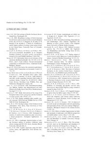
Preparation of the diluted bacterial solution, wounding of the explants, dipping in bacterial solution and co-cultivation in the presence of kanamycin -Centrifuge the bacterial culture at 5000 g for 10 minutes and re-suspend in 10 ml of sterile deionised water. Dilute to obtain 10 ml of a bacterial solution at OD600=0.1. The following steps are all performed in a horizontal laminar flow hood. -Pour the bacterial solution into a small sterile glass Petri dish (5.5 cm diameter). -For transformation, choose young healthy well-expanded leaves from the 2-3 week-old in vitro plantlets. Transfer 4 trifoliate leaves at a time to a large glass Petri dish (14.4 cm diameter) containing sterile water. After cutting off the three folioles at their base, each foliole is wounded with 3 to 4 scalpel cuts (see figure). Manipulating the explants in water avoids dessication. Transfer the wounded explants to the bacterial solution and leave them in the solution for several minutes. Then transfer the leaflets, abaxial side up, to a Petri dish containing solid CIM medium (Callus-Inducing-Medium) + kanamycin 50mg/l (12 folioles/dish). Seal the dishes with parafilm and co-cultivate the leaflets with the bacteria for 3 days in the culture room (25°C, 16h/8h photoperiod).
2-17
Regeneration and transformation methods
Figure : Explant wounding.
1 - cut off each leaflet
2 - wound with scalpel
Comment : The choice of the leaflets is very important to insure good transformation efficiency. Only young green expanded leaves should be used (usually a maximum of one to two leaves per plant). The other leaves will be either too old, or insufficiently developed. If the plants are not well developed do not use them! Controls involve dipping leaflets in water instead of bacterial solution. In routine experiments, 24 leaflets are wounded and used for controls. 12 are used to test the regeneration media (without kanamycin selection) and 12 are placed on media containing kanamycin to control for efficient selection. De-contaminating the explants in liquid medium after co-cultivation (day 3) -Prepare sterile 100 ml Erlenmeyer flasks containing 20 ml of liquid CIM medium with kanamycin 50mg/l and augmentin 400mg/l (5 parts amoxicillin/1 part clavulanic acid, Beecham Laboratories, France). -To eliminate bacteria attached to the explants, these are first dipped in sterile water, and then blotted on sterile filter paper. Excess bacteria should remain stuck to the filter paper. -Explants are then transferred to the flasks (12 leaflets/flask) -Washing is continued for 3 days with continual agitation (150t/mn) at 25°C, 16h/8h photoperiod. Comment : This decontamination step is essential to remove excess bacteria. If this washing step is not performed the bacteria will overgrow the explants often leading to subsequent death of the plant tissue (Chabaud et al., 1996). Note : AGL1 is resistant to ampicillin, so carbenicillin cannot be used to decontaminate the explants following co-cultivation. For this reason we use the antibiotic mix augmentin.
2-18
Regeneration and transformation methods
Callogenesis (day 6 onwards) -After washing, the entire content of each Erlenmeyer flask (liquid medium + leaflets) is poured into a sterile glass Petri dish and the leaflets transferred abaxial side up to a Petri dish containing 20 ml of solid CIM medium + kanamycin (50mg/l) + augmentin (400 mg/l) for callogenesis (12 leaflets/dish). -Every 3 weeks, explants are subcultured on fresh CIM medium containing the same antibiotics. Note: The leaflets can be rapidly blotted onto sterile filter paper before transfer to CIM medium but this is not essential. Comment : The first kanamycin-resistant calli are seen within 3 weeks of culture on CIM. They appear as green spots on the pale-brown non-transformed tissue of the leaflets. Within 1 month of culture approximately 40 % of the leaflets should develop Km-resistant calli. This reaches 90% after 2.5 months, and no new calli are observed after 3 months. After 1 to 2 weeks the green calli turn brown, as they become embryogenic. Embryogenesis (4-6 weeks onwards) -Once a week, calli which are sufficiently well-developed (around 1cm diameter) are transferred for embryogenesis to solid EIM medium (Embryo-Inducing-Medium) + kanamycin (50 mg/l) + augmentin (200mg/l). Each callus is transferred independently to a small Petri dish (5.5 cm diameter) containing 9 ml medium. Embryogenic calli are then subcultured every 3 weeks on the same medium. Comment : Embryogenesis is improved on EIM medium but it is not rare to see embryos already on CIM medium. Embryos are very easily distinguished from the brown callus because they are green and with a round smooth shape. Embryo development to plantlets - Once a week, embryos which have developed on calli (from globular to torpedo stages), are removed and transferred for further development to EDM medium (Embryo-Development-Medium) + kanamycin (50mg/l) + augmentin (200 mg/l). Comment : This step can be very long. Some embryos go through secondary embryogenesis instead of developing into a plantlet. Secondary embryos have to be transferred every 3 weeks onto fresh EDM medium until a few of them develop shoots. In a few cases, embryos develop directly to an entire plantlet with shoot and root (these are transferred directly to PDM medium). Leaf development is indicated by the presence of trichomes (these are not present on embryos).
2-19
Regeneration and transformation methods
Rooting of shoots - As soon as leaves are visible, leafy stems can be transferred for rooting to a Petri dish containing PDM medium (Plant-Development-Medium), without kanamycin and with augmentin 200mg/l. When plantlets are completely developed (with shoot and root), they are transferred to a Magenta box (1 plant/box) containing PDM. Transgenic plants are maintained in vitro by taking cuttings every 2 months. Note : It is advised to retain only one kanamycin resistant plant/explant to insure that the collection of regenerated transgenic plants derive from independent transformation events. Comment : Within 4 to 5 months of culture, 25 % of explants give rise to entire kanamycin resistant plantlets. More clones can be obtained if necessary over a longer period. For each construct, we usually examine 15-20 independent transgenic clones and retain a small number of representative clones. About half of the transgenic plants contain only one copy of the transgene. The chosen clone(s) is(are) grown for seed production and homozygous lines can be selected in the selfed progeny. Controls -A single Petri dish with leaflets dipped in water and cultured on CIM (with kanamycin) which shouldn't develop any calli even if they swell slightly. -A single Petri dish with leaflets dipped in water and cultured on CIM (without kanamycin), which should regenerate into plantlets more rapidly than the kanamycinresistant calli. In 3 weeks, calli are already well-developed, and are often embryogenic on CIM. This control is useful to get used to the regeneration process (in the absence of kanamycin selection), and plantlets can be obtained within 1.5 months. References : 2HA : genotype 2HA3-9-10-3 : Nolan et al.(1989) Plant Cell Reports 8:278-281. Send your seed requests to Dr Ray Rose :
[email protected] AGL1 : Lazo et al. (1991) Biotechnology 9: 963-967. Protocol : Chabaud et al. (1996) Plant Cell Reports 15:305-310. The improvements detailed in this protocol have not yet been published. They can be referenced either as Chabaud M., personal communication or Thesis, 1998, University of Paul Sabatier, Toulouse, France. For additional questions :
[email protected]
2-20
Regeneration and transformation methods
Specific material : - Centrifuge for preparation of the Agrobacterium suspension. - Horizontal laminar flow hood. - Culture room at 25°C with a 16h/8h photoperiod. - Shaker for 100ml Erlenmyer flasks (at least 10 flasks). - Scalpel and fine forceps. - Small (5.5 cm ) and large (14.4 cm ) sterile glass Petri dishes. - Sterile filter papers for blotting the explants. - Sterile Erlenmeyer flasks (100ml) with cotton seals. - Small (5.5 cm) and standard (9 cm)) sterile plastic Petri dishes. - Sterile Magenta boxes
2-21
Regeneration and transformation methods
Media : Optimised regeneration media for Medicago truncatula cultivar Jemalong: - CIM : Callus-Inducing Medium - EIM : Embryo-Inducing Medium - EDM : Embryo-Development Medium - PDM : Plant-Development Medium -SHb10 : Shenk and Hildebrandt, for plant growth (the same as PDM except lackingAIB and with agar instead of Phytagel) The following are the final compositions of the various media, which are prepared from the stock solutions listed further below :
Macroelements KNO3 NH4NO3 NH4H2PO4 MgSO4, 7H2O MnSO4, H2O ZnSO4, 7H2O CaCl2, 2H2O KH2PO4 H3BO3 KCl Microelements CuSO4,5H2O KI CoCl2, 6H2O Na2MoO4, 2H2O Fe EDTA FeSO4, 7H2O Na2 EDTA Vitamins Nicotinic acid Pyridoxin HCl Thiamin HCl Glycin Myo inositol Miscellaneous Casein Hydrolysate Bacto-tryptone MES Growth hormones 2,4-D trans-zeatin I.A.B. Sucrose Agar-agar Phytagel pH
CIM mg/l
EIM mg/l
EDM mg/l
PDM mg/l
1900 1650 370 16.9 8.6 440 170 6.2 -
1900 1650 370 16.9 8.6 440 170 6.2 -
1875 600 225 10 2 300 131 3 225
2500 300 400 10 1 200 5 -
0.025 0.830 0.025 0.250
0.025 0.830 0.025 0.250
0.025 0.750 0.025 0.250
0.2 1 0.1 0.1
27.85 37.25
27.85 37.25
9.19 12.29
15 20
5 10 10 2 100
5 10 10 2 100
1 1 10 -
5 0.5 5 1000
2000
2000
250
-
250 3mM
3mM
-
-
1 2 g/l 30 6 5.8
1 g/l 30 6 5.8
g/l 30 2 5.8
2-22
0.2 g/l 10 2 5.8
Regeneration and transformation methods
Note : All the media are prepared from stock solutions of macroelements, microelements and vitamins. These stock solutions are stored at -20°C in 50 ml aliquots. The growth regulators, trans-zeatin and IAB, and the antibiotics, kanamycin and augmentin, are added after autoclaving. Stock solutions : Macro elements : 20 X For 500 ml : Macro KNO3
UM 19 g
P4 18.75 g
NH4NO3
16.5 g
6g
MgSO4,7H2O
3.7 g
2.25 g
4g
KH2 PO4
1.7 g
1.31 g
-
CaCl2, 2H2O
4.4 g
3g
2g
KCl NH4, H2PO4
-
2.25 g -
3g
UM 680 mg
P4 500 mg
SH 500 mg
ZnSO4, 7H2O
430 mg
100 mg
50 mg
KI H3BO3
41.5 mg 310 mg
37.5 mg 150 mg
50 mg 250 mg
Na2MoO4, 2H2O
12.5 mg
12.5 mg
5 mg
CuSO4, 5H2O
1.25 mg
1.25 mg
10 mg
CoCl2, 6H2O
1.25 mg
1.25 mg
5 mg
P4 50 mg 50 mg 500 mg 5g
SH 250 mg 25 mg 250 mg -
SH 25 g
Microelements : 100 X For 500 ml : Micro MnSO4, H20
Vitamins : 100 X For 500 ml : Vitamins Nicotinic acid pyridoxin thiamin glycine Myo-inositol
UM 250 mg 500 mg 500 mg 100 mg 5g
Other stock solutions : MES 1M pH 5.8 : Dissolve 29.3g MES in 100ml water. Adjust pH with KOH 10 M. Add water to a final volume of 150 ml. Autoclave and stock at 4 °C. FeEDTA 100 X : Dissolve separately 1.4 g FeSO4, 7H20 in 100 ml and 2.1 g Na2 EDTA, 2H2O in 100 ml. Mix and make up to 500 ml water. Stock at –20°C in 50 ml aliquots. 2-4D (2-4 dichlorophenoxyacetic acid) 0.4 mg/ml : dissolve 40 mg in 5 ml EtOH. Make up to 100 ml with water. Store at 4°C. (Added before autoclaving).
2-23
Regeneration and transformation methods
Trans-zeatin 0.4 mg/ml : dissolve 40 mg in 5 ml KOH 1M. Make up to 100 ml in water. Filter sterilise and store at 4°C for 3 months maximum. (Added after autoclaving) IAB (indol butyric acid) 0.4 mg/ml : dissolve 40 mg in 5 ml KOH 1M and make up to 100 ml with water. Filter-sterilise and store in dark at 4°C. (Added after autoclaving) Kanamycin 50 mg/ml : 1000 X stock solution, filter-sterilized, 1 ml aliquots stored at –20°C. (Added after autoclaving.) Augmentin is weighed immediately before use and poured directly into cooled medium after autoclaving. (The stock solution in water is not stable). Reference: 1g amoxicillin/200 mg clavulanic acid, I.V. injectable, SmithKline Beecham Laboratoires Pharmaceutiques, Nanterre, France (usually available in your local chemist).
Supprimé : .)
pH is adjusted to 5.8 before autoclaving with 1M KOH. Preparation of media from stock solutions : For 1 L CIM Macroelements 20 X UM microelements 100 X UM vitamins 100 X UM FeEDTA stock solution myo inositol casein hydrolysat bacto-trypton 2-4 D stock sucrose MES 1M pH 5.8 pH agar-agar phytagel
EIM 50 ml UM 10 ml UM 10 ml UM 10 ml 2g 250 mg 2,5 ml 30 g 3 ml 5.8 6g -
50 ml 10 ml 10 ml 10 ml 2g 30 g 3 ml 5.8 6g -
added after autoclaving zeatin stock IAB stock
5 ml -
2.5 ml -
EDM P4 50 ml P4 10 ml P4 10 ml 3.3 ml
PDM SHb10 SH 50 ml SH SH 10 ml SH SH 10 ml SH 5.5 ml 1g
50 ml 10 ml 10 ml 5.5 ml 1g
250 mg
30 g
10 g
10 g
5.8 2g
5.8 2g
5.8 6g -
-
0.5 ml
-
Note : Media containing Phytagel as the gelling agent must be poured just after autoclaving. This medium cannot be re-melted after it has solidified. TY medium : Bacto-tryptone 5g/l Yeast extract 3g/l CaCl2 6mM (added after autoclaving) pH =7.2 agar-agar 15g/l
2-24