Phytochemistry Reviews (2004) 3: 371–379
© Springer 2005
Monitoring defensive responses in macroalgae – limitations and perspectives S.L. La Barre1,∗ , F. Weinberger1 , N. Kervarec2 & P. Potin1 1 UMR
7139 CNRS/GOEMAR/UPMC, Végétaux Marins et Biomolécules, B.P. 74, Station Biologique, 29682 Roscoff Cedex, France; 2 Laboratoire de Résonance Magnétique Nucléaire, Faculté des Sciences, B.P. 809 Université de Bretagne Occidentale, 29285 Brest Cedex, France; ∗ Author for correspondence (Phone: +33 (1) 9829 2360; Fax: +33 (1) 9829 2324; E-mail:
[email protected]) Key words: elicitor, macrophytic algae, mass spectrometry, metabolomics, nuclear magnetic resonance spectroscopy
Abstract As part of an ongoing research program aiming at monitoring molecular changes in the tissues and metabolite trafficking in the hydrosphere of algae subjected to chemical stresses, we are discussing the various analytical techniques that have been employed to characterize, and sometimes to quantity these metabolites. High-field multinuclear and solid-state nuclear magnetic resonance (NMR) spectroscopies are powerful tools for metabolite characterization from extracts and in vivo, but quantification and kinetic aspects show some limitations. Modern MS (mass spectrometry) is extremely useful for fingerprinting samples against databases and when dealing with very low concentrations of metabolites, the limitations being set by the type of chromatographic separation and mode of detection coupled with the mass spectrometer. Regarding chemical communication, optimization in terms of resolution and efficiency of hydrosphere chemical analysis can theoretically be achieved in a system which integrates (i) a multiparametric incubation chamber, (ii) a gasphase or a liquid-phase separation system and (iii) mass spectrometer(s) equipped with one or two detectors responding to the analytical and quantitative needs. This text reviews some of the techniques that have been employed in various types of plant metabolic studies, which may serve as a basis towards an integrative analytical strategy directly applicable to the metabolomics of selected marine macrophytes. Abbreviations: APCI – Atmospheric Pressure Chemical Ionization; cITP – Capillary Isotachophoresis (cITP); DAD – Diode Array Detector; ECD – Electron Capture Detection; EI-MS – Electron-Impact Mass Spectroscopy; ESI – Electro-Spray Ionization; GC – Gas Chromatography; 1 H NMR – Proton Nuclear Magnetic Resonance; HPLC – High-Performance Liquid Chromatography; HR-MAS – High-Resolution Magic Angle Spin; HVOC – Halogenated Volatile Organic Compound; HXO – Hypohalous Acid; ICP-MS – Inductively Coupled Plasma Mass Spectrometry; LC-MS – Liquid Chromatography coupled with Mass Spectroscopy; LMWHC – Low-Molecular Weight Halocarbon; MAA – Mycosporin-like Amino Acids; MIP-AED – Microwave-Induced Plasma Atomic Emission Detection; MS – Mass Spectroscopy; MS/MS – refers to two tandem-coupled mass spectrometers; NMR – Nuclear Magnetic Resonance; 31 P NMR – Phosphorus Nuclear Magnetic Resonance; PAR – Photosynthetic Active Radiation; SBSE – Stir Bar Sorptive Extraction; SIM – Single Ion Monitoring; SPME – Solid-Phase Micro Extraction; UV – Ultra-Violet; UVR – Ultra-Violet Radiation; VHC – Volatile HaloCarbon; VHOC – Volatile Halogenated Organic Compounds; X- – Halide Ions.
Introduction Marine algae are continuously being challenged by micro-organisms, including viruses, bacteria and other
algae as well as grazers, all of which may cause wounds and diseases. In addition, underwater competition for settlement and epiphytism may affect growth by reducing light availability or by increasing hy-
372 drodynamic drag. The combination of competition and predation pressures represents a constant threat to the fitness of macrophytic algae throughout their life, equivalent to that encountered by sessile autotrophs in terrestrial ecosystems. Macroalgae normally grow in shallow or eulittoral environments where the hydrodynamic regime (currents, tides, turbidity) is fast-changing. In addition to biotic stress, they may have to adjust to rapid and dramatic physical or chemical changes, e.g., UV radiation (UVR), high Photosynthetic Active Radiation (PAR), desiccation, salinity and nutrient limitation. To survive in such a seemingly hostile and unpredictable environment, marine plants obviously had to evolve survival strategies, under the form of so-called escape responses e.g. habitat refuges, species association, rapid growth etc., reviewed by Biber (2002), in combination with the production of chemical deterrents and growth inhibitors, UV screens or osmolytes, each representing a metabolic investment on behalf of the individual. A rapid response likely to represent a chemical defence against biotic stress in seaweeds is the emission of halogenated volatile organic compounds (HVOC’s). An increased production of such iodinated, brominated or chlorinated carbon skeletons is associated with oxidative stress from various origins, whether caused by carbon deprivation, high light intensity or mechanical injuries (Mtolera et al., 1996). HVOC’s biogenesis involves vanadium-haloperoxidases, which catalyse the oxidation of halides (X− ) into hypohalous acids (HXOs). As highly electrophilic agents, hypohalous acids can react with a variety of organic compounds and as such are considered as potent natural biocides (Wever et al., 1991). In brown algae, phlorotannins are induced by exposure to UV radiation and in response to grazing (Cronin and Hay, 1996; Pavia and Toth, 2000; Toth and Pavia, 2000). Another class of secondary metabolites, the mycosporine-like amino acids (MAAs) may be viewed as antioxidants in the rhodophytes (Dunlap and Shick, 1998). MAAs are synthesised via the shikimic acid pathway, a metabolic route which leads to the production of the higher plant antibiotics known as phytoalexins (Sommsich and Halbrock, 1998), and shikimic acid may be a central element of defence activation in these algae. In higher plants low molecular weight hydrocarbons (LMWHCs) such as isoprene (2-methyl-1,3-butadiene) and ethene are potent signaling molecules which are synthesised during environmental stress or upon grazing (Kimmerer and
Kozlowski, 1982; Abeles et al., 1992; Kesselmeier and Staudt, 1999). In marine brown algae LMWHCs are so far recognized as sexual pheromones (Pohnert and Boland, 2002) or herbivore deterrents (Hay et al., 1998; Schnitzler et al., 2001). These compounds may help to ward off infectious or fouling microorganisms (Hay, 1996) and they are very useful for broadcasting warning messages, i.e. informing the population of the presence of pathogens or grazers. Recent bioassayguided separations of crude bioactive algal extracts have proved the power and usefulness of such approaches to decipher the function of algal metabolites Previous investigations of the role of seaweed chemicals against herbivores in fucoid brown algae assumed that phlorotannins (Ragan et al., 1986) act as antifeedants (see Hay and Fenical, 1988). However, the deterrence of Fucus vesiculosus chemical extracts was recently reinvestigated using herbivore bioassays to guide chemical investigations (Deal et al., 2003). Although crude extracts from F. vesiculosus strongly deterred feeding by the sea urchin Arbacia punctulata, phlorotannins from this extract did not affect feeding at 2-fold or 4-fold natural concentration by dry mass. Feeding deterrence was instead due to: (1) one polar galactolipid in the ethyl acetate soluble extract and (2) at least one non-phenolic compound in the water-soluble extract. Given the high polarity of these chemical deterrents, they could co-occur with phlorotannins and confound bioassays performed on polar extracts before separation. The hypothesis that many marine plants use chemical defences selectively against microbial pathogens, epiphytes, and saprophytes was recently tested using a similar approach. Lobophorolide [1], a polycyclic macrolide with subµM antifungal activity, appears to function as an antifungal chemical defence, protecting the brown alga Lobophora variegata from at least one pathogenic and one saprophytic fungus, while being inactive against a thrausochytrid fungus, a pathogenic bacterium as well as against herbivorous fishes (Kubanek et al., 2003). Most researchers studying environmental or genetic effects on secondary metabolite production tend to focus on a particular class of compounds for practical reasons. Analyzing several groups of secondary compounds with different chemical properties is timeconsuming and often requires the use of individual extraction procedures and chromatographic methods. This functional diversity stems from the fact that many different secondary metabolic pathways are coordinately regulated by biotic elicitors and environmental stresses, as now evidenced by the abundant lit-
373
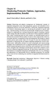
Figure 1. Diagram representing a circulating incubation chamber for the time-lapse analysis of compounds released in the medium, and further analyses of algal samples (1) Time-lapse analysis of compounds trapped on SPME fibers (2) Analysis of algal tissues and of their extracts (3) Alternative analysis of seawater samples collected in sealed vials and processed using a cold purge and trap system. (boldface): analytical methods of choice for the metabolic profiling and the chemical characterization
erature in terrestrial plants, (Sommsich and Halbrock, 1998; Scheel, 1998). When prompted, sessile marine organisms release communication (lato sensu) metabolites as bundles or sequentially in their immediate hydrosphere. The analysis of these metabolites, together with that obtained classically from tissue extracts, is essential for a complete ‘metabolomic picture’. It follows that different sampling procedures must be followed when operating in vivo and in vitro (i.e. between molecules highly diluted in seawater and metabolites concentrated in an organic extract). This also concerns to some extent the separation steps where appropriate chromatographic equipment must be used for online treatment, and of course, the choice of detection sensitivity. Here we briefly review recent methodological development s in the chemical ecology and biochemistry of marine algae, from which we can begin to outline an integrated methodology for a broad-spectrum metabolite fingerprinting of induced compounds, with optimal interactivity between the extraction and separation (HPLC, GC) steps, and the pertinent spectroscopic analysis and profiling of selected metabolites. Mass spectrometry analysis The recent developments of mass spectrometry in biology were initially motivated by medical research targeted towards high throughput screening as well as key analytical applications, e.g. understanding
cellular processes and the discovery of disease biomarkers. The use of mass spectrometry (MS) as the principal method for the detection of bioactive molecules presents an alternative way of performing natural products screening, especially in the absence of specific biomarkers (review by Van Elswijk and Irth, 2003). In particular, the need for dereplication of structures at initial stages of the analysis of ‘unknown’ natural samples, calls for outstandingly discriminating technology. In this regard, LC-MS, thanks to the use of appropriate interfaces e.g. electrospray (ES) or atmospheric pressure chemical ionization (APCI) is now capable of analyzing a tremendous range of molecules of all sizes. Database search using the nominal molecular mass of a compound then becomes highly efficient (Jemal et al., 2003). Ecotoxicological applications are presently targeted towards analysis of anthropogenic wastes and their fate in the atmosphere, water and soil, with climatic and toxicology concerns stimulating stateof-the-art technological advances allowing detection and identification of metabolites at picomolar levels. In this respect, the modularity of MS systems, and the miniaturization of most components often allows on-site analysis. Metabolomic studies, in which the molecular repertoire of a sample serves as a basis to the elucidation of biosynthetic processes as well as for the description of osmolyte trafficking, coud indeed benefit from all these technological advances, as proposed hereunder
374 for the study of the defences of macrophytic algae undergoing experimental stress. Coupling MS with GC Increase in instrument sensitivity and versatility of MS spectrometry through couplings with appropriate detectors and chromatographic systems, as used for the analysis of environmental pollutants, opens new perspectives for the detection of metabolites released by the algae. Sample preparation must accordingly respond to the needs of each experiment, i.e. retrieve meaningful information and comply with the level of performance of the equipment. In seaweeds, an increased production of iodinated, brominated or chlorinated, low-molecular weight carbon skeletons, referred to as halogenated volatile organic compounds (HVOCs), is associated with oxidative responses from various causes (Potin et al., 2002). The most popular strategy to determine volatile organic compounds is the coupling of a powerful separation technique, such as capillary gas chromatography (GC), with a sensitive detection method. Usually, the determination of HVOCs has been performed in the past by coupling GG with EI-MS (electron impact mass spectrometry) or ECD (electron capture detection) as explained in Abrahamsson and Klick (1990) and Urhahn and Ballschmiter (2000). Due to the poor selectivity of the electron capture mode of detection, comprehensive chromatographic techniques, i.e. columns with long retention times, packed with various stationary phases are needed for the determination of co-eluting compounds. Dual detection by tandem MS-MS is often necessary, the first detector providing the sensitivity and the molecular ion information useful for checking target analytes against an internal database, the second system providing a further fragmentation source for individual structure identification. A detector providing element-selective information like in ICP-MS (inductively coupled plasma mass spectrometry) represents a significant improvement in this respect, as recently tested by Schwarz and Heumann (2002) in tandem to an ECD. A two-dimensional on-line detection system was coupled with capillary GC to utilize the element selective detection by ICPMS but also the high sensitivity of an ECD for HVOC determination. This GC-ECD/ICP-MS system had been designed especially for the determination of brominated and iodinated HVOCs in aquatic and air samples. Prior to the measurements, the HVOCs must therefore be isolated from the corresponding samples.
For enrichment of HVOCs from aquatic samples a recently developed stir bar sorptive extraction technique (SBSE) was applied. This technique is based on the principles of solid-phase micro-extraction (SPME), which is often used to extract organic compounds from aqueous samples. Measurements were also carried out by coupling capillary GC with an EI-MS (electronimpact mass spectrometer) detector and a MIP-AED (microwave-induced plasma atomic emission detection), respectively, often used for HVOC determinations (Schwarz and Heumann, 2002). As well as information on elemental composition, quantitative data of selected molecules from GC-MIP-AED screening analysis after compound independent calibration can be obtained more easily than from target analysis (eg GC-MS in the SIM-mode, see Frischenschlager et al., 1997 and Jemal et al., 2003) Marine algae also feature a variety of nonvolatile, halogenated secondary metabolites, including terpenes, acetogenins and indoles, very few of which have been quantified at or near the surface of seaweeds and then tested at the same concentrations against ecologically relevant challengers. Perhaps the most detailed study is that of halogenated furanones produced by the Australian red alga Delisea pulchra. The presence of these metabolites at the surface of the alga was demonstrated (Dworjanyn et al., 1999), and methods for extracting the compounds from the surface with minimal damage to the thallus were also developed (de Nys et al., 1998). Levels of compounds in these extracts were quantified by gas chromatographymass spectrometry (GC-MS), and variation in levels of furanones among different parts of the thallus and different individuals were characterized (de Nys et al., 1998; Dworjanyn et al., 1999; Dworjanyn, 2001). Though thallus surface concentrations were low, typically around 10−1 to 10−2 ng/cm2 for individual compounds, strong inhibition of the settlement of a variety of ecologically relevant eukaryote and prokaryote propagules were observed in the laboratory and field (de Nys et al., 1995; Maximilien et al., 1998; Dworjanyn, 2001). Coupling MS with HPLC When interfaced with a diode array detector (DAD), HPLC allows an experienced analyst to identify known compounds by comparison with their HPLC retention times and UV spectra (Van Elswijk and Irth, 2003). In the application of liquid chromatography (LC)-mass spectrometry (MS) to biological samples,
375 electrospray ionization (ESI) has shown to desolvate and ionise ‘fragile’ species e.g. protein, peptides, nucleic acids and bioactive macrolides. The interests of this technique are non-destructive soft ionization and suitability for compounds with a wide range of molecular masses. However, few applications to the analysis of algal metabolites have been reported. MAAs are one of nature’s sunscreens, with about 20 structurally distinct MAAs presently identified in marine organisms (Dunlap and Shick, 1998). The commonly used method for MAA detection and quantification is separation by high-performance liquid chromatography (HPLC) followed by chromophore detection at characteristic UV wavelengths (generally ∼310 and ∼340 nm) or obtaining entire UV, λ scans via diode array detection (DAD, see Dunlap et al., 1986). A common difficulty has been the lack of commercially available MAA standards, the preparation of which is costly and time consuming. Therefore, much of the characterization of MAAs has been accomplished using the distinctive nature of their absorption spectra. While such spectral methods are generally suitable, difficulties arise in the identification and quantification of MAAs for which standards are not available or in the examination of MAAs altered by dietary, chemical, or bacterial degradation. In addition, there are instances where closely related compounds share similar absorption maxima and an isocratic HPLC retention time (e.g., shinorine [λmax = 334 nm, RT= 3.5 min] and porphyra-334 [λmax = 334 nm, RT= 3.7 min]), which further complicates the identification process. Lastly, characterization of unknown UV-absorbing compounds is initially limited to obtaining the retention time and wavelength maxima, without additional information relating to their structural features and classification. Recently, a mass spectral approach was applied to MAA characterization utilizing liquid chromatography coupled with electrospray ionization mass spectrometry (LC/MS) (Whitehead and Hedges, 2002). Quantitative mass spectral analysis of MAAs with respect to a L-tyramine hydrochloride internal standard was found to have a precision of better than 3% and a detection limit of approx. 2.0 pg. Overall, this method adds another level in MAA elucidation by providing the molecular weight and/or fragmentation pattern of each constituent. Using a data matrix of retention time, absorption maximum, and molecular weight, the identity of the MAAs can be unambiguously established. Mass spectrometry also provides a means to better charac-
terize novel MAAs and MAA degradation products in general. Activated defences, i. e., which consist in the rapid conversion of defensive precursor into harmful molecules following cell damage, are also found in both macro- and micro-algae. (Cetrulo and Hay, 2000; Potin et al., 2002). A rapid transformation of an acetylated terpene has been found in the green alga Caulerpa taxifolia. This alga reacts upon wounding with the rapid, esterase-mediated transformation of its major secondary metabolite caulerpenyne into highly reactive 1,4-dialdehydes (Jung and Pohnert, 2001). In LC/APCI-MS measurements, a complex series of closely related hydrazones bearing one or two derivatised aldehyde groups could be assigned by their characteristic fragmentation pattern. Since these transformations occur immediately after wounding or complete tissue disruption, a de novo expression of the required esterases is highly unlikely. It was suggested instead that the disruption of an existing intracellular segregation leads to the transformation of the terpenoid substrates after getting into contact with pre-stored esterases. The demonstration that fatty acid oxidative pathways are involved in the immunity of marine algae was lacking until very recently. Following the oxidative burst in C. crispus (Bouarab et al., 1999), various lipid hydroperoxides are produced, which were identified by LC/ACPI-MS. They were not only derived from arachidonic acid, the ‘animal-type’ eicosanoic fatty acid already well documented in red algae (Gerwick, 1999), but also from linolenic and linoleic acids, precursors common in higher plants (Bouarab et al., 2004).
Nuclear magnetic resonance spectroscopy Classical spectroscopic methods have been applied to the identification, and occasionally to the quantification of metabolites produced or destroyed as a result of an experimental event inflicted to a plant, in particular the action of a chemical elicitor, i.e. an extra-cellular signal mimicking attack by an aggressor. This methodology has been applied to metabolic studies of macrophytic algae undergoing various experimental stresses. Progress in solid-state NMR spectroscopy now allows chemical fingerprinting of tissues without the need to extract the metabolites, with severe limitations when it comes to quantification. NMR spectroscopy is
376 the choice method for the study of such metabolites, in vitro from solvent extracts, and in vivo from cell suspensions or from ‘intact’ tissues (Ratcliffe, 1994; Ratcliffe and Sachard-Hill, 2001). Soluble metabolite profiles can be established and compared before and after an experimental perturbation capable of inducing a metabolic stress, for example by presenting an elicitor. Whereas 1 H NMR profiles can get quite complex with many overlapping signals, detection of other nuclei may sometimes prove very useful in specific detections. 31 P NMR is an attractive method, in combination with 13 C NMR, for detecting changes in phosphorylated compounds, organic and inorganic, which are observed in aqueous extracts during the first stages of the perception of an elicitor (Bligny et al., 2001). We attempted to evaluate early metabolic changes following the elicitation of young thalli of Laminaria digitata by oligoguluronate fractions. Phosphorylated fractions were prepared using a protocol designed for plant cell cultures (Pugin et al., 1977), but the yield was too low to obtain useful NMR information. The same problems arose with 31 P NMR experiments on the red alga Gracilaria conferta exposed to oligoagar fractions (unpublished results). Whereas 1 H NMR profiles on neutralized perchloric acid extracts proved quite informative, peak-shifting was significant between corresponding 31 P NMR spectra, even with minimal differences in pH during the buffering process. Care must be taken in the preparation and conditioning of the extracts, so that i) sufficient material is available for phosphorus-proton correlation studies and ii) pH-related drifting of chemical shifts is kept minimal. In vivo (solid-state) 31 P NMR is said to discriminate between metabolites from different subcellular compartments, allowing comparisons between vacuolar and cytoplasmic contents. Under HR-MAS (High Resolution Magic Angle Spin) NMR, it is possible to analyse minute amounts of intact plant material using both 1D and 2D spectra of low molecular mass compounds in solution inside the organisms, as well as spectra of structural polymers (Defernez and Colquhoun, 2003). We have been able to observe significant profile differences in both soluble and insoluble material between i) holdfast, ii) stipe iii) meristematic and iv) thallus region of the brown kelp Laminaria digitata. The HR-MAS technique is also suited to the monitoring the accumulation or disappearance of various classes of metabolites or of their intermediates following stress, provided identically treated samples are replaced between runs. The usefulness of HR-MAS
NMR as an analytical tool, both in the study of the content of low-molecular-weight compounds in seaweeds, as well as in the study of metabolism was first exemplified in the red alga Gracilariopsis lemaneiformis (Broberg et al., 1998). Some metabolites, such as the major heterosides digeneaside and isofloridoside, other components such as isethionic acid and the amino acids taurine and citrulline were identified in situ, using HR-MAS NMR, and some metabolic events, in the same red alga, following a change in salinity, were monitored using the same technique (Broberg et al., 1998). In our laboratory, using HRMAS, we compared the 1 H NMR metabolic profiles of living tissues of the brown kelp Laminaria digitata infested by the endophytic parasite Laminarionema elsbetiae to unexposed material, and observed the emergence of new proton signals in the δ = 2 − 4 ppm region, presumably corresponding to the biosynthesis of defence compounds (study under way). Ecotoxicologists are now able to detect trace amounts of known or novel pollutants, and to quantify them in complex mixtures from highly polluted samples, the new challenge being to characterize the contaminant-degradation products at the molecular level (Cardoza et al., 2003). This is achieved online by optimizing the separation of analytes by capillary isotachophoresis (cITP), using high-field magnets (800 MHz), and by complementing this system with HPLC-MS/MS analysis, while high-field NMR systems are rarely accessible, which may pose a problem with sample conservation. Monitoring algal defence responses: towards an integrated approach? Chemical elicitors are capable of selectively inducing the production and emission of chemically aggressive and of bioactive metabolites, yet there is no evidence to date of their implication in the tissue storage of toxins in macrophytic algae. The complete metabolomic ‘scan’ of a model e.g. a young brown kelp Laminaria following elicitation by oligoguluronates (Potin et al., 1999; Küpper et al., 2001) would require both the time-lapse analysis of compounds released in the medium, and that of the tissue themselves, against same from naive samples. Incubation and sampling devices Any in vivo experiment involving metabolic kinetics or biosynthesis monitoring on cell cultures or whole
377 organisms ideally requires an incubation chamber operating under a constant physico-chemical environment (Fig. 1, this paper). Macrophytic algae can be kept well alive under a reduced volume of seawater unlike many marine invertebrates, provided medium changes can be made regularly. The size of a chamber should be sufficient to accommodate a seaweed sample around 10–20 g, in a water volume not exceeding 100 ml. At least two chambers (experimental and control) are needed. Circulation must be stopped during coincubation (about 30 min) with the elicitor (usually 0.01% concentration for oligoguluronates), but medium sampling can be made in circulating or in still conditions, depending on whether metabolite built-up is necessary (e.g., for HVOC) or not for analysis. In over-confined medium, the consequence is that the alga may react to increased concentrations of its own excreta, or to CO2 or nutrient deprivation, in addition to the elicitor. The purged volume may be partially or totally retrieved using sealed ampoules or SPME fibers or other such traps, for subsequent separation and spectroscopic analysis using one of the methods described above. Under circulating conditions, a cup-like collector, through which interfacial water may be drawn by a peristaltic pump through a solid-phase trapping device can be positioned against algal tissue. Automation of sampling procedure is only necessary for online analysis, but if sampling is done manually, great care must be taken to avoid contamination of medium samples. This certainly applies if algal internal fluids are sampled by e.g. SPME. Whether an arithmetic or a geometric time scale is to be used for sampling must be determined experimentally, knowing that (i) the release of allelochemicals is unlikely to be qualitatively nor quantitatively uniform throughout the duration of the experiment, up to 48 hrs or more, and ii) that some metabolites may absorbed by the tissues at the same time. Separation, detection and analysis Complete metabolic profiles of water-borne metabolites that are likely to be up or down regulated following an elicitation or an infestation, are not easily obtainable by mass spectroscopy. The co-occurrence of polymers with small metabolites that mayor may not require positive or negative ionization by chemical derivatization for detection, supposes that there is no single mass spectroscopy method that is amenable to general metabolic profile monitoring. Thus MS metabolic profiles must be limited to the range of molecules
and fragments defined by the detection limits of a particular MS system with its chromatographic couplings. Bearing this in mind, semi-automated systems linking one or a series of algal incubation chambers to a dedicated analytical suite should provide almost realtime monitoring of metabolites trafficking following experimental stress. The simplest illustration is that of volatile organic species in seawater analyzed on GC MS/MS suites (discussed earlier), provided time intervals between samples allow complete processing. Since very high field magnets are not easily accessible, (800 MHz and up), the use of NMR spectroscopy is more or less confined to biological samples or their extracts, and excludes external medium analysis. Going back to the Laminaria incubation chamber, a metabolic profile by e.g. HR-MAS NMR of tissue samples can be run concurrently in order to complete the ‘metabolomic picture’. Alternatively, NMR spectra can be continuously recorded on living biological material, in especially designed non-rotating incubation cells (in 25-mm NMR tubes), which are fed with appropriately oxygenated nutrient media. Such systems are particularly useful with long acquisition times, provided spectral complexity and peak shifting remain manageable (Roby et al., 1987) using 31 P NMR on plant cell cultures). Such arrangements are not compatible with HR-MAS-NMR, in which small solid samples are rotated at up to 15,000 Hz. Thanks to reduced acquisition times, however, this technique appears better suited to one-time sample analysis.
Conclusions NMR and MS recent developments allow chemical fingerprinting in liquid and solid samples, and it becomes gradually possible to monitor biosynthetic processes in a variety of sources, with detection levels accurate down to nanomolar or even picomolar level. Lengthy extraction and separation procedures, often detrimental to the sample, appear no longer necessary. When introducing experimental stresses such as an eliciting agent to a living sample, in order to better characterize chemical responses, it is now technically possible to design fully integrated incubators which allow microsampling of internal or external fluids, automated or not, with little or no damage to the organism, and to couple this system to the appropriate analytical equipment.
378 We now have the possibility of experimentally mimicking attacks by pathogens in marine algae and of exploring the nature and the transduction pathways of the induced defences (Bouarab et al., 1999, 2004; Potin et al., 1999; Weinberger et al., 1999, 2001; Küpper et al., 2001). Establishing whether the key points underlined above actually are involved in chemical defence against pathogens and grazers can be now studied by elicitation with oligosaccharides followed by monitoring the induced responses and by assays of acquired resistance. Combining investigations in both laboratory cultures and in the field will give information on the threshold concentrations for defensive activity and on the biotope levels under stressed or normal conditions and contribute major insights in the regulation mechanisms that control the environmental level of these metabolites. Systematic investigation of gene expression in the presence or absence of signals will identify genes involved in these defences. In this context analytical chemists will be asked to provide rapid screens of many different classes of metabolites. When chemical or gene defence markers are available, the search will be open for waterborne, distance communication molecules.
References Abeles FB, Morgan PW & Saltveit ME Jr (1992) Ethylene in Plant Biology, Academic Press, New York/Boston. Abrahamsson K & Klick S (1990) Determination of biogenic and anthropogenic volatile halocarbons in sea water by liquid-liquid extraction and capillary gas chromatography. J. Chrom. 513: 39– 45. Biber PD (2002) The effects of environmental stressors on the dynamics of three functional groups of algae in Thalassia testudinum habitats of Biscayne Bay, Florida: a modelling approach. PhD. dissertation, University of Miami, Coral Gables FL, 350 p. Bligny R, Gout E, Aubert S, Van Der Rest B & Douce R (2001) Utilisation de la RMN du 13 C et du 31 P pour les analyses métaboliques in vivo et in vitro chez les cellules et les tissus végétaux. Ecole Thématique de Biologie Végétale du CNRS (pp. 1–8). Bouarab K, Adas F, Gaquerel E, Kloareg B, Salaun J-P & Potin P (2004) The innate immunity of a marine red alga involves oxylipins from both the eicosanoid and octadecanoid pathways. Plant Physiol., 135: 1838–1848. Bouarab K, Potin P, Correa J & Kloareg B (1999) Sulfated oligosaccharides mediate the interaction between a marine red alga and its green algal pathogenic endophyte. Plant Cell 11: 1635–1650. Broberg A, Kenne L & Pedersén M (1998) In situ identification of major metabolites in the red alga Gracilariopsis lemaneiformis using high-resolution magic angle spinning nuclear magnetic resonance spectroscopy. Planta 206: 300–307. Cardoza LA, Almeida VK, Carr A, Larive CK & Graham D (2003) Separations coupled with NMR detection. Trends Analyt. Chem. 22: 766–775.
Cetrulo GL & Hay ME (2000) Activated chemical defenses in tropical versus temperate seaweeds, Mar. Ecol. Prog. Ser. 207: 243–253. Cronin G & Hay M E (1996) Induction of seaweed chemical defences by amphipod grazing. Ecology 77: 2287–2301. De Hoffmann E, Charette J & Stroobant V (1999) Quantitative information. In : Spectrometrie de masse, De Hoffmann E, Charette J & Stroobant V (eds) ch. 5 (pp. 162–173), Dunod, Paris. De Nys R, Steinberg PO, Willemsen P, Dworjanyn SA, Gabalish CL & King RJ (1995) Broad spectrum effects of secondary metabolites from the red alga Delisea pulchra in antifouling assays. Biofouling 8: 259–271. De Nys R, Dworjanyn SA & Steinberg PD (1998) A new methodology for determining surface concentrations of marine natural products on seaweeds. Mar. Ecol. Prog. Ser. 162: 79–87. Deal Michael S, Hay ME, Wilson D & Fenical W (2003) Galactolipids rather than phlorotannins as herbivore deterrents in the brown seaweed Fucus vesiculosus. Oecologia 136: 107–114. Defernez M & Colquhoun IJ (2003) Factors affecting the robustness of metabolite fingerprinting using 1 H NMR spectra. Phytochemistry 62: 1009–1017. Dunlap WC, Chalker BE & Oliver JK, (1986) Bathymetric adaptations of reef-building corals at Davies Reef, Great Barrier Reef, Australia: III. UV-B absorbing compounds. J. Exp. Mar. Biol. Ecol. 104: 239–248. Dunlap WC & Shick JM (1998) Ultraviolet radiation-absorbing mycosporine-like amino acids in coral reef organisms: A biochemical and environmental perspective. J. Phycol. 34: 418–430. Dworjanyn SA (2001) Chemically mediated antifouling and the cost of producing secondary metabolites in a marine alga. PhD Thesis. University of New South Wales, Sydney. Dworjanyn SA, De Nys R & Steinberg PD (1999) Localisation and surface quantification of secondary metabolites in the red alga Delisea pulchra. Mar. Biol. 133: 727–36. Frischenschlager H, Mittermayr C, Peck M, Rosenberg E & Grasserbauer M (1997) The potential of gas chromatography with microwave-induced plasma atomic emission detection (GCMIP-AED) as a complementary analytical technique in environmental screening analysis of aqueous samples. Anal. Bioanal. Chem. 359: 213–221. Gerwick WH (1999) Eicosanoids in nonmammals In: Comprehensive Natural Products Chemistry, Vol. 1. Edited by Sankawa U, Barton DHR, Nakanishi K, Meth-Cohn New York: Elsevier, pp 207–254. Hay M & Fenical W (1988) Marine plant-herbivore interactions: the ecology of chemical defense. Ann. Rev. Ecol. Syst. 19: 111–145. Hay ME (1996) Marine chemical ecology: what’s known and what’s next? J. Exp. Mar. Biol. Ecol. 200: 103–134. Hay ME, Piel J, Boland W & Schnitzler I (1998) Seaweed sex pheromones and their degradation products frequently suppress amphipod feeding but rarely suppress sea urchin feeding. Chemoecology 8: 91–98. Jemal M, Ouyang Z, Zhao W, Zhu M & Wu WW (2003) A strategy for metabolite identification using triple-quadrupole mass spectrometry with enhanced resolution and accurate mass capability. Rapid Commun. Mass Spectrom. 17: 2732–2740. Jung V & Pohnert G (2001) Rapid wound-activated transformation of the green algal defensive metabolite caulerpenyne Tetrahedron 57: 7169–7172. Kesselmeier J & Staudt M (1999) Biogenic volatile organic compounds (VOC): an overview on emission, physiology and ecology. J. Atmos. Chem. 33: 23–88.
379 Kimmerer TW & Kozlowski TT (1982) Ethylene, ethane, acetaldehyde and ethanol produced by plants under stress. Plant Physiol. 69: 840–847. Kubanek et al. (2003) Seaweed resistance to microbial attack: A targeted chemical defense against marine fungi. Proc. Nat. Acad. Sci. USA 100: 6916–6921. Küpper FC, Kloareg B, Guern J & Potin P (2001) Oligoguluronates elicit an oxidative burst in brown algal kelp, Laminaria digitata. Plant Physiol. 125: 278–291. Maximilien R, De Nys R, Holmstrom C, Gram L, Givskov M, Crass K, Kjelleberg S & Steinberg PD (1998) Chemical mediation of bacterial surface colonisation by secondary metabolites from the red alga Delisea pulchra. Aquat. Microb. Ecol. 15: 233–246. Mtolera MSP, Collen J, Pedersen M, Ekdahl A, Abrahamson K & Semesi AK (1996) Stress-induced production of volatile halogenated organic compounds in Eucheuma denticulatum (Rhodophyta) caused by elevated pH and high light intensities. Eur. J. Phycol. 31: 89–95. Pavia H & Toth G (2000) Inducible chemical resistance in the brown seaweed Ascophyllum nodosum. Ecology 81: 3212–3225. Pohnert G & Boland W (2002) The oxylipin chemistry of attraction and defense in brown algae and diatoms. Nat. Prod. Rep. 19: 108–122. Potin P, Bouarab K, Küpper FC & Kloareg B (1999) Oligosaccharide recognition signals and defence reactions in marine plant-microbe interactions. Curr. Opin. Microbiol. 2: 276–283. Potin P, Bouarab K, Salaün JP, Pohnert G & Kloareg B (2002) Biotic interactions of marine algae. Curr. Opin. Plant Bioi. 5: 1–10. Pugin A, Frachisse JM , Tavernier E, Bligny R, Gout E, Douce R & Guerne J (1997) Early events induced by the elicitor cryptogein in tobacco cells: involvement of a plasma membrane NADPH oxydase and activation of glycolysis and the pentose phosphate pathway. Plant Cell 9: 2077–2091. Ragan MA, Glombitza KW (1986): Phlorotannins, brown algal polyphenols. Prog. Phycol. Res. 4: 129–241. Ratcliffe GR & Sachard-Hili Y (2001) Probing plant metabolism with NMR. Ann. Rev. Plant Physiol. Plant Mol. Biol. 52: 499– 526. Ratcliffe GR (1994) In vivo NMR studies of higher plants and algae. In: Callow JA (ed) Advances in Botanical Research, Vol. 20 (pp. 43–123). Academic Press, London.
Roby C, Martin JB, Bligny R & Douce R (1987) Biochemical changes during sucrose deprivation in higher plant cells. Phosphorus-31 nuclear magnetic resonance studies. J. Biol. Chem. 262: 5000–5007. Scheel, D. (1998) Resistance response physiology and signal tranduction. Curr. Opin. Plant Biol. 1: 305–310. Schnitzler I, Pohnert G, Hay M & Boland W (2001) Chemical defence of brown algae (Dictyopteris spp.) against the herbivorous amphipod Ampithoe longimana, Oecologia 126: 515–521. Schwartz Heumann (2002) Two-dimensional on-line detection of brominated and iodinated volatile organic compounds by ECD and ICP-MS after GC separation. Anal. Bioanal. Chem. 374: 212–219. Sommsich IE & Halbrock K (1998) Pathogen defense in plants: a paradigm of biological complexity. Trends Plant Sci. 3: 86–90. Toth GB & Pavia H (2000) Water-borne cues induce chemical defence in a marine alga Ascophyllum nodosum. Proc. Natl. Acad. Sci. USA 97: 14418–14420. Urhahn T & Ballschmiter K (2000) Analysis of halogenated C1 /C2 trace compounds in marine atmosphere. Fresenius J. Anal. Chem. 366: 365–367. Van de Velde F, Utsen SH , Usov AI., Rollema HS & Cerezo AS (2002) 1 H and 13 C high resolution NMR spectroscopy of carrageenans: application in research and industry. Trends Food Sci. Tech. 13: 73–92. Van Elswijk DA & Irth H (2003) Analytical tools for the detection and characterization of biologically active compounds from nature. Phytochem. Rev. 1: 427–439. Weinberger F, Friedlander M & Hoppe H-G (1999) Oligoagars elicit a physiological response in Gracilaria conferta (Rhodophyta). J. Phycol. 35: 747–755. Weinberger F, Richard C, Kloareg B, Kashman Y, Hoppe HG & Friedlander M (2001) Structure-activity relationships of oligoagar elicitors towards Gracillaria conferta (Rhodophyta). J. Phycol. 37: 418–426. Wever R, Tromp MGM, Krenn BE, Marjani A & Van Tol M (1991) Brominating activity of the seaweed Ascophyllum nodosum: impact on the biosphere. Environ. Sci. Technol. 25: 446–449. Whitehead K & Hedges JI (2002) Analysis of mycosporine-like amino acids in plankton by liquid chromatography electrospray ionization mass spectrometry. Mar. Chem. 80: 27–39.