MOLECULAR MEDICINE REPORTS 9: 1359-1364, 2014
Morphological study of the TK cholangiocarcinoma cell line with three-dimensional cell culture KOHEI AKIYOSHI1, MINORI KAMADA2, NOBUTAKE AKIYAMA3, MASAFUMI SUZUKI4, MICHIKO WATANABE5, KOUKI FUJIOKA1, KEIICHI IKEDA1, SHUICHI MIZUNO6 and YOSHINOBU MANOME1 1
Department of Molecular Cell Biology, 2Institute of DNA Medicine, 3Department of Molecular Immunology, Research Center for Medical Sciences, and 4Department of Pathology, Jikei University School of Medicine, Minato, Tokyo 105-8461; 5Institute of DNA Sciences, Yokohama, Kanagawa 231-0862, Japan; 6Department of Orthopedic Surgery, Brigham and Women's Hospital, Harvard Medical School, Boston, MA 02115, USA Received May 27, 2013; Accepted January 23, 2014 DOI: 10.3892/mmr.2014.1939 Abstract. Cholangiocarcinoma is an intractable carcinoma originating from the bile duct epithelium. To gain an understanding of the cell biology of cholangiocarcinoma, in vitro cell culture is valuable. However, well‑characterized cell lines are limited. In the present study, the morphology of the TK cholangiocarcinoma cell line was analyzed by three‑dimensional culture. Dispersed TK cells were injected into a gelatin mesh scaffold and cultivated for 3‑20 days. The morphology of the TK cells was investigated by phase‑contrast microscopy, optical microscopy, scanning electron microscopy (SEM) and transmission electron microscopy (TEM). TK cells were observed to proliferate three-dimensionally in the scaffold. The cells exhibited a globoid structure and attached to the scaffold. The SEM observation demonstrated typical microvilli and plicae on the surface of the structure. Light microscopy and TEM confirmed intercellular and cell‑to‑scaffold attachment in the three‑dimensional mesh. The culture also exhibited the formation of a duct-like structure covered by structured microvilli. In conclusion, three‑dimensional culture of TK cells demonstrated the morphological characteristics of cholangiocarcinoma in vitro. Production of high levels of carbohydrate antigen (CA)19‑9, CA50 and carcinoembryonic antigen was previously confirmed in the TK cell line. As a characteristic morphology was demonstrated in the present study, the TK cholangiocarcinoma cell line may be useful as an experimental model for further study of cholangiocarcinoma.
Correspondence to: Professor Yoshinobu Manome, Department of
Molecular Cell Biology, Jikei University School of Medicine, 3‑25‑8 Nishishimbashi, Minato‑ku, Tokyo 105-8461, Japan E-mail:
[email protected]
Key words: cholangiocarcinoma, cell line, three-dimensional culture, morphology
Introduction Cholangiocarcinoma is an intractable malignant tumor originating from the bile duct, and the incidence of this carcinoma is increasing worldwide. Regardless of recent advances in medical treatment, surgical resection is the only potentially curative treatment for the disease. However, less than one‑third of patients recommended for surgical resection as the tumor rapidly progresses to the perivascular or organ systems and is often only diagnosed at an advanced stage. Although chemotherapy, including a combination of molecular targeting‑therapies, is the therapy of choice for the majority of patients, the prognosis remains poor at the present time (1,2). While most patients do not have a specific background underlying the occurrence of cholangiocarcinoma, several risk factors have been demonstrated in the disease pathogenesis (3). These factors include the presence of gallstones, chronic ulcerative colitis, infection by liver flukes, congenital biliary cysts, primary sclerosing cholangitis (PSC) and nitrosamine exposure (4,5). Recently, cholangiocarcinoma has been frequently highlighted as a high incidence was observed amongst industrial workers in Japanese printing companies (6). Five cholangiocarcinoma cases were identified among 33 workers who were employed for >1 year in a printing factory in Osaka. Further investigation increased the total case load to 18. In this instance, the onset of the disease was at a relatively young age, arising between the ages of 25‑40 years. Similar cases were also identified in the Tokyo, Fukuoka and Miyagi provinces in Japan. Etiology of the disease has been attributed to long‑term exposure to chemical substances, such as dichloromethane and 1,2‑dichloropropane (7). However, further investigation is required to clarify the involvement of these chemicals in the pathogenesis of cholangiocarcinoma, including tumor initiation, promotion and/or progression. In order to understand the mechanism of pathogenesis of malignant tumors, cellular biological methods, with morphological, biochemical and molecular approaches utilizing cell culture, are valuable. With regard to cholangiocarcinoma, studies have been conducted using several cell lines (8‑12). However, available cholangiocarcinoma cell lines are limited
1360
AKIYOSHI et al: MORPHOLOGICAL STUDY OF THE TK CHOLANGIOCARCINOMA CELL LINE
and results from these lines were predominantly intended for molecular applications. Thus, these accessible cell lines are not accompanied by detailed biochemical and morphological data sets and are insufficient for full understanding of the pathogenic mechanisms. Previously, we established a cholangiocarcinoma cell line from a 78‑year‑old female patient diagnosed with extrahepatic cholangiocarcinoma. This line was designated as TK and was demonstrated to express biochemical markers, such as carbohydrate antigen (CA) 19‑9, CA50 and carcino‑embryonic antigen (CEA) (13). The cell line is able to be implanted into nude mice to produce xenograft animal models. However, in addition to these biochemical characterizations, morphological study is required for a greater understanding of the nature of the cell line, particularly its utilization as an experimental cholangiocarcinoma model. A three‑dimensional cell culture method was also developed with an originally‑devised collagen mesh. In the mesh, cells are able to attach freely to the scaffold and proliferate without space limitation from the anchors. One application of this method is in the investigation of cell differentiation. For example, when fibroblasts were cultured three‑dimensionally in the presence of demineralized bone powder, the cells produced an extracellular matrix similar to that deposited around chondrocytes (14,15). This effect was confirmed by kinetic analysis of gene expression throughout the chondroinduction period (16). Furthermore, this method of cell culture is able to be combined with several bioreactors. The effects of medium perfusion (17), hydrostatic pressure (18,19) and exposure to low oxygen tension (20) were analyzed by this method. In addition, the culture was particularly useful for a morphological study, simulating the in vivo behavior of a malignant tumor. The method enabled individual distinction of the four representative malignant glioma cell lines used for cell biological experiments, along with their different characteristics. Behaviors associated with cell differences, migration, attachment and patterns of proliferationbecame evident only following three‑dimensional cell culture (21). The use of three‑dimensional cell culture is becoming a more widespread and is being applied to a broader range of cell lines (22). However, there are few studies regarding three‑dimensional cell culture of human cholangiocarcinoma cell lines. To add to the biochemical data obtained in our previous study, in the present study the morphology of the TK cell line was analyzed in three‑dimensional cell culture to determine whether or not the cell line may be used for a wide range of experimental studies of cholangiocarcinoma in vitro.
bio‑absorbable, degradable gelatin. In brief, dispersed TK cells (1x10⁴ cells/100 µl complete RPMI medium) were injected into the scaffolds of three‑dimensional meshes and left to stand for 4‑6 hours at 37˚C in a 5% CO2 incubator. Following the attachment of the cells to the scaffolds, the meshes were transferred onto a 10‑cm dish, immersed in 10 ml culture medium and further cultivated for 3‑20 days (21).
Materials and methods
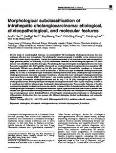
Morphology of cultured cells in the scaffold. Following injection of dispersed TK cells into the three‑dimensional mesh, the cells attached to the scaffold. Inoculated cells initially adhered to the material and then started to grow at the attached spots. Plating efficiency was not determined, as unattached cells did not remain attached to the mesh and instead diffused into the culture medium and were removed from further cultivation. After five days, proliferated cells were detected in clusters and the TK cells had aggregated and formed globoid structures (Fig. 1A). Fig. 1A obtained by phase contrast microscopy demonstrates the appearance of the cholangiocarcinoma
TK cell line and three‑dimensional cell culture. TK cells were cultured with RPMI‑1640 complete medium (Gibco Life Technologies Japan, Tokyo, Japan), supplemented with 15% fetal bovine serum (Lot no. SFB30‑1478, Equitech‑Bio, Kershville, TX, USA), 2 mM glutamine and 1 mM sodium pyruvate (Gibco Life Technologies Japan). The three‑dimensional culture method for the experiments was a modification of the method described by Mizuno et al (15). The scaffold material used for three‑dimensional culture was a
Morphological examinations Phase contrast microscopy. Cell attachment and proliferation in three-dimensional culture were observed with a phase contrast microscope (CK2, Olympus Corporation, Tokyo, Japan). The meshes were directly subjected to microscopy without fixing or staining during the culture. Light optical microscopy. Cells in the culture mesh were fixed with 10% phosphate‑buffered formalin and subjected to an automatic paraffin embedding system (ETP‑150CV, Sakura Finetek Japan Co., Ltd., Tokyo, Japan). Paraffin‑embedded specimens were sliced into 6‑µl‑thick sections using a microtome and stained with haematoxylin and eosin. These sections were examined under a light microscope (IX71, Olympus Corporation). Images were captured using a charge‑coupled device image sensor (VB‑7010, Keyence Japan, Osaka, Japan). Scanning electron microscopy (SEM). The cells attached to the mesh were fixed by treatment with 1.2% glutaraldehyde in 0.1 M phosphate buffer (pH 7.3, 400 mOsm). The specimens were dehydrated with a graded series of ethanol ranging from 50% through 70, 80, 90 and 100%. Following further treatment with 100% iso‑amylacetate, the samples were dried by a critical point dryer (Hitachi High‑Technologies Corporation, Tokyo, Japan) and sprayed with Au-Pd. Cells in the mesh were examined at 15 kV under a JSM‑5800LV scanning electron microscope (JEOL Ltd., Tokyo, Japan). Transmission electron microscopy (TEM). For transmission electron microscopy, the tissues were fixed with 2% glutaraldehyde in 0.1 M phosphate buffer and dehydrated by serial dilution of ethanol. Subsequent to treatment with a substituting agent, the tissues were infiltrated prior to polymerization in epoxy resin and sectioning with an ultramicrotome (Leica, Vienna, Austria). Ultra‑thin sections were further treated with uranyl acetate and lead citrate, and observed by the Hitachi H‑7500 transmission electron microscope (Hitachi High‑Technologies Corporation, Tokyo, Japan). Results
MOLECULAR MEDICINE REPORTS 9: 1359-1364, 2014
A
1361
B
C
Figure 1. Three‑dimensional culture of TK cells in the scaffold. (A) Appearance of the cholangiocarcinoma cells by phase contrast microscopy on day 5 of culture (magnification, x20). (B) Optical micrograph of the cholangiocarcinoma on day 14 of culture. TK cells aggregated and formed a duct‑like structure at x40 maginifcation and at (C) x100 magnification.
A
B
C
D
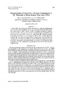
Figure 2. Scanning electron micrographs on day 10 of the three‑dimensional culture. (A) TK cells attached to the scaffold and proliferated (magnification, x400). (B) Cells aggregated and formed a globoid structure. Although the cells conglomerated and formed a balloon-like structure, the boundary of each cell was relatively clear. (magnification. x1,400). (C) The surface of the cells was covered with dense microvilli or sparse plicae (magnification, x2,700). (D) Although the cultured cells originated from the same TK cell line, the cells exhibited different morphologies at the surface (magnification, x7,500).
cluster in three‑dimensional culture. The structure grew relative to the culture duration. On day 14 of culture, light optical microscopy demonstrated that the TK cells had aggregated and formed duct‑like assemblies (Fig. 1B). At higher magnification, cells were observed to be filled with deposits of a secretory substance in the cytoplasm (Fig. 1C). These deposits were periodic acid schiff‑positive as demonstrated in
a previous study (13). Fig. 1C also demonstrated the organization of the attachment of the cell to the scaffold. SEM shows cell processes on TK cell surfaces. The cells aggregated and formed globoid shapes in the scaffold. The overall architecture was clearly demonstrated by scanning electron microscopy on day 10 of culture (Fig. 2A). The
1362
AKIYOSHI et al: MORPHOLOGICAL STUDY OF THE TK CHOLANGIOCARCINOMA CELL LINE
A
B
C
D
E
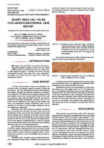
Figure 3. Transmission electron micrographs on day 7 of three‑dimensional culture. (A) A cross‑section of the structure. The extracellular space inside the structure is shown. (B) The surface of the structure is covered with microvilli and each cell exhibits a different electron density. (C) The cultured cell had a granular or duct-like structureand the lumen was covered with microvilli. (D) Higher magnification revealed that the structure was composed of more than one cell and a desmosome was observed between cells. (E) Attachment of the cell to the scaffold.
surface of the structure consisting of TK cells was covered with numerous floral‑shaped microvilli. Closer observation revealed that the pattern was not homogeneous, despite the cells originating from the same cell line (Fig. 2B). Higher magnification of another region of the culture demonstrated that certain cells possessed relatively sparse plicae whereas others possessed dense microvilli (Fig. 2C). These two types of processes were observed on the same globoid structure; however, were segregated and distributed differently. This was confirmed by examination at higher magnification (x7,500; Fig. 2D). TEM observation of the three‑dimensional culture. A cross-section of the globoid aggregate on day 7 of the three‑dimensional culture was observed by TEM (Fig. 3A), showing the internal arrangement of the cells to be semi‑irregular. Cells of varying electron‑density were observed in the microscopy image. Numerous microvilli were identified on the surface of the structure and also protruded into the extracellular space on the inner side of the aggregate. The cells exhibited irregularly‑shaped nuclei and the endoplasmic reticula and mitochondria were not well‑developed. On the outside of the structure, microvilli were observed only on the surface layer of the cells and distribution of the microvilli was demonstrated to be dense when observed under higher magnification (x5,000; Fig. 3B). When cultured three‑dimensionally, certain cells formed gland‑like structures and the lumen were covered by
microvilli (Fig. 3C). These structures consisted of multiple cells attached to each other by a cell adherent apparatus, such as a desmosome (Fig. 3D). The scaffold demonstrated bio‑adaptability and cultured cells attached to the scaffold via cell processes and/or microvilli (Fig. 3E). Discussion In this study, the established human TK cholangiocarcinoma cell line was cultivated three‑dimensionally. Morphological observations demonstrated characteristics of cholangiocarcinoma that are not observed by ordinary two‑dimensional culture. The observations of the morphological characteristics of the cultured TK cells add to the biochemical characteristics demonstrated in a previous study (13). Cholangiocarcinoma is one of the most intractable human diseases (23). The majority of cases are inoperable and only 30% of patients qualify for surgical treatment (24). While total numbers of patients are small compared with those with more common carcinomas of the colon or lung, the rising incidence (25) and high mortality rate (26) require the development of more effective therapeutic strategies. The median survival time is