FEBS 25415
FEBS Letters 508 (2001) 85^89
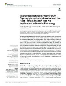
Multiple sites of interaction between the intracellular domains of an inwardly rectifying potassium channel, Kir6.2 Phillippa A. Jones, Stephen J. Tucker*, Frances M. Ashcroft University Laboratory of Physiology, Parks Road, Oxford OX1 3PT, UK Received 21 September 2001; accepted 4 October 2001 First published online 23 October 2001 Edited by Maurice Montal
Abstract The amino-terminal and carboxy-terminal domains of inwardly rectifying potassium channel (Kir) subunits are both intracellular. A direct physical interaction between these two domains is involved in the response of Kir channels to regulatory factors such as G-proteins, nucleotides and intracellular pH. We have previously mapped the region within the N-terminal domain of Kir6.2 that interacts with the C-terminus. In this study we use a similar in vitro protein^protein interaction assay to map the regions within the C-terminus which interact with the N-terminus. We find that multiple interaction domains exist within the C-terminus: CID1 (amino acids (aa) 279^323), CID2 (aa 214^222) and CID3 (aa 170^204). These domains correlate with regions previously identified as making important contributions to Kir channel assembly and function. The highly conserved nature of the C-terminus suggests that a similar association with the N-terminus may be a feature common to all members of the Kir family of potassium channels, and that it may be involved in gating of Kir channels by intracellular ligands. ß 2001 Published by Elsevier Science B.V. on behalf of the Federation of European Biochemical Societies. Key words: Inwardly rectifying; potassium channel; ATP-sensitive; Kir6.2; KATP
1. Introduction Inwardly rectifying potassium (Kir) channels are found in a wide variety of tissues and cell types where they regulate the resting membrane potential and transmembrane K £uxes. Fifteen Kir subunits have now been identi¢ed, comprising seven di¡erent subfamilies [1^3]. Some of these channels open and close (gate) spontaneously, whereas the gating of several subfamilies is tightly regulated by intracellular ligands. For example, Kir3.1 and Kir3.4 are opened by binding G-proteins, Kir6.2 is closed by binding ATP, and Kir1.1 and Kir4.1 are inhibited by protons [1^3]. All Kir channels investigated to date are activated by phosphatidylinositol bisphosphate (PIP2 ). Regulation of Kir channels by intracellular ligands is of major physiological signi¢cance. G-protein activation of IKACh (a heterotetramer of Kir3.1 and Kir3.4) mediates the slowing of the heart rate in response to vagal nerve stimula-
*Corresponding author. Fax: (44)-1865-272469. E-mail address:
[email protected] (S.J. Tucker).
tion [1^3]. The KATP channel of the pancreatic L-cell is formed by coassembly of Kir6.2 and the sulphonylurea receptor SUR1 (a member of the ABC-transporter superfamily) [4,5]. KATP channels are sensitive to intracellular adenine nucleotide concentrations and thereby couple the metabolic status of the cell to its electrical activity. In the L-cell, this provides the link between changes in blood glucose and insulin secretion [5]. In renal epithelial cells, the pH-sensitive channels Kir1.1 and Kir4.1^Kir5.1 are thought to play a key role in the pH-dependent regulation of K £uxes [6,7]. Kir subunits possess two transmembrane domains linked by a pore loop. The N- and C-termini are intracellular and contain the binding sites for intracellular ligands. Several recent studies have indicated that functional interactions between the N- and C-termini participate in ligand gating. Thus, a direct physical interaction between the N- and C-terminal domains of the G-protein-gated Kir3.0 subunits has been shown to enhance GLQ binding, supporting the idea that both domains contribute to the binding site for GLQ [8^10]. The ability of Kir1.1 and Kir2.3 to respond to changes in intracellular pH involves conformational changes in both the N- and C-termini [11,12]. Likewise, the pH-sensitivity of Kir1.1 is primarily de¢ned by the anomalous titration of a lysine residue within the N-terminus of Kir1.1, which results from its close proximity to two highly conserved arginine residues, one of which lies in the C-terminus [13]. This indicates that the N- and C-termini of Kir1.1 must be closely associated. Similarly, residues in both the N- and C-termini of Kir6.2 have been implicated in inhibition of the KATP channel by intracellular ATP [14,15]. Several studies have demonstrated that multiple regions in both the N- and C-termini are also involved in Kir channel assembly and subunit speci¢city [16^18]. Although interactions between the N- and C-termini of Kir channels are implicated in both their structural and functional organisation, direct biochemical evidence de¢ning these interactions is sparse. We have previously shown that the N- and C-termini of Kir6.2 physically associate [19], and that inhibition of the channel by ATP is in£uenced by residues in both of these domains [14,15]. We have mapped the N-terminal interaction domain and shown this is a highly conserved region within the proximal N-terminus [19]. In this study, we use a similar in vitro protein^protein interaction assay to map the regions within the C-terminus which physically interact with the N-terminus. We ¢nd three interaction domains, which lie within the proximal two-thirds of the C-terminus. Interestingly, these C-terminal interaction domains (CIDs) turn out to be important determinants of Kir channel assembly and gating.
0014-5793 / 01 / $20.00 ß 2001 Published by Elsevier Science B.V. on behalf of the Federation of European Biochemical Societies. PII: S 0 0 1 4 - 5 7 9 3 ( 0 1 ) 0 3 0 2 3 - X
FEBS 25415 5-11-01
86
P.A. Jones et al./FEBS Letters 508 (2001) 85^89
2. Materials and methods 2.1. Molecular biology Constructs were prepared as previously described [19]. C-terminal truncations of Kir6.2 were generated by PCR and subcloned in-frame between the EcoRI and SalI sites of the pET28a vector (Novagen). This vector directs protein expression under the control of the T7 promoter. In order to assist with detection of relatively short [35 S]methionine-labelled C-terminal proteins an additional two methionine residues were engineered into all constructs just before the stop codon. 2.2. Protein production The method was essentially that described previously [19]. Brie£y, N-terminal glutathione-S-transferase (GST) (6UHis) fusion constructs were transformed into the BL21(DE3) Escherichia coli strain, proteins were induced with 0.25 mM IPTG and cultures grown for 3^ 4 h at 20³C. Cultures were harvested by centrifugation, resuspended in bu¡er S (150 mM Tris pH 7.8, 50 mM NaCl, 25 mM imidazole, 1% NDSB-256, 0.5% CHAPS, 0.2% Tween 20), lysed by sonication and the insoluble material precipitated by centrifugation at 10 000Ug for 15 min. N-terminal fusion proteins were then puri¢ed from the supernatant on a Ni2 -agarose column and eluted with 100 mM EDTA. Constructs were synthesised using the TNT T7 quick-coupled transcription translation system (Promega) according to the manufacturer's instructions. After synthesis, the reaction was stopped by adding 500 Wl bu¡er S per 50 Wl reaction volume and insoluble material was precipitated by centrifugation at 100 000Ug for 30 min before use in the binding assay. 2.3. Binding assay In vitro binding assays were carried out in a 1.5 ml microcentrifuge tube by adding 20 Wl GST fusion protein (20 Wg), 10 Wl bovine serum albumin (10 mg/ml), 15 Wl glutathione-agarose beads (60% slurry; Pharmacia), 200 Wl bu¡er S and 250 Wl of the relevant radiolabelled C-terminus (prepared as above). Tubes were then mixed by constant rotation for 40 min at room temperature and the beads then washed three times for 15 min in 1 ml bu¡er S at room temperature. After the ¢nal wash, all supernatant was removed and the beads were resuspended in 15 Wl 2Uprotein sample bu¡er. A 10 Wl aliquot was then subjected to 10% SDS^PAGE and autoradiography. All binding assays were repeated at least four times and the ¢gures show representative examples of each construct tested.
Fig. 1. Mapping of CID1. Individual GST fusion proteins (as indicated) were tested for their ability to interact with di¡erent in vitro translated [35 S]Met-labelled Kir6.2 C-terminal fragments (aa 279^ 391, aa 295^391 and aa 279^323). GST-Con = GST alone, GSTNterm = Kir6.2 N-terminus residues 14^53. Due to the di¡erent sizes of each C-terminal fragment a sample of each in vitro translated [35 S]Met-labelled Kir6.2 C-terminal fragment (IVT Con) was run alongside to enable size comparison with the interacting C-terminal proteins.
Fig. 2. Mapping of CID2. Individual GST fusion proteins (as indicated) were tested for their ability to interact with di¡erent in vitro translated [35 S]Met-labelled Kir6.2 C-terminal fragments (aa 203^ 280, aa 214^280 and aa 222^280). GST-Con = GST alone, GSTNterm = Kir6.2 N-terminus residues 14^53. A sample of each in vitro translated [35 S]Met-labelled Kir6.2 C-terminal fragment was run alongside to enable size comparison (IVT Con).
3. Results We used an in vitro protein^protein interaction assay that is based upon the ability of recombinant GST fusion proteins to interact with in vitro translated proteins labelled with [35 S]methionine [19]. If the two proteins interact then the radiolabelled protein can be puri¢ed using glutathione Sepharose beads. We sought to determine those regions within the C-terminus which interact with the N-terminus by making serial truncations of the C-terminus of Kir6.2. Di¡erent length fragments of the C-terminus were generated by PCR and in vitro translated incorporating [35 S]methionine. They were then tested for their ability to physically associate with a GST fusion protein containing residues 14^53 of the N-terminus of Kir6.2. This region encompasses the highly conserved N-terminal interaction domain that is principally responsible for interacting with the C-terminus. We screened the entire
Fig. 3. Mapping of CID3. Individual GST fusion proteins (as indicated) were tested for their ability to interact with di¡erent in vitro translated [35 S]Met-labelled Kir6.2 C-terminal fragments (aa 170^ 214 and aa 170^204). GST-Con = GST alone, GST-Nterm = Kir6.2 N-terminus residues 14^53. A sample of each in vitro translated [35 S]Met-labelled Kir6.2 C-terminal fragment was run alongside to enable size comparison (IVT Con).
FEBS 25415 5-11-01
P.A. Jones et al./FEBS Letters 508 (2001) 85^89
87
Fig. 4. Identi¢cation of the CIDs. Schematic representation of the C-terminus of Kir6.2 and the C-terminal fragments tested: (Y) interacting; (N) non-interacting. Residues 170^204, 214^222 and 279^ 323 are critical for interaction with the N-terminus of Kir6.2. These domains are labelled as `C-terminal interaction domains' or CID1, CID2 and CID3, respectively.
C-terminus of Kir6.2, as we have previously shown that this domain (residues 170^391) speci¢cally associates with the N-terminal interaction domain [19]. Fig. 1 shows that removal of residues 170^278 from the
C-terminus does not a¡ect the ability of the remaining protein (amino acids (aa) 279^391) to bind to the N-terminus GST fusion protein, and that GST itself does not interact. However, truncation of a further 16 residues produces a protein (aa 295^391) which is unable to bind to the N-terminus. To narrow down this region further, a protein spanning residues 279^323 was generated and tested. Fig. 1 shows that this protein is capable of binding to the N-terminus. We call this region of Kir6.2 CID1. CID1 is not the only domain capable of interacting with the N-terminus. Fig. 2 shows that proteins spanning residues 203^ 280 and 214^280 are also capable of interacting, but that deletion of a further 8 aa produces a protein that will no longer bind (aa 222^280). This suggests that there is a second region contained within this area (aa 214^222 or CID2) that either interacts directly with the N-terminus, or is necessary for the interaction of CID2. Further examination of the proximal C-terminus reveals that residues 170^214 and 170^204 are also capable of binding to the N-terminus, suggesting that a third interaction domain is contained within this region (Fig. 3). Attempts to further narrow down this region were unsuccessful because the resulting truncated proteins were too small to detect reproducibly (not shown). Fig. 4 is a schematic representation which summarises our results. There appear to be three main `interaction domains'
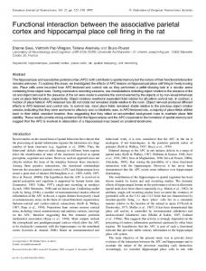
Fig. 5. Highly conserved nature of the interaction domains. The C-termini of di¡erent Kir subunits were aligned as indicated. Areas of identity are shaded dark grey and areas of similarity light grey. Residue numbers refer to Kir6.2. The relative positions of the CIDs are indicated by solid black lines above the alignment.
FEBS 25415 5-11-01
88
P.A. Jones et al./FEBS Letters 508 (2001) 85^89
lying within the proximal two-thirds of the C-terminus of Kir6.2 that are involved in interaction with the N-terminus. These regions encompass aa 279^323, aa 214^222 and aa 170^ 204 and we have called them CID1, CID2 and CID3, respectively. The distal C-terminus does not appear to contribute to binding. Fig. 5 shows that the three CIDs are in highly conserved regions of the C-terminus. 4. Discussion Our results de¢ne three structural elements within the C-terminus of the inwardly rectifying K channel Kir6.2 which determine its physical association with the N-terminus. The presence of multiple interaction sites is in agreement with earlier studies that have implicated multiple regions within the N- and C-termini in homo- and heteromeric assembly of Kir channels [16^18,20]. In contrast, a single common structural motif appears to de¢ne assembly of Kv channels [16,20]. The interaction domains we describe will not only facilitate structural associations between the N- and C-termini, but may also participate in functional interactions such as ligand binding and gating. Indeed, there is evidence that ligand binding and gating of several Kir channel family members involves contributions from both N- and C-termini. 4.1. The interaction domains of Kir6.2 The proximal interaction domain (CID3, aa 170^204) of Kir6.2 contains residues that have been shown to be involved in ATP inhibition (K185) [14,21], PIP2 binding (R176, R177) [22^25], pH-sensitivity (H175) [26] and channel gating (T171) [14]. Thus it appears to be of key importance in the regulation of channel function by intracellular ligands. CID2 and CID1 also contain residues which have been associated with PIP2 activation (K222, R301 and R314) [25]. Although the region de¢ned as CID2 is not as highly conserved between Kir subunits as CID1 and CID3 it is worth noting that it is directly adjacent to a highly conserved segment (P226^Q235, Fig. 5). It is therefore possible that whilst deletion of aa 214^222 (CID2) abolishes binding of the aa 222^280 fragment (Fig. 4) it is not actually the deleted residues which directly interact with the N-terminus, but rather those adjacent to the deleted segment. Attempts to map the CID2 domain further and to reconstitute binding with a smaller protein encompassing this region were unsuccessful due to the small size of the in vitro translated products (not shown). There is now growing evidence that an interaction between the N- and C-termini is critical for inhibition of Kir6.2 by ATP. First, mutations that a¡ect the channel ATP-sensitivity are found in both of these regions and lie in two main clusters, between residues 46^51 and 179^186 [14]. Second, mutations in both the N-terminus (R50G) and C-terminus (K185Q) of Kir6.2 have been shown to reduce the photoa¤nity labelling of Kir6.2 by 8-azido-ATP [15]. These results suggest that either residues in both the N- and C-termini contribute to inhibitory ATP binding site, or that the ATP binding site lies solely in one of these regions but is allosterically regulated by physical association with the other. The interaction domains we have identi¢ed are therefore likely to provide the structural framework for such functional interactions. 4.2. Relevance to other Kir channels Fig. 5 shows that the interaction domains of Kir6.2, in
particular CID1 and CID3, lie within regions of the C-terminus that are conserved between Kir channels. It is therefore likely that similar interaction domains are to be found in other Kir channels. Indeed, there is signi¢cant evidence to support an important role for these domains in many other Kir channels. The regions of Kir2.1 equivalent to CID2 and CID3 in Kir6.2 have been found to be involved in both subunit assembly and heteromeric compatibility of this channel [16]. Interestingly, these authors also found that C-terminal deletions of more than 95 residues caused a loss of biochemical association and functional expression. The equivalent truncation limit corresponds to D323 in Kir6.2 and truncations beyond this region would disrupt the interaction of CID1 (aa 279^323) with the N-terminus. Thus, these results suggest that participation of the CIDs and NID in subunit assembly may be a common feature of Kir channels. There is also evidence that interaction domains equivalent to those we describe are involved in gating of other Kir channels. Schulte et al. identi¢ed two highly conserved arginine residues in Kir1.1 that are responsible for the anomalous titration of a lysine residue in the N-terminus [13]. One of these arginines is the equivalent of R34 in Kir6.2, which is contained within the N-terminal interaction domain (aa 29^46), whilst the other (R301) lies within CID1 (aa 279^323). Likewise, GLQ interactions with Kir3.0 subunits involve both the N- and C-terminus. Biochemical studies have identi¢ed residues within CID3 as minimally necessary for GLQ binding by Kir3.1 (aa 318^462 of Kir3.1) [8]. A residue within this region (L333) was also critical for agonist-induced sensitivity of Kir3.4 to GLQ subunits [27]. Other studies suggest the interaction between these two domains is state-dependent. For example, in Kir1.1, several cysteine residues in the intracellular domains are accessible to thiol-reactive reagents only in the closed state and not in the open state [11]. Interestingly, two of these cysteines (C80 and C303) lie within the equivalents of the N-terminal interaction domain (aa 24^53; C80) and CID1 (aa 279^323; C303) of Kir6.2. Furthermore, Qu et al. have shown that the physical association between the N- and C-termini of Kir2.3 can be in£uenced by protons [12]. The region responsible for the pH-dependent interaction they identi¢ed (aa 196^230 of Kir2.3) encompasses CID2 of Kir6.2 (aa 214^222). As is the case for Kir6.2, the proximal interaction domain (CID3, aa 170^204) of other Kir channels also plays a key role in the modulation of channel activity by intracellular ligands. For example, CID3 is identical to one of the PIP2 binding sites identi¢ed in Kir2.1 [28]. Indeed, the three PIP2 binding regions identi¢ed in Kir2.1 correspond to domains 163^194, 195^234 and 313^354 of Kir6.2 and thus overlap with the three CIDs identi¢ed in this study. Residues equivalent to R176 and R177 have also been shown to contribute to PIP2 binding in other Kir channels [22^24] 4.3. Conclusion It is tempting to speculate that the physical interactions between the N- and C-termini we describe here are not simply static but also possess dynamic properties. Indeed, they may contribute to a common Kir channel gating mechanism that has been di¡erentially adapted to respond to a variety of intracellular signals such as ATP, G-proteins, pH and protein kinases. However, further studies will be necessary to determine precisely how this is achieved.
FEBS 25415 5-11-01
P.A. Jones et al./FEBS Letters 508 (2001) 85^89
89
Acknowledgements: This work was supported by the Wellcome Trust and The Royal Society. S.J.T. is a Royal Society University Research Fellow. F.M.A. is the Royal Society GlaxoSmithKline Research Professor. We also gratefully acknowledge the assistance of Heinz Neumann during the initial stages of the project.
References [1] Reimann, F. and Ashcroft, F.M. (1999) Curr. Opin. Cell Biol. 11, 503^508. [2] Jan, L.Y. and Jan, Y.N. (1997) Annu. Rev. Neurosci. 20, 91^ 123. [3] Jan, L.Y. and Jan, Y.N. (1997) J. Physiol. 505, 267^282. [4] Inagaki, N., Gonoi, T., Clement, J.P., Namba, N., Inazawa, J., Gonzalez, G., Aguilar-Bryan, L., Seino, S. and Bryan, J. (1995) Science 270, 1166^1170. [5] Seino, S. (1999) Annu. Rev. Physiol. 61, 337^362. [6] Tucker, S.J., Imbrici, P., Salvatore, L. and Pessia, M. (2000) J. Biol. Chem. 275, 16404^16407. [7] Wang, W., Hebert, S.C. and Giebisch, G. (1997) Annu. Rev. Physiol. 59, 413^436. [8] Huang, C.L., Jan, Y.N. and Jan, L.Y. (1997) FEBS Lett. 405, 291^298. [9] Slesinger, P.A., Reuveny, E., Jan, Y.N. and Jan, L.Y. (1995) Neuron 15, 1145^1156. [10] Tucker, S.J., Pessia, M. and Adelman, J.P. (1996) Am. J. Physiol. 271, H379^H385. [11] Schulte, U., Hahn, H., Wiesinger, H., Ruppersberg, J.P. and Fakler, B. (1998) J. Biol. Chem. 273, 34575^34579. [12] Qu, Z.Q., Yang, Z.J., Cui, N.R., Zhu, G.Y., Liu, C.X., Xu, H.X., Chanchevalap, S., Shen, W.Z., Wu, J.P., Li, Y.J. and Jiang, C. (2000) J. Biol. Chem. 275, 31573^31580.
[13] Schulte, U., Hahn, H., Konrad, M., Jeck, N., Derst, C., Wild, K., Weidemann, S., Ruppersberg, J.P., Fakler, B. and Ludwig, J. (1999) Proc. Natl. Acad. Sci. USA 96, 15298^15303. [14] Tucker, S.J., Gribble, F.M., Proks, P., Trapp, S., Ryder, T.J., Haug, T., Reimann, F. and Ashcroft, F.M. (1998) EMBO J. 17, 3290^3296. [15] Tanabe, K., Tucker, S.J., Matsuo, M., Proks, P., Ashcroft, F.M., Seino, S., Amachi, T. and Ueda, K. (1999) J. Biol. Chem. 274, 3931^3933. [16] Tinker, A., Jan, Y.N. and Jan, L.Y. (1996) Cell 87, 857^868. [17] Fink, M., Duprat, F., Heurteaux, C., Lesage, F., Romey, G., Barhanin, J. and Lazdunski, M. (1996) FEBS Lett. 378, 64^68. [18] Koster, J.C., Bentle, K.A., Nichols, C.G. and Ho, K. (1998) Biophys. J. 74, 1821^1829. [19] Tucker, S.J. and Ashcroft, F.M. (1999) J. Biol. Chem. 274, 33393^33397. [20] Tinker, A. and Jan, L.Y. (1999) Curr. Top. Memb. 46, 143^158. [21] Reimann, F., Ryder, T.J., Tucker, S.J. and Ashcroft, F.M. (1999) J. Physiol. 520, 661^691. [22] Baukrowitz, T., Schulte, U., Oliver, D., Herlitze, S., Krauter, T., Tucker, S.J., Ruppersberg, J.P. and Fakler, B. (1998) Science 282, 1141. [23] Shyng, S.L. and Nichols, S.G. (1998) Science 282, 1138^1141. [24] Fan, Z. and Makielski, J.C. (1997) J. Biol. Chem. 272, 5388^ 5395. [25] Shyng, S.L., Cukras, C.A., Harwood, J. and Nichols, C.G. (2000) J. Gen. Physiol. 116, 599^607. [26] Xu, H., Cui, N., Yang, Z., Wu, J., Giwa, L., Abdulkadir, L., Sharma, P. and Jiang, C. (2001) Biol. Chem. 276, 12898^12902. [27] He, C., Zhang, H.L., Mirshahi, T. and Logothetis, D.E. (1999) J. Biol. Chem. 274, 12517^12524. [28] Soom, M., Schonherr, R., Kubo, Y., Kirsch, C., Klinger, R. and Heinemann, S.H. (2001) FEBS Lett. 490, 49^53.
FEBS 25415 5-11-01