Constructions containing the Drosophila white gene and dif- ferent amounts and ...... white in the developing eye of the pupa and, according to. Bingham and ...
The EMBO Journal vol.4 no.13A pp.3501 -3508, 1985
Multiple upstream regulatory elements control the expression of the Drosophila white gene
V.Pirrottal, H.Steller2 and M.P.Bozzetti3 Europe Molecular Biology Laboratory, Postfach 10.2209, D-6900 Heidelberg, FRG, and 'Department of Cell Biology, Baylor College of Medicine, One Baylor Plaza, Houston, TX 77030, USA
2Present address: Department of Biochemistry, University of California at Berkeley, Berkeley, CA 94720, USA 3Present address: Istituto di Genetica, Universita degli Studi, Via G.Amendola 165a, 70126 Bari, Italy Communicated by V.Pirrotta
Constructions containing the Drosophila white gene and different amounts and arrangements of its regulatory region were introduced into the germ line of white mutant flies by P-mediated transformation. The results obtained with the different transposon constructions show that different parts of the 1.8-kb region preceding the transcription start are required for the expression of the gene in different tissues and at different developmental stages. Different sequences independently control the expression of the gene in the adult testes, in the larval and adult Malpighian tubules and in the eye. Another sequence located > 1080 bp upstream of the transcription start is the target of zeste interaction. The results also suggest that sequences required for dosage compensation are contained between -216 and the transcription start site. We show that at least some of these regulatory elements are equally functional if their distance from the promoter is varied or if their orientation is inverted. Their properties suggest that they act as enhancer-like elements to regulate the activity of the white promoter and, at least in the case of the zeste regulatory site, that they can act also in 'trans' on a white promoter locked in close physical proximity by homologous chromosome pairing. Key words: tissue-specific enhancers/zeste interaction/dosage compensation/P-transposons
Introduction The activity of the white gene of Drosophila is necessary for the deposition of pigment in several body structures of the larva and adult fly. White-dependent pigmentation is found in the larval Malpighian tubules, in the adult Malpighian tubules, in the adult testes, ocelli and, most conspicuously, in the adult eyes. Although no other structures or activities are known to depend on white expression, the possibility of its participation in other non-essential functions cannot be excluded. As is apparent from the catalogue of pigmented structures, the white gene must be expressed in a variety of different tissues and at different stages in the life cycle. Pigmentation of the larval Malpighian tubules is detectable by the second instar, but eye pigmentation begins to be laid down in the early pupa while the testes sheaths only become pigmented several days after eclosion. Fjose et al. (1984) have recently shown that at least some of this tissue and developmental specificity is reflected by the pattern of in situ hybridisation to RNA in thin sections of emIRL Press Limited, Oxford, England
bryos and larvae. White RNA begins to be detectable in 10 h embryos in the primordia from which the Malpighian tubules will develop. A second spurt of white activity was observed in the eye-antenna disc and in the subpharyngeal ganglion of late third instar larvae. The gene is therefore regulated both with respect to the developmental time and to the tissues or structures in which it is expressed. It is likely that this regulatory complexity is reflected in the complexity of the DNA region immediately upstream of the white gene. We know that the upstream region is also responsible for two other regulatory aspects. (i) Dosage compensation by which white, an X chromosome gene, has the same overall expression in females (two copies) as in males (one copy). (ii) Interaction with the zeste locus, whereby the z1 mutation causes two homologously paired copies of the white gene to be specifically underexpressed with respect to two unpaired copies or a single copy of white (Gans, 1953). This underexpression is apparently tissue specific: it decreases eye pigmentation by >90%, but it has little effect on the pigmentation of the ocelli or testes. Using P-mediated gene transfer, transposons containing the vvhite gene and three or more kilobases of its 5'-flanking region have been reintroduced in various genomic sites (Hazelrigg et al., 1984; Gehring et al., 1984). The transformed flies showed, in general, that the expression of the white gene was both quantitatively and developmentally correct. The gene was also dosage compensated and interacted with zeste, indicating that these transposons included all the relevant control sequences. The simplest explanation for the various regulatory effects exhibited by the white gene is that they are executed by specific cis-acting regulatory elements that modulate the rate of transcription of the gene. To test this hypothesis and to identify the sequence elements involved in the different regulatory aspects, we altered the white gene and reintroduced it in the germ line of the fly by P-mediated transformation (Rubin and Spradling, 1982; Spradling and Rubin, 1982). In another paper we studied the effects of removing the promoter and regulatory region of the gene and substituting it with the hps-70 heat-shock promoter (Steller and Pirrotta, 1985b). In the work reported here, we constructed transposons lacking different parts of the region immediately preceding the white promoter and/or in which most of the major intron had been deleted. The performance of these transposons when reintroduced in the fly shows the existence of multiple, independent regulatory elements in the 5'-flanking region of the gene, responsible for different regulatory aspects. Our results agree with a similar study by Levis et al. (1985b) related in the accompanying paper.
Results Figure 1 shows the structure of a series of transposons containing the white gene with various amounts of the 5'-flanking region in various arrangements. These constructions were assembled in the Carnegie-I vector (Rubin and Spradling, 1983) except for BmA-w which utilised the pUChsneo vector (Steller and Pirrotta, 1985a). When reintroduced into the Drosophila genome, all 3501
V.Pirrotta, H.Steller and M.P. Bozzetti -1000
0
+1000
+ 2000
+ 3000
.
H2
WSPI
Table I. Ps
rgIsm
RV
Start
.1
H3
Sm
I
T Z Mt E DC
HPst-W PstH-W
HBgZ-W B--W Pst-W
-BBmA-W )- --(
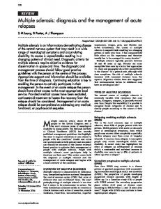
Fig. 1. Map of the proximal part of the white
)-
+ + + -
+ + + n.t.
+ + + + + + + + + + + + + + + +++
HABgRVA-W + + - + + B4hsp-W + - + + -
and of the transposons used. The black boxes indicate the first and part of the second exon connected by the major intron. The coordinate origin is taken at the site of transcription initiation (Steller and Pirrotta, 1985b) which corresponds approximately to position +3692 in the sequence of O'Hare et al. (1984). Restriction sites referred to in the text are indicated as H2: Hindll; Bg: BglII, Ps: PstI, Bm: BamHI, RV: EcoRV, H3: HindlII, Xb: XbaI. The position of the wsP1 insertion is also indicated. The structure of the white gene in the transposons used is shown below with arrows indicating the orientation of the Hindlfl-PstI segment. The HBgZ-w transposon contains an insertion of 670 bp from the bacterial lacZ gene. Parentheses indicate sequences deleted and hsp stands for the hsp-70 heat-shock promoter fused to position +10 of the transcribed sequence (Steller and Pirrotta, in preparation). The symbols on the right tabulate the activity of the different constructions in the testes (T), in the eye (E), in the Malpighian tubules (MT), the ability to interact with zeste (Z) and dosage compensation (DC). gene
of them expressed white gene function, as detected by eye pigmentation, but, for a given transposon, to different degrees depending on the site of integration. In general, eye pigmentation was stronger and less variable from one transformed line to another, with transposons containing the 5'-flanking region upstream of position -1081. The same effect was noted if this upstream sequence was inverted with respect to the promoter (PstH-w) or placed further apart by the insertion of 670 bp of foreign DNA (HBg Z-w). Different levels of pigmentation in newly eclosed flies raised under the same conditions can only be explained by different levels of activity of the white gene in the different lines. However, pigment concentration apparently responds in a non-linear way to the expression of the gene. Clearly, at high levels of activity, the eye reaches a maximum level of pigmentation. Lower gene activities result in intermediate levels of pigmentation, but the relative amounts of different eye pigments also vary, resulting in brown coloration in some lines but orange-red in others. Both the red (drosopterins) and the brown (ommochromes) whitedependent pigments are deposited, although in different proportions in the different lines (Figure 2). The variable response to the presumed level of white activity is illustrated by the effect of gene dosage. Table I shows the pigment concentration, expressed in percentage of wild-type, of flies containing one copy of the transposon and flies homozygous for that transposon. Pigment levels are not only highly non-linear with gene dosage, but they also respond differently in different lines transformed with the same transposon, suggesting that the effect of homozygosity on white gene expression can vary from one chromosomal site to another. In some of the earlier transformation experiments, the helper P element used to contribute transposase function for the integration of the white transposon was itself able to integrate. In consequence, some of the transformed lines obtained contained P elements and were therefore unstable for some generations. Among the offspring produced during this time, we obtained flies in which the white transposon has been mobilised and occupied 3502
Pigment assays of selected
mutants and
transformed lines
Xb
Strain
Bg-w Bg-w Bg-w Bg-w Bg-w Bg-w Bg-w z
ZIp6
41p1 w"
Het. o0
line line line line line line line
5A 5B
IOC 13A
131 13R 16B
Hom. oa
24.7 i 0.5 Lethal 19.5 ± 1 81.2 ± 11.7 ± 0.5 19.5 ± 1.2 1 22.1 ± 1 69.5 ± 4.5 ± 0.5 Lethal 3.2 ± 0.5 23.4 4 4O 105 5 5.2 + 0.7 9.1 ± 0.7 2.4 ± 0.3
3 3 1
2
Het. 9
Hom. 9
5.8 7.8 3.6 1.3 9.1 4.5 1.3
Lethal 74.7 ± 2 25.3 ± 2
1 ± 0.5 ± 0.5 0.5 ± 1 ± 0.5 ± 0.3
-
66.2 Lethal 0.4 ± 4.1 4.6 ± 5.7 ± 2.7
I1
0.5 I1 0.7 0.3 0.3
Pigment concentration is expressed is percentage of the value for Canton S flies. The values given for the zl, Z'P6, WP1 and vw males are of course for the hemizygous condition. The Bg-w lines all contained autosomal insertions. The values were averaged from three assays each using 10 flies. new sites. In some of these cases the white gene at the new chromosomal location gave rise to a gradient of pigmentation in the eye: in three cases, the anterior part of the eye was pigmented while the posterior part was progressively whiter. In the fourth case the posterior border of the eye was strongly pigmented while the rest of the eye was white.
Dosage compensation All the transposons shown in Figure 1, with the exception of B4
hsp-w respond visibly to dosage compensation: males with one copy of the transposon have a higher level of pigmentation than
females with one copy when inserted into the X chromosome in autosomal sites. However, the site of integration affects the degree of dosage compensation, as illustrated quantitatively in Table I for a series of lines containing the Bg-w transposon in autosomal sites. As measured by pigment quantity, most lines exhibit various degrees of overcompensation. In this respect and in others, they resemble w5P mutants, in which the 5'-untranscribed flanking sequences contain insertions or deletions. We call it overcompensation because males with one copy produce more than twice as much pigment as females with one copy of the transposon but, in another sense, the males are undercompensated because, in all our lines, a male with one copy is significantly less pigmented than a female with two copies. Part of this effect may be due to a non-linear response of pigmentation to the rate of transcription, but other flanking sequences may influence the mechanism that stimulates transcription of X chromosome genes in the male. Replacement of the entire promoter and upstream sequences causes a loss of dosage compensation. In the B4 hsp-w transposon the hsp-70 heat-shock promoter has been fused to the beginning of the white untranslated leader region (Steller and Pirrotta, 1985b). Although lines transformed with this transposon are strongly pigmented even when raised at room temperature, gene dosage effects are noticeable since individuals with one copy of the transposon are detectably less pigmented than flies with two copies. In these lines, however, both males and females carrying an autosomally integrated transposon have the same degree of pigmentation, corresponding to 75-80% of wild-type levels. This finding argues against the presence of sequences responsible for dosage compensation in the transcribed part of the gene. Furthermore, since transposons like Bg-w and HABgRVA-w are both dosage compensated, we should expect to find the sequences responsible in the 216-bp region between the EcoRV site and the start of transcription. or
Multiple regulatory elements of the white gene
a
b
e
f
d
9
h
s
t
r.
q
r
this was the host used in all transformation experiments; (b) Bg-w line 13A, heterozygous 9; Fig. 2. Eye color of selected transformed lines: (a) y V7123 (c) Bg-2 line 13A, heterozygous a; (d) y w+ wild-type control; (e) Bg-w line 16A, heterozygous a; (f) Bg-w line 16A, homozygous o; (g) Bg-w line 16A heterozygous 9; (h) Bg-w line 16A homozygous 9; (q) Pst-w, heterozygous a; (r) Pst-w homozygous a; (s) Pst-w heterozygous 9; (t) Pst-w homozygous 9. -
Fig. 3. Testes pigmentation. Testes and Malpighian tubules (d) PstH-w line 5; (e) HABgRVA-w line o-2.
were
dissected from 10-day-old male flies. (A) y wv7"23; (b) Canton S; (c) Bg-w line 16A;
Fig. 4. Zeste interaction. Different lines transformed with the HABgRVA-w transposon show different levels of eye pigmentation and different degrees of interaction with zeste. In each case, the picture shows a male with one autosomal copy of the transposon (right) and a male from the same line with one copy of the transposon and the e°P6 wsn chromosome. (a) line 9-1; (b) line 9 -P; (c) line o-2; (d) line 9-4. 3503
V.Pirrotta, H.Steller and M.P. Bozzetti
I
Fig. 5. W1ite-dependent fluorescence of Malpighian tubules. Malpighian tubules dissected from flies transformed with different transposons were photographed with a 10 x phase contrast neofluar objective. In each case, the upper picture was taken with white light, the lower with fluorescence optics. (a) y w67C23; (b) BmA-w; (c) PstH-w; (d) HABgRVA-w line 9-2; (e) Canton S.
Testes pigmentation We examined our transformed flies and a number of other mutants for their degree of testes pigmentation in the adult male. This pigmentation develops gradually over several days after eclosion and clearly corresponds to a different tissue- and developmentally specific activity of the white gene. In sharp contrast with eye pigmentation, it is not affected by zeste since z1 males and particularly Z0p6 males have strongly pigmented testes under our conditions. Mutations of the WsP class affect the white regulatory region and strongly decrease eye pigmentation but have different effects on the testes. We found that the wsP3 mutation, a deletion starting approximately at -900 and removing sequences further upstream (Davison et al., 1985), gives unpigmented testes. In contrast, wsP1 an insertion at -1229 (O'Hare et al., 1984) and WsP2, a small deletion from - 1181 to -1292 (Davison et al., 1985) both appear to enhance testes pigmentation. It is possible that in these cases pigment precursors synthesised elsewhere in the body and not utilized in the eye accumulate and result in stronger testes pigmentation. However, the quantitative aspects of testes pigmentation are difficult to establish for lack of an assay other than visual inspection and because of the influence of diet, age, temperature and environmental conditions. When our transformed lines were examined qualitatively we found that none of the transposons lacking the region upstream of the BglII site (position -1081) conferred testes pigmentation even if the eye color was strong and approaching wild-type. In contrast, all transposons that include the region upstream of the BglH site in either orientation produced testes pigmentation even when the eye color was much lighter than wild-type (Figure 3). For a given transposon of this class there was, however, some correlation between the intensity of eye pigmentation and that of the testes: lines with ligher eye color also had more lightly pigmented testes. These observations suggest that a regulatory sequence upstream of position -1081 is required to activate the expression of the white gene in the testis sheath but is not essential for its function in the eye. As might be expected, the B4 hsp-w transposon confers testes pigmentation. Without heat shock, the coloration is weak and develops only several days after eclosion. Heat shock in the pupal stage causes earlier appearance of pigmentation. Zeste interaction The normal white gene responds to the presence of the z1 mutation by greatly decreasing its phenotypic expression in the eyes if two homologously paired copies of white are present. Since 3504
white is in the X chromosome, z1 females have yellow eyes while z1 males have red eyes. Two kinds of observations suggested that the target of the zeste effect is the proximal part of the white locus. One is that a tandem duplication of the proximal part of white renders the intact white locus sensitive to zeste even in the male (Green, 1961; Judd, 1961). The other is that mutations of the wsP class, which alters the untranscribed proximal part of the gene, render it insensitive to zeste (Gans, 1953; Green, 1959). We tested our transposons for their ability to interact with zeste using one or both of two methods. One was to introduce the zlwllE4 chromosome in transformed lines made homozygous for an autosomally integrated transposon. The other and more rapid method utilised the z0p6 mutant. This mutant, isolated by Lifschytz and Green (1984) starting from a z1 chromosome, contains a second mutation in the zeste gene that renders it able to interact even with a single copy of white. The eye color of flies with the constitution z1 wllE4/zl w"lE4, T/T was compared with that of z+ w67c231Z+ w67c23, T/T and Zop6 w/Y, T/+ flies were compared with z+ w67C23, T/+ where T represents the transposon. In all cases in which both were carried out on the same line, the two tests gave equivalent results. Zeste interaction, revealed by a ligher eye color was never observed with any of the 14 lines carrying transposons lacking the region upstream of the BglII site at position -1081. For all transposons containing this upstream region, at least one transformed line could be found that interacted with zeste. The interaction was, in many cases, much weaker than that observed in wild-type flies and five of a total of 16 lines did not give a visible interaction. Notably, most of the lines transformed with HABgRVA-w interacted with zeste, including some with relatively light eye pigmentation (Figure 4), while strongly pigmented lines transformed with Bg-w or with B4 hsp-w did not. We conclude that, independently of the strength of expression in the eye, the target of the zeste interaction resides in the region - 1850 to -1081, that more proximal sequences, at least between -1081 and -216, have little effect but that sequences further downstream of -1850 may contribute to the strength of the zeste effect. Furthermore, the segment -1850 to -1051 is equally capable of promoting the zeste effect in either orientation (transposons HPst-w2 and PstH-w25) or when placed at a greater distance from the rest of the gene by the insertion of 760 bp of foreign DNA (transposon HBgZ-w) or at a shorter distance from the promoter (transposon HABgRVA-w). Malpighian tubule pigmentation Both adult and larval Malpighian tubules of the different
Multiple regulatory elements of the white gene
transformed lines were examined for white-dependent pigmentation. This pigmentation is easily detected by eye in the cases in which the white gene is strongly expressed but is less obvious when the expression is weak. To detect more reliably even low levels of pigmentation, we made use of the fact that the whitedependent pigments fluoresce strongly while the unpigmented tubules have very low background fluorescence (Figure 5). The results, summarised in Figure 1, show that the ability to accumulate pigment in the Malpighian tubules is best correlated not with the intensity of eye pigmentation but with the presence in the transposon of the region -743 to -216. The Bg-w transposon, containing the - 1081 to 0 region and Pst-w (-836 to 0) both produce pigmented tubules while transposon HABgRVA-w does not, even in those lines that express white strongly in the eye and testes. HABgRVA-w contains in addition a deletion of 2757 nucleotides, removing most of the first intron of the white gene. To determine whether the activity in Malpighian tubules is dependent on intron sequences, we examined transposon BmA-w, which has the same intron deletion but includes the -743 to 0 region. Unfortunately, the only line transformed with Bm-w that we obtained expressed weakly even in the eye (yellow-orange eye color). Nevertheless, tubule pigmentation was clearly detectable and was confirmed by fluorescence analysis (Figure 5). These results indicate that expression of the white gene in the Malpighian tubules requires a specific regulatory sequence that is not necessary for expression in the eye or in the testes. In all cases, the larval and adult tubules behaved exactly the same, suggesting that the same region and probably the same sequence is involved in directing gene expression in the tubules at both stages. Discussion In summary, the results show that the 5'-flanking sequences of the white gene contain a complex regulatory region. The fact that at least part of this region has the same regulatory effect in either orientation shows that it does not contain previously undetected tissue-specific promoters but rather that it regulates the activity of a single promoter. Elements of this regulatory region affect different properties and can be separated from one another functionally. The interval - 1850 to -1081 contains determinants necessary for the expression of the gene in the adult testes and for interaction with zeste. These two are functionally distinct since mutations like wsPl and WsP2 eliminate zeste interaction but not testes-specific expression. In particular, VoP2 indicates that the sequences necessary for zeste interaction lie partly or entirely in the interval - 1181 to -1292, while those required for testes pigmentation are either between -1081 and - 1181 or between -1292 and -1850. The interval - 1081 to -216 contains determinants required for the expression in the larval and adult Malpighian tubules. None of the constructions we have examined discriminates between the larval and adult Malpighian tubule pigmentation and it is likely that the same sequence is involved in both cases. In the accompanying paper, Levis et al. show that transposons containing only up to -400 of the regulatory region are still able to express the white gene in the Malpighian tubules. Combined with our results, this indicates that the sequences responsible reside in the interval from -400 to +216. According to Davison et al. (1985), the interval -960 to -600 also includes another regulatory site that responds to the su (wsP) + allele causing a depression of white RNA levels in the adult head tissues. All of our transposons express the white gene in the eye. While those transposons cloned in the pUChsneo vector were identified
by the independent criterion of G418 resistance, most of the construction made use of the Carnegie 1 vector and were therefore selected for eye pigmentation. Nevertheless, the fact that pigmented transformants could be found for all the constructions indicates that none of the deletions or rearrangements systematically abolishes expression in the eye. These results imply either that determinants necessary for expression in the eye pigment cells are localised in the interval -216 to 0 or, alternatively, that they are multiple and present in more than one interval. Another possibility that cannot be entirely excluded is that eye-specific expression depends on regulatory elements present in the transcribed part of the gene or is stimulated by the P element sequences present in the transposon. Position effects Changes in the level of gene activity or its distribution within a tissue are known to derive from position effects. When a normally euchromatic gene such as white is transposed to the vicinity of heterochromatin, its expression can be depressed to different extents in different cells of a tissue and their clonal descendants (Becker, 1960; Spofford, 1976). Some of our transformant lines, under dysgenic conditions, frequently gave rise to non-uniform eye phenotypes. These were not of the spotty or clonal variety but rather in the form of a gradient of pigmentation. In several independent cases, pigmentation increased from the posterior to the anterior border of the eye. In one extreme case, most of the eye was white with only a few pigmented facets at the anterior edge. Similar cases have been observed by Hazelrigg et al. (1984) and Levis et al. (1985a). The reverse gradient is also possible. One variant, originating from a HABgRVA-w line, had strongly pigmented facets at the posterior edge of the eye, fading off rapidly in the middle and anterior portions. These gradient distributions are clearly not clonal but rather related to the antero-posterior position of facets in the compound eye, suggesting that the activity of the gene is controlled either by positional information along this axis or by other events that occur progressively along it. One possible interpretation is that the gradient reflects a narrowed time specificity of the expression of the gene. Steller and Pirrotta (1985b), using a heat shock-dependent white gene, have shown that eye pigmentation requires white expression during a fairly narrow time window in the first to second day of pupation. In the development of the eye, cell patterning proceeds along a sharply defined boundary, a dorso-ventral morphogenetic furrow that moves from the posterior to the anterior border of the eye imaginal disc (Ready et al., 1976; Campos-Ortega and Hofbauer, 1977). It is possible therefore that the time window for pigment deposition in the eye pigment cells also occurs in a temporal gradient from posterior to anterior. If the activity of the white gene is shut off prematurely, this would result in pigmentation only of the earliest maturing cells, in the posterior region of the eye. If, on the other hand, the white gene is activated late, only the later maturing cells in the anterior portion of the eye would be pigmented. Dosage compensation Like many, if not all, X chromosome genes the white gene is dosage compensated so that one copy of the gene in males is expressed approximately twice as much as one copy in females. Evidence that this compensation occurs at the transcriptional level comes from the rates of [3H]uridine incorporation measured by autoradiography of polytene chromosomes (Mukherjee and Beermann, 1965; Korge, 1970; Holmquist, 1972; Maroni and Lucchesi, 1980). That dosage compensation depends on local regulatory elements is indicated by the fact that X chromosome 3505
V.Pirrotta, H.Steller and M.P. Bozzetti
genes translocated to autosomes (Tobler et al., 1971) or, as shown here, white transposons integrated in autosomes are still dosage compensated hence they carry with them the dosage compensation determinants (Hazelrigg et al., 1984; Gehring et al., 1984). These determinants might be located very close to the gene since mutations in the white-proximal region like we or wSP cause defects in dosage compensation. Furthermore, relatively large autosomal segments translocated to the X chromosome do not appear to acquire dosage compensation (Roehrdanz et al., 1977). On the other hand, the effect of such regulatory elements cannot be narrowly local since autosomal gene like rosy and ddc, inserted in the X chromosome by P-mediated transformation, acquire at least some degree of dosage compensation (Spradling and Rubin, 1983; Scholnick et al., 1983). These considerations point to the conclusion that sequence elements responsible for compensation are widely dispersed on the X chromosome but that their effect reaches over distances of several kilobases, beyond promoters in their immediate vicinity. Accordingly, it may be possible for such sequences to be placed at some distance 5' or 3' to the gene or even, conceivably, within the gene itself. Our results show that all of our transposons but B4 hsp-w exhibit a sort of dosage compensation in that one copy of the gene in the male is expressed more than one copy of the gene in the female. This is not a fully normal dosage compensation effect because the degree of male-specific over-expression is generally more than a factor of two, resembling the abnormal dosage compensation observed in the wsP mutant. In spite of this, the pigmentation due to a single copy in the male never reaches the level of a female with two copies, as it should for it to be compensated. Part of this discrepancy may be due to the non-linearity of the pigmentation response to doubling the gene dosage. Moreover, for a given transposon, the effect varies from one site of integration in the autosomes to another, suggesting that the genomic context is to some degree also involved. By the same argument as that advanced above for eye specificity, our results suggest that the dosage compensation determinant is located in the interval -216 to 0 or that multiple determinants are involved. The fact that the B4 hsp-w transposon is not dosage compensated suggests that the determinant is not in the transcribed region of the gene. It is possible to argue that the heat shock promoter is, by its nature, insensitive to modulation by the dosage compensation mechanism even in the uninduced state. We note, however, that X-linked heat-shock genes are found in D. pseudoobscura and that they are dosage compensated (Pierce and Lucchesi, 1980). Nature of the regulatory sequences The regulatory elements identified by these experiments are located in the region preceding the start of transcription and hence presumably affect the transcriptional activity of the gene rather than the processing or the stability of the RNA. They can be viewed as sequences responsible for the activation of the promoter in the various specific tissues or developmental stages. Their distance from the transcription start site implies that they do not require close proximity of the actual promoter they control. In the case of the testes specificity and of the zeste-interacting region, we have shown that their distance from the promoter can be artificially increased by the insertion of 673 nucleotides of foreign DNA or decreased by deleting 825 nucleotides and that their orientation relative to the promoter can be inverted without affecting their function. In other words, they fulfill many of the criteria defining enhancer elements such as are found in many viral and vertebrate genes. The mechanisms by which enhancer sequences potentiate the 3506
activity of promoters placed in their general vicinity are not known, but recent evidence indicates that they increase the frequency of transcriptional starts at the promoter affected (Weber and Schaffner, 1985; Treisman and Maniatis, 1985). It is surprising to find that the different tissue specificities of the white gene depend on a number of distinct sequences. One might suppose that a simpler solution would have been to have one regulatory sequence activated by a trans-acting factor present in the different tissues. Multiple sequences require instead each its own tissue-specific regulatory factor whose production in turn presupposes another tissue-specific regulatory mechanism. Aside from the fact that what appears to us to be a simpler solution is not necessarily that arrived at by evolution, the multiple regulatory factor hypothesis could in fact be more economical. The investment in multiple factors, each with a given tissue specificity, would be well repaid if each factor controlled not just the white gene but a set of genes with common tissue
specificity. Zeste interaction The properties of the zeste interacting element indicate that it too can be understood as an enhancer-like sequence. The evidence for the interaction of the zeste product with white derives from two types of genetic effects. One involves the partial complementation by which a wSP allele heterozygous with wa or some other mutation in the distal part of white partially restores eye pigmentation levels (Green, 1959). This interaction, by which the intact regulatory region of the wa gene is able to control the intact coding region of the WSP gene, appears to be dependent on the zeste+ product (Babu and Bhat, 1980). The other and more dramatic effect is that by which the z1 mutant product depresses the activity of two paired copies of the white gene (Gans, 1953). This effect manifests itself in the eye by a strong decrease in pigmentation but does not alter the pigmentation of the ocelli, testes or Malpighian tubules. While overall white RNA levels are not appreciably decreased in zeste mutants (O'Hare et al., 1983; Pirrotta and Brockl, 1984), Bingham and Zachar (1985) have recently shown that the accumulation of white RNA in the fly head is dramatically reduced. It is important to note that the zeste interaction does not absolutely require the somatic pairing of two white alleles since, as Lifschytz and Green (1984) have shown, additional mutations in the z locus render it able to give the zeste eye phenotype even in the presence of a single copy of white. The properties of these mutations indicate that the close physical proximity of more than one copy of white simply enhances the zeste effect. Hazelrigg et al. (1984) have also shown that in the presence of zl, a white tranposon inserted at a particularly favorable site gives a strong zeste effect even on a single, unpaired copy of the transposon. Jack and Judd (1979) reported that although the z1 mutation appears to be recessive with respect to the z+ allele, it is in fact weakly dominant. In zl/z+ heterozygotes which carry tandem duplications of white, the zeste effect becomes increasingly strong in w+/Dpw+ and in Dpw+/Dpw+ configurations. They proposed that the zeste effect depended on the local concentration of white RNA in the vicinity of the white genes. Our results show that transposons with HABgRVA-w can give a zeste interaction even in lines in which the transposon is weakly expressed in the eye (Figure 4). This suggests that the ability to interact with zeste is independent of the level of transcription of the white gene and is rather correlated with the presence of a specific regulatory region in the transposon. The results reported here indicate that the target (direct or indirect) of the zeste product resides in the interval -1850 to
Multiple regulatory elements of the white gene
- 1081 of the white locus, that it can act in either orientation relative to the white gene and that its distance from the white transcription start can be both increased and decreased without abolishing the zeste effect although its magnitude is strongly influenced by the site of integration of the transposon. These results suggest the possibility that the unusual properties of the zeste interaction could be explained if the zeste target sequence acts as a specific enhancer to stimulate the expression of white in the eye pigment cells. Davison et al. (1985) have recently put forth a similar suggestion based on the transcriptional effect of several wsP mutations. Their results would place the zeste target in the interval 1181 to -1292. The model we propose would further require the involvement of the zeste product in some aspect of the function of this enhancer. The pairing-dependent effects could be explained if we allow the enhancer to act not only on a promoter present on the same DNA molecule but also on a promoter locked in very close physical proximity by the synaptic pairing of homologous chromosomes. The presence of two or more copies of the enhancer element would then enable them to act synergistically. Similarly, if one gene copy lacks an active enhancer, it could be at least partially activated by the enhancer present in an intact, synaptically paired copy of the gene as, for example, in the wsP/wa heterozygote (Green, 1959). The z1 mutation results in a zeste product that has partial activity in that it is still capable of interacting with the target sequence and is stimulated by pairing, but has been mutationally altered so that, at least in the white gene, it has the inverse effect, decreasing the transcription of white in the developing eye of the pupa and, according to Bingham and Zachar (1985), in the adult head. Pairing-dependent effects have been demonstrated in at least two other loci in Drosophila. Lewis (1954) first detected them in the Ubx unit of the bithorax complex and called the phenomenon transvection. Gelbart (1982) and Gelbart and Wu (1982) showed that transvection effects occur also in the decapentaplegic complex (dpp). Both in Ubx and in dpp, transvection requires the activity of the zeste gene (Kaufman et al., 1973; Gelbart and Wu, 1982) but, while the z1 mutant is equally competent as the z+ gene with respect to transvection at Ubx, it is inactive with respect to dpp. Effects that are formally analogous to transvection have been reported in two other loci: sgs-4 (Korge, 1977) and vg6-64C (Ashburner, 1967, 1969) but a requirement for zeste in these cases has not yet been demonstrated. Trans-acting factors have been shown to be required for the activity of vertebrate enhancers (Banerji et al., 1983; Borrelli et al., 1984; Wildemann et al., 1984; Ephrussi et al., 1985). Although the function of these factors has not been elucidated, it is unlikely that the zeste product plays a similar role in the activity of white, Ubx or dpp genes. If this were the case, we would expect zeste to be an essential gene. While completely zeste-defective mutants have not been conclusively demonstrated, zeste was not found to be a lethal locus in an intensive screen for lethal mutations in that region of the Drosophila genome (Judd et al., 1972). The zeste gene has now been cloned (Mariani et al., 1985) and the molecular analysis of its product and its activity should soon be possible.
Materials and methods Drosophila strains As host for the transformation experiments we used y w67c23(2), a particularly vigorous line with a bleached-white eye caused by a deletion of the proximal part of the white locus (Lefevre and Green, 1972; Pirrotta et al., 1983). C(J)DX w cv, and lines carrying SMS and TM3 chromosomes in a y w67c23 background
were used as balancers in the analysis of the various transformed lines. Tests of zeste interaction were done using z1 w11sE4 or wP6W sn (obtained from E.Lifschytz). Construction of transposons To assemble the various transposons used in this work we made use of many different subclones and intermediate constructions. The following is an abbreviated outline of the steps followed. A 9-kb EcoRI-BglII fragment containing the white gene was isolated from clone lambda w 1I (Pirrotta et al., 1983). The EcoRI site in this fragment is a synthetic one derived from the lambda EMBL4 vector (Frischauf et al., 1983) and corresponds to position -4200 in the map of the white locus, while the BglII site is at position +4774, according to the coordinates of O'Hare et al. (1984). This fragment was ligated to the Carnegie-I vector (Rubin and Spradling, 1983) that had been cut with EcoRI + BamHI and treated with calf intestinal phosphatase, to produce transposon Bg-w. Cleavage with PstI and religation produced transposon Pst-w in which the segment between the PstI site at position +4528 and the PstI site in the polylinker has been deleted. Transposons HPst-w and PstH-w were produced by opening Pst-w at the PstI site and ligating in either orientation the HindlI-PstI segment (coordinates +5543 to +4528) provided with PstI sites at both ends by excision from a polylinker sequence. Transposon HPst-w was linearised with BglII and ligated to a 673-bp BamHI-BglII fragment from phage ml3mp8 (Messing et al., 1981) containing mostly lacZ sequences. The resulting transposon, HBgZ-w, contains therefore an insertion of 673 bp of lacZ DNA at the BglIH site. To construct BmA-w, we isolated from Bg-w the XbaI-HindJII fragment containing the distal portion of the white gene (+441 to -4200) and ligated it to the HindIII site of pUCHbr, a subclone containing the segment BamHI-Hindill (+4436 to +3169). The entire white region contained in the resulting clone was excised with EcoRI and inserted in the pUChsneo vector (Steller and Pirrotta, 1985a) cut with the same enzyme. To produce an internal deletion of the white regulatory region, we cut subclone pUCH2H3, containing the segment +5543 to +3169, with BglII and EcoRV, filled in the BglII end and religated, producing a deletion from +4774 to +3910. The EcoRV site at +3910 is present in the Oregon R white gene but absent in the Canton S strain. The fragment containing the deletion was then excised from the subclone and ligated to the XbaI site at position +441 of the white gene cloned in Carnegie-1. The resulting transposon, HABgRVA-w, contains the upstream sequences from the Hindll site (+5543) to the BgllI site (+4774), lacks the interval BglII-EcoRV (+4774 to +3910), continues from the EcoRV site to the HindIII site (+3910 to +3169), is deleted again between the HindlII site and the XbaI site (+3169 to +441) and continues again with the distal part of the white gene. Note that the coordinates refered to in the text are measured from the approximate transcription start site determined by Steller and Pirrotta (1985b) and corresponding to position +3692 + 2 in the sequence of O'Hare et al. (1984). All transposons contained the white gene in the same orientation relative to the P element sequences in the vector: the direction of white transcription was opposite to that of the P promoter.
Microinjection of embryos Embryos from strain w6C723 were collected at 30-min intervals and injected as previously described (Steller and Pirrotta, 1985a). The DNA concentrations were 500 pLg/ml of the transposon and 100 Ag/ml of the helper DNA. In the earlier experiments we used as helper p7r25.1 (O'Hare and Rubin, 1983) or Icarus DNA (Steller and Pirrotta, in preparation) both of which contain autonomous transposons able to integrate. To avoid integration of the helper and consequent instability of the transformed lines, later experiments made use of the phs-ir helper (Steller and Pirrotta, in preparation) in which the P transposase gene is transcribed from the hsp- 70 heat-shock promoter and in which the 3 '-terminal repeat of the P element is deleted to prevent integration. The GO adults were mated to uninjected wv67"23 partners and transformants were identified in the GI progeny by eye pigmentation. When the P vector used was pUChsneo, the GO crosses were allowed to lay eggs on food containing 1 mg/ml G418 to select the transformed progeny. Transformed flies were individually mated to partners carrying w and balancer chromosomes to map the position of the transposon and establish stable lines. Testis and Malpighian tubule pigmentation Males from the different lines or mutants were allowed to age at least 10 days before dissection and visual inspection of the testes and Malpighian tubules. Larval Malpighian tubules were examined in climbing third instar larvae. For more sensitive and reliable detection of white-dependent tubule pigmentation, dissected Malpighian tubules were examined under a Zeiss photomicroscope equipped with epifluorescence optics and photographed through a standard FITC blue filter set. Assay of eye pigmentation Flies were raised at 25°C, progeny collected 4-8 days after eclosion and separated according to sex. The heads were removed with a razor blade and the pigments were extracted with 0.1 % HCI in absolute methanol (Ephrussi and Herold, 1944) and measured as described by Hazelrigg et al. (1984). The measurements were standardised to the values for corresponding Canton S flies raised under the same conditions.
3507
V.Pirrotta, H.Steller and M.P. Bozzetti
Acknowledgements We thank Bob Levis, Tulle Hazelrigg and Gerry Rubin for sharing their results with us before publication. We are grateful to E.Lifschytz and P.Bingham for mutant flies, to Christa Garber and Helene Cambier for technical assistance and to Evelyn Stuart for typing the manuscript. H.S. was supported by an EMBL pre-doctoral fellowship and M.-P.B. was the recipient of EMBL and EMBO shortterm fellowships. Part of this work was funded by a grant to V.P. by the U.S. National Institutes of Health.
References Ashburner,M. (1967) Nature, 214, 1159-1160. Ashburner,M. (1969) Chromosoma, 27, 64-85. Babu,P. and Bhat,S.G. (1980) in Siddiqi,O., Babu,P., Hall,L.M. and Hall,J.C. (eds.), Developmentbnd Neurobiology of Drosophila, Plenum Press, NY, pp. 35-44.
Banerji,J., Olson,L. and Schaffner,W. (1983) Cell, 33, 729-740. Becker,H.J. (1960) Genetics, 45, 519-534. Bingham,P.M. and Zachar,Z. (1985) Cell, 40, 819-825. Borrelli,E., Hen,R. and Chambon,P. (1984) Nature, 312, 608-612. Campos-Ortega,J.A. and Hofbauer,A. (1977) Wilhelm Roux Arch. Entw. Org., 181, 227-245. Davison,D., Chapman,C.H., Wedeen,C. and Bingham,P.M. (1985) Genetics, 110, 479494. Ephrussi,B. and Herold,J.L. (1944) Genetics, 29, 148-175. Ephrussi,A., Church,G.M., Tonegawa,S. and Gilbert,W. (1985) Science (Wash.), 227, 134-140. Fjose,A., Polito,L.C., Weber,V. and Gehring,W.J. (1984) EMBO J., 3, 2087-2094.
Frischauf,A.-M., Lehrach,H., Poustka,A. and Murray,N. (1983) J. Mol. Biol., 170, 827-842. Gans,M. (1953) Bull. Biol. France Belg., 38 Suppl., 1-90. Gehring,W.J., Klemenz,R., Weber,V. and Kloter,V. (1984) EMBO J., 3, 2077-2085.
Gelbart,W.M. (1982) Proc. Natl. Acad. Sci. USA, 79, 2636-2640. Gelbart,W.M. and Wu,C.-T. (1982) Genetics, 102, 179-189.
Green,M.M. (1959) Heredity, 13, 303-315. Green,M.M. (1961) Genetics, 46, 1555-1560. Hazelrigg,T., Levis,R. and Rubin,G.M. (1984) Cell, 36, 469481. Holmquist,G. (1972) Chromosoma, 36, 413452. Jack,J.W. and Judd,B.H. (1979) Proc. Natl. Acad. Sci. USA, 76, 1368-1372. Judd,B.H. (1961), Proc. Natl. Acad. Sci. USA, 47, 545-550. Judd,B.H., Shen,M.W. and Kaufman,T.C. (1972) Genetics, 71, 139-156. Kaufman,T.C., Tasaka,S.E. and Suzuki,D.T. (1973) Genetics, 75, 299-321. Korge,G. (1970) Nature, 225, 386-388. Korge,G. (1977) Chromosoma, 62, 155-174. Lefevre,G. and Green,M.M. (1972) Chromosoma, 36, 391412. Levis,R., Hazelrigg,T. and Rubin,G.M. (1985a) Science, 229, 558-561. Levis,R., Hazelrigg,T. and Rubin,G.M. (1985b) EMBO J., 4, 3489-3499. Lewis,E.B. (1954) Am. Nat., 88, 225-239. Lifschytz,E. and Green,M.M. (1984) EMBO J., 3, 999-1002. Mariani,C., Manet,E. and Pirrotta,V. (1985) EMBO J., 4, 2045-2052. Maroni,G. and Lucchesi,J.C. (1980) Chromsoma, 77, 253-261. Messing,J., Crea,J. and Seeburg,P.H. (1981) Nucleic Acids Res., 9, 309-321. Mukherjee,A.S. and Beermann,W. (1965) Nature, 207, 785-786. O'Hare,K. and Rubin,G.M. (1983) Cell, 34, 25-35. O'Hare,K. Levis,R. and Rubin,G.M. (1983) Proc. Natl. Acad. Sci. USA, 80, 6917-6921.
O'Hare,K., Murphy,C., Levis,R. and Rubin,G.M. (1984) J. Mol. Biol., 180, 437455.
Pierce,D.A. and Lucchesi,J.C. (1980) Chromosoma, 76, 245-254. Pirrotta,V. and Brockl,Ch. (1984) EMBO J., 3, 563-568. Pirrotta,V., Hadfield,C. and Pretorius,G.H.J. (1983) EMBO J., 2, 927-934. Ready,D.F., Hanson,T.E. and Benzer,S. (1976) Dev. Biol., 53, 217-240. Roehrdanz,R.L., Kitchens,J.M. and Lucchesi,J.C. (1977) Genetics, 85, 489496. Rubin,G.M. and Spradling,A.C. (1982) Science (Wash.), 218, 348-353. Rubin,G.M. and Spradling,A.C. (1983) Nucleic Acids Res., 11, 6341-6351. Scholnick,S.B., Morgan,B.A. and Hirsh,J. (1983) Cell, 34, 3745. Spofford,J.B. (1976) in Ashburner,M. and Novitski,E. (eds.), The Genetics and Biology of Drosophila, Vol. ic, Academic Press, NY, pp. 955-1018. Spradling,A.C. and Rubin,G.M. (1982) Science (Wash.), 218, 341-347. Spradling,A.C. and Rubin,G.M. (1983) Cell, 34, 47-57. Steller,H. and Pirrotta,V. (1985a) EMBO J., 4, 167-171. Steller,H. and Pirrotta,V. (1985b) EMBO J., 4, in press. Tobler,J., Bowman,J.T. and Simmons,J.R. (1971) Biochem. Genet., 5, 111-117. Treisman,R. and Maniatis,T. (1985) Nature, 315, 72-75.
3508
Weber,F. and Schaffner,W. (1985) Nature, 315, 75-77. Wildemann,A.G., Sassone-Corsi,P., Grundstrom,T., Zenke,M. and Chambon,P. (1984) EMBO J., 3, 3129-3133. Received on 16 September 1985