AND ROBERT ANDERSON 1. 1 Department of Microbiology and Infectious Diseases, University of Calgary Health Sciences. Centre, Calgary, Alberta T2N 4N1, ...
J. gen. Virol. (1989), 70, 3335-3346.
Printed in Great Britain
3335
Key words: MHV/E2 glycoprotein/mutant L-2 ceils
Mutation of Host Cell Determinants which Discriminate between Lytic and Persistent Mouse Hepatitis Virus Infection Results in a Fusion-resistant Phenotype By M A L E K I D A Y A , 1 F R E D W O N G , 1 M A R G U E R I T E C E R V I N , 1 GERALD EVANS, 1 HARRY VENNEMA, 2 WILLY SPAAN 2 AND R O B E R T A N D E R S O N 1.
1Department of Microbiology and Infectious Diseases, University of Calgary Health Sciences Centre, Calgary, Alberta T2N 4N1, Canada and 2Institute of Virology, Department of Infectious Diseases and Immunology, Veterinary Faculty, State University, Yalelaan l, 3508 TD Utrecht, The Netherlands (Accepted 15 August 1989) SUMMARY
The expression of mouse hepatitis virus (MHV) E2-specific m R N A , the E 2 polypeptide and its associated cell fusing activity was monitored in various cell types inoculated with a recombinant vaccinia virus, designated vMS containing the Ez gene. The results suggest that host cell permissiveness to MHV infection correlates with cellular susceptibility to membrane fusion mediated by the MHV E2 glycoprotein. In addition, we utilized a genetic approach to the analysis of host cell functions involved in determining permissiveness to MHV. By using the chemical mutagen ethyl methanesulphonate, mouse fibroblast cell mutants were generated and selected for their resistance to cell killing by MHV. When challenged with MHV, all five mutants examined gave rise to persistent infections, in contrast to wild-type L-2 cells which were rapidly killed by the virus. The results provide genetic evidence in support of a previous correlation proposed between MHV permissiveness and two host determinants, namely susceptibility to MHV infection and to MHV-mediated cell fusion. Fusion resistance was specific to fusion mediated by the MHV E2 glycoprotein as shown in contact fusion assays between uninfected cells and cells infected either with MHV or with an E2-expressing recombinant vaccinia virus. In contrast, mutant cells were not resistant to fusion after treatment with polyethylene glycol. The observed high rate of generation of these mutants suggests that the conversion of a fully MHVsusceptible cell to a semi-resistant one is a fairly common event, possibly involving a single mutation. In this case, resistance to MHV infection and to E2-mediated membrane fusion may depend on a common host function. This result provides prospects for the precise genetic and biochemical characterization of the steps involved in host cell permissiveness to MHV infection.
INTRODUCTION Mouse hepatitis virus (MHV) produces infections ranging from persistent to acute, depending on the host cell type as well as other factors. Despite recent advances in the molecular biology of coronaviruses such as MHV, the mechanisms which determine the outcome of infection remain largely unclear. MHV gives rise to certain cytopathic functions, such as cell fusion and inhibition of host cell protein synthesis, which, if differentially expressed in a cell-specific manner, could determine the severity of MHV infection. In fact, cell differences in susceptibility to MHV-induced fusion have been implicated as host cell factors which, at least in part, determine the degree of permissiveness to MHV infection (Mizzen et al., 1983, 1987a). 0000-9030 © 1989 SGM Downloaded from www.microbiologyresearch.org by IP: 54.210.20.124 On: Wed, 21 Oct 2015 00:01:49
3336
M. DAYA AND OTHERS
In addition to host cell genotype, there is also evidence that cellular susceptibility to M H V infection is modulated by events associated with differentiation (Beushausen et al., 1987). Thus the elucidation, by genetic or biochemical means, of the specific host functions involved in virus replication will be a key to the understanding not only of mechanisms of M H V permissiveness/restriction but also of the tissue tropisms, age dependence, and other variables associated with MHV infection. One of the most susceptible cell types to cytocidal M H V infection is the mouse L-2 fibroblast (Lucas et al., 1977; Mizzen et al., 1983). In contrast to the strongly fusogenic infection observed in L-2 cells, M H V infection of the closely related LM (Merchant & Hellman, 1962) and L M - K (Kit et al., 1963) cell lines is less fusogenic and usually results in a state of virus persistence (Mizzen et al., 1983; Kooi et al., 1988). Since all three of these mouse fibroblast sublines were originally derived from a common parent, the L-929 cell, it is probable that their degree of genetic divergence is small. As a consequence, the n u m b e r of distinct host cell gene functions which determine whether M H V infection is acute or persistent may be amenable to dissection by methods of somatic cell genetics. We chose to pursue this idea by generating and selecting mouse fibroblast L-2 cell mutants which were able to survive a normally cytocidal M H V infection. The generation of virus-resistant cell mutants has proven to be useful in the identification of host cell functions involved in virus replication, particularly in prokaryotic systems (Friedman et al., 1984). Although analogous studies in eukaryotic cell-virus interactions have not yet reached a comparable level of sophistication, initial indications (Toyama et al., 1977; Tufaro et al., 1987; Hara et al., 1989; K a p l a n et al., 1989) confirm the feasibility of this approach. METHODS Cells and viruses. Monolayer cultures of L-2 (Rothfels et al., 1959),LM-K (Kit et al., 1963)and Veto (Yasumura & Kawakita, 1963) cells were grown in MEM supplemented with 5~ foetal calf serum (FCS). The A59 strain of MHV (Manaker et al., 1961) and the WR strain of vaccinia virus (Parker et al., 1941) were obtained from the American Type Culture Collection. The vMS strain of vaccinia virus is a recombinant virus containing the E2 gene of MHV(A59) (H. Vennema et al., unpublished results). Cells were inoculated with virus by adsorption for 30 min at room temperature and subsequent incubation at 37 °C in MEM containing 5~ FCS for various times as indicated below. Plaque assays were performed using standard techniques (Lucas et al., 1977)employing L-2 cells for MHV and Veto cells for WR and vMS. Phase contrast photomicrography of Giemsa-stained cultures was used to monitor c.p.e. RNA dot blot analysis. RNA was extracted (Cheley & Anderson, 1984) from vMS-infected cell cultures at various times post-inoculation (p.i.), quantified by measuring absorbance at 260 nm, and aliquots of either 1 or 5 ~tgwere spotted onto a Hybond-N membrane (Amersham) according to the manufacturer's specificationsusing a Schleicher & Schtill dot blot apparatus. Ez-specific sequences were detected by hybridization with a probe prepared by oligolabelling,in the presence of [32p]dATP, of a cloned fragment (T25) of the MHV(A59) E2 gene. For controls, 1 p.g aliquots of the RNA samples were subjected to treatment with RNase-free DNase (1 unit, Pharmacia) for 10 min at 37 °C in buffer according to the manufacturer's specifications (followed by heat inactivation) or with mild alkali (0.6 M-NaOH) for 1 h at 37 °C (followed by addition of one half volume of 0.2 MTris-HC1 pH 7-5 and neutralization with 3 M-HC1),prior to reconstitution to the appropriate concentrations of formaldehyde and SSPE. Immunoprecipitation of 3sS-labelledpolypeptides. Monolayer cultures of mock-, MHV-, vMS- and WR-infected cells were labelled with [35S]methionineat 6 h p.i. (for MHV) or 9 h p.i. (for vMS or WR) for 30 or 120 min respectively. Cells were harvested in immunoprecipitation buffer (Mizzen et al., 1983), clarified and immunoprecipitated with mouse anti-MHV antiserum. Immunoprecipitates were dissolved in dissociation buffer and resolved by fluorograph!c SDS-PAGE. Generation of M H V semi-resistant L-2 cell mutants. Confluent monolayers of L-2 cells in three 75 cm2 flasks (5 x 10⁷ cells/flask)were incubated for 18 h at 37 °C in MEM supplemented with 5~o FCS and containing ethyl methanesulphonate (EMS; 300 p-g/ml).Cell monolayers were washed with citrate-saline and incubated in EMSfree medium for a further 48 h. The cells were then trypsinized and incubated for a further 5 days. Cultures were inoculated with MHV (Manaker et al., 1961)at an approximate m.o.i, of 0.1 and incubated for 36 h. After this time most of the monolayer was destroyed, although a few isolated cells remained attached to the plastic substrate. The medium was replaced and these cells were allowed to grow for 10 days after which they had formed individual colonies. An average of eight to 10 colonies was observed per 5 x 10⁷ MHV-challenged cells. When nonmutagenized L-2 cells were subjected to a similar regimen of MHV challenge and subsequent regrowth, a smaller
Downloaded from www.microbiologyresearch.org by IP: 54.210.20.124 On: Wed, 21 Oct 2015 00:01:49
Cellular permissiveness to M H V infection
3337
number (one or two colonies) was observed. Thus the rate of generation of spontaneous MHV-resistant cell mutants is apparently increased by EMS treatment. Cultures were trypsinized and cloned by limit dilution in 96well plates. A total of five independent clones were grown up and subjected to analysis as described below. Immunofluorescence. To assess cellular susceptibility to MHV or vMS infection, percentages of MHV antigenexpressing cells were determined by immunofluorescence. Cell cultures (2 x 10⁶ cells) were inoculated with MHV or vMS (8 x 10⁶ p.f.u, as assayed on L-2 ceils), allowed to adsorb virus for 30 min at room temperature, washed three times with medium containing 59/00FCS, and incubated at 37 °C. In order to prevent cell fusion and secondary spread of infection, cultures were treated at 3 h p.i. with fusion-inhibiting anti-E 2 monoclonal antibody (MAb) (Mizzen et al., 1987b). At 6 h (post-MHV infection) or 9 h (post-vMS infection) cultures were fixed with 5 ~ acetic acid/95 ~ ethanol for 3 min, washed, blocked with 30 ~ goat serum, incubated overnight with polyclonal anti-MHV antiserum, washed and treated 1 h with goat anti-mouse Ig conjugated to fluorescein isothiocyanate (FITC). After washing and mounting, the cells were examined by immunofluorescence. Contact fusion assay. (Mizzen et al., 1983, 1987a.) Sparsely seeded coverslip cultures (3 x 10⁵ cells) of L-2, LM-K, Vero or L-2 mutant cells were either mock-infected or infected with MHV or vMS. At either 4 h (MHV) or 8 h (vMS) p.i. the cultures were supplemented with a 10-fold excess of uninfected L-2, LM-K, Vero or L-2 mutant cells and incubated at 37 °C for a further 4 h. Cultures were fixed and stained with Giemsa and examined by phase contrast microscopy. RESULTS
Expression of M H V E2-mediated fusion in cells differing in permissiveness to M H V infection In terms of relative efficiency of infectious centre formation, cells m a y be classed as being permissive, semi-permissive or non-permissive to M H V infection (Mizzen et al., 1987 a; Kooi et al., 1988). Despite differences in the relative infectibility of such cell lines, which m a k e standardized studies of viral gene expression difficult, previous findings have suggested a correlation between cellular permissiveness to M H V infection and susceptibility to m e m b r a n e fusion mediated by the E2 glycoprotein (Mizzen et al., 1983, 1987a). In order to verify this correlation directly, cultures of L-2 (MHV-permissive), L M - K (MHV-semi-permissive) and Veto (MHV-non-permissive) cells were inoculated with E2-expressing vMS as well as wild-type W R vaccinia viruses which are able to infect all three cell lines. A t 12 h p.i., cultures were monitored for c.p.e., as shown in Fig. 1. Cells infected with wild-type vaccinia virus W R showed some cell rounding and clumping, particularly in the case of the Veto cell infection. U p o n infection with vMS, extensive cell fusion was observed only in L-2 cells; no fusion was evident in Veto cells and much smaller syncytia were observed in L M - K cells. Despite the smaller syncytial size observed in vMS-infected L M - K cells, numbers of syncytia were similar to those seen in vMS-infected L-2 cells. Veto cells infected with vMS showed cell rounding and aggregation similar to that observed from W R infection. These results thus suggest a preliminary correlation between permissiveness to M H V infection and susceptibility to Ez-mediated fusion.
Detection of M H V E2-speci~c m R N A synthesis in vMS-infeeted cell lines To ensure that all three cell lines were capable of supporting the synthesis of E2-specific m R N A , R N A samples were prepared at various times p.i. and subjected to dot blot hybridization using a radiolabelled M H V E2-specific c D N A . As shown in Fig. 2 (a), all three cell lines showed detectable amounts of E2-specific m R N A by 4 to 6 h p.i. Interestingly, Veto cells which showed no cell fusion had reproducibly higher levels of Ez-encoding m R N A than the L-2 and L M - K cells which were much more similar in this regard. As a control to ensure that the hybridization signal represented m R N A specific to E2 rather than replicated D N A sequences, aliquots of the 8 h samples were treated with either D N a s e or alkali. As shown in Fig. 2 (b), it is evident that the dot blot hybridization procedure is specific for R N A .
Detection of M H V E2 glycoprotein synthesis in vMS-infected cells To determine the presence of M H V E2 glycoprotein specifically in vMS-infected cells, immunoprecipitation was performed on cultures of L-2, L M - K or Veto cells which were either mock-infected, or infected with either the vMS or W R strain of vaccinia virus. As shown in Fig. 3, an immunoprecipitable protein comigrating with M H V E 2 (p180) was detected in all three cell lines infected with vMS but was not detected in mock-infected cells or in cells infected with
Downloaded from www.microbiologyresearch.org by IP: 54.210.20.124 On: Wed, 21 Oct 2015 00:01:49
3338
M. D A Y A AND OTHERS
Mock
r:
: : . .
vMS
:. r " :~
~
WR
' 7
LM-K
Fig. 1. Cytopathic effect in L-2, LM-K and Vero cells, either mock-infected or infected with the vMS or WR strains of vaccinia virus. Cultures were fixed and stained with Giemsa at 12h p.i. and photographed under phase contrast microscopy. (a) 0
2
Time p.i. (h) 4
(b)
6
Control
DNase
NaOH
L-2
LM-K i~ : i ~ i "
i. . . .
~i~ ~
Vero Fig. 2. Dot blot hybridization of E~-specific RNA in L-2, LM-K and Vero cells infected with vMS. (a) RNA was extracted from vMS-infected cultures at various times p.i. and 5 ~tg aliquots were subjected to dot blot hybridization (Cheley & Anderson, 1984) using a 32p-labelled MHV E2-specific cDNA (see Methods). (b) To demonstrate that the hybridization signal was due to RNA and not DNA, samples (1 Ixg)of the RNA extracted at 8 h p.i. were either untreated or treated with DNase or mild alkali. W R . It should be noted that the levels of i m m u n o p r e c i p i t a t e d E2 from vMS-infected cells were considerably lower t h a n those f o u n d in M H V - i n f e c t e d L-2 cells; consequently, longer autoradiographic exposure times were required, giving rise to higher b a c k g r o u n d b a n d s in the mock-, vMS- a n d W R - i n f e c t e d cell samples (Fig. 3). Also of note is the relative lack of i m m u n o p r e c i p i t a b l e N protein in M H V - i n f e c t e d L-2 cells, for w h i c h we have no good explanation. It is nevertheless clear that E2 is the m a j o r vMS-specific polypeptide expressed in
Downloaded from www.microbiologyresearch.org by IP: 54.210.20.124 On: Wed, 21 Oct 2015 00:01:49
Cellular permissiveness to M H V infection L-2
L-2 I
MHV
Vero
LM-K II
11
Mock
vMS
WR
3339
II
Mock
vMS
WR
Mock
vMS
WR
E1/PE Fig. 3. Immunoprecipitation of [35S]methionine-labelled polypeptides synthesized in L-2, LM-K or Vero cells, either mock-infected or infected with the vMS or WR strain of vaecinia virus. As a reference an immunoprecipitated sample extract of [3SS]methionine-labelled MHV(A59)-infected L-2 cells is included in lane 1. all three cell types and that the major form of the E~ is the 180K polypeptide, rather than its proteolytically derived product of Mr 90K which is observed in some cell lines infected with M H V (Frana et al., 1985).
Detection of Jusogenic E2 glycoprotein on the surface of vMS-infected cells To demonstrate the ability of various cell lines to express fusogenic E2 at the cell surface, a 'contact fusion' assay was performed. We have shown previously that cells which express E2 at their surface can induce contact fusion with appropriate uninfected cell neighbours (Mizzen et al., 1983, 1987a). In this assay, 'donor' cells (potentially expressing E2) are placed in contact with a 10-fold excess of uninfected 'target' cells and fusion is monitored after a 4 h incubation at 37 °C. Using vMS-infected donor cells, obvious fusion was observed that was highly dependent on the target cell used. It is evident from Fig. 4 that all three cell lines, when infected with vMS, show functional expression of membrane fusion when placed in contact with L-2 cell targets. In contrast, no fusion was observed when Vero cell targets were used and only occasional fusion was seen when LM-K cells functioned as target cells. As a control experiment, no fusion was observed when either uninfected or WR-infected L-2, L M - K and Vero cells were used as 'donor' cells (data not shown).
Generation of L-2 cell mutants selected for survival against M H V infection As described in Methods, a number of mutant L-2 cell clones were obtained that survived a normally cytocidal infection by MHV. By using limiting dilution none of the five mutant L-2 cell clones tested were found to be producing virus or viral antigen (by immunofluorescence) indicating that they had escaped infection by MHV. Several cell lines in which M H V infection is persistent show a restricted susceptibility to infection, as shown by the infectious centre assay (Lucus et al., 1978 ; Mizzen et al., 1983; Kooi et al., 1988). Therefore one possible explanation for the ability of L-2 cell mutants to survive M H V infection, is a partial resistance to the establishment of the infectious process. To examine this
Downloaded from www.microbiologyresearch.org by IP: 54.210.20.124 On: Wed, 21 Oct 2015 00:01:49
3340
M. DAYA AND OTHERS L-2
Donor cells LM-K
Vero
t'q
2
Fig. 4. Contact fusion assay between uninfected or vMS-infected (and E2-expressing)donor ceils and uninfected target cells. Donor cells were seeded sparsely on coverslips and were subsequently (8 h p.i.) overlaid with a 10-fold excess of target cells. Following 4 h incubation at 37 °C, cultures were fixed, stained with Giemsa and examined by phase contrast microscopy. (Arrows indicate syncytia.) Table 1. Susceptibility of mutant L-2 cells to infection by M H V and vMS* Cell
Percentage of cells showing immunofluorescence after infection A
L-2 LM-K M2 M10 M12 M22 M26
MHV
vMS
100 0.5 + 0.4 11 + 4 10 + 5 3+ t 11 + 5 3+ 1
100 100 78 65 83 88 73
* Monolayer cutures (2 x 10⁶ cells) were inoculated with MHV (8 x 10⁶ p.f.u, as assayed on L-2 cells)or vMS (8 × 10 6 p.f.u.). In order to prevent cell fusion and secondary infection, cultures were treated with fusion-
inhibiting anti-E~ MAb after 3 h p.i. At 6 h (post-MHV infection) or 9 h (post-vMS infection), cultures were examined by immunofluorescence using MHV polyclonal antiserum and goat anti-mouse FITC. Results for MHV-infected cells are expressed as the means + standard deviations for three experiments. possibility, cultures of wild-type L-2, L M - K and m u t a n t L-2 cells were inoculated with the same stock of pre-titrated MHV and subsequently monitored for infection by immunofluorescence. Because of the risk of secondary infection arising from cell-cell fusion, cultures were incubated in the presence o f a M A b which inhibits fusion (Mizzen et al., 1987b). By this method we could assure that only cells infected by the original inoculum would be scored as positive. The results shown in Table 1, demonstrate a diverse ability among the five m u t a n t cell lines to permit M H V infection. W h e n exposed to the same M H V inoculum, m u t a n t L-2 cells showed reduced numbers of infected cells, as indicated by immunofluorescence. For comparison, L M - K cells also show relatively lower levels of M H V infectious centres, in agreement with previously reported studies (Mizzen et al., 1983). The results thus suggest a relative resistance of the L-2 cell mutants (as compared to wild-type L-2 cells) to become infected by M H V and consequently escape its cytocidal effects. This fact is likely to be of critical importance to the success of generating the m u t a n t L-2 cells by the selection procedure employed. I n contrast to the results
Downloaded from www.microbiologyresearch.org by IP: 54.210.20.124 On: Wed, 21 Oct 2015 00:01:49
Cellular permissiveness to M H V infection
108
I
I
I
I
I
3341
I
107
106
10511
104~ /
2
4 6 8 10 12 Time after infection (days)
14
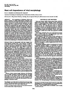
Fig. 5. Persistent MHV infection of L-2 cell mutants. Confluentcultures (approx. 107 cells) of LM-K and L-2 (wild-type and mutant) cells were inoculated with MHV (106 p.f.u, as determined on L-2 cells) and incubated at 37 °C. Media were changed daily and titrated for infectious virus by a plaque assay. On day 7, all surviving cultures were subpassaged by trypsinization (arrow). Symbols:L-2, O; M2. Q; M10, n; M12, II; M22, A; M26, A; LM-K, O.
with MHV, infection of mutant L-2 cells with vMS did not show such severe restriction of infection (Table 1). Mutant L-2 cells replicate M H V in a persistent fashion In order to study the long-term behaviour of MHV infection in the mutant cells, cultures were inoculated with virus and maintained for a period of 2 weeks. As shown in Fig. 5, wild-type L-2 cells produced progeny MHV for only 2 days, at which time the cell monolayer had been completely fused and detached from the plastic substrate. All mutant cells tested showed a remarkably similar pattern of virus production, characterized by continued, fluctuating levels of MHV, over the experimental period of 14 days. Although the monolayers showed evidence of syncytial development, this was much reduced compared to that of the wild-type L-2 cells. For comparison, MHV production in LM-K ceils, previously shown to support a persistent infection of MHV (Mizzen et al., 1983), was also monitored in parallel over the 14 day period and was found to follow a pattern similar to that observed with the mutant L-2 cells (Fig. 5). Immunofluorescence analysis of MHV-infected L-2 cell mutant cultures, maintained for 14 days at 37 °C showed fluctuating numbers of MHV-infected cells which generally ranged from 0.5 to 5 ~ of the total cells in each culture (data not shown). No evidence was found that progeny MHV obtained from persistently infected L-2 cell mutants might be attenuated or less cytopathic than the wild-type MHV used as inoculum. Both progeny as well as inoculum virus produced similar numbers and sizes of plaques when inoculated onto L-2 cells. Mutant L-2 cells show resistance to MHV-induced fusion As it was noted that the progression of MHV-induced cell fusion appeared more slowly in the mutant cell lines as compared to wild-type L-2 cells, it seemed likely that the cell mutants might
Downloaded from www.microbiologyresearch.org by IP: 54.210.20.124 On: Wed, 21 Oct 2015 00:01:49
3342
M. DAYA AND OTHERS
L-2 Mutants
Fig. 6. L-2 cell mutants are resistant to MHV-mediated cell fusion. A contact fusion was performed in which sparsely seeded cultures of MHV-infected L-2 cells were overlaid (at 4 h p.i.) with a 10-fold excess of uninfected L-2, LM-K or L-2 mutant (M2, M10, M12, M22 or M26) cells. Following 4 h incubation at 37 °C cultures were stained with Giemsa and examined by phase contrast photomicrography. be relatively more resistant to fusion and therefore able to restrict virus spread and accompanying cytopathology throughout the cultures. To test this idea directly a contact fusion assay was performed (Mizzen et al., 1983) in which sparsely seeded MHV-infected wild-type L-2 cells were overlaid with a 10-fold excess of cells (either mutant or wild-type), and the resultant cell-cell fusion was monitored after 3 h at 37 °C. As shown in Fig. 6 syncytial development was markedly reduced in the mutant cell lines, confirming the idea that they are relatively resistant to M H V - i n d u c e d fusion. Although it is clear that the mutant L-2 cells are resistant to E2-mediated fusion, they do not show a generalized resistance to fusogenic agents. This was tested by determining the numbers of multinucleate cells formed by treatment of wild-type and m u t a n t L-2 (M2, M10, M12, M22 and M26) cells with polyethylene glycol; no difference was observed among the cells tested (data not shown). T a k e n together, the results are compatible with the hypothesis that the mutant L-2
Downloaded from www.microbiologyresearch.org by IP: 54.210.20.124 On: Wed, 21 Oct 2015 00:01:49
Cellular permissiveness to M H V infection
3343
Fig. 7. L-2 cell mutants express fusion-active E 2 protein at the cell surface. A contact fusion assay was performed in which sparsely seeded cultures of L-2 (a, b) or L-2 cell mutants (M2, c; M10, d; M12, e; M22, f o r M26, g) were mock-infected (a) or infected with vMS (b to g). At 8 h p.i. cultures were overlaid with a 10-fold excess number of uninfected L-2 cells. Following a further 4 h incubation at 37 °C, cultures were stained with Giemsa and examined by phase contrast photomicrography.
cells are defective in an E2-specific cell surface recognition factor (possibly a receptor) which is required for efficient membrane fusion to occur. Mutant L-2 cells express fusogenic E2 at the cell surface Although the L-2 cell mutants show resistance to MHV-mediated fusion, they are not themselves deficient in expressing fusogenic E2 protein at their cell surfaces. This was demonstrated by a contact fusion procedure in which sparsely seeded cultures of vMS-infected L-2, LM-K or L-2 cell mutant cells were overlaid with an excess of uninfected L-2 cells at 8 h p.i. and incubated for 4 h at 37 °C. As shown in Fig. 7, all cells which were infected with vMS induced fusion with the uninfected L-2 cell neighbours indicating that the former were expressing fusogenic E2 at their outer surfaces. DISCUSSION
Cell fusion mediated by the E2 glycoprotein plays an important role in the pathogenesis of MHV infections. As a contributing factor to intercellular dissemination of virus, cell fusion may facilitate persistent infections, as demonstrated in the presence of continued neutralizing antibody response (Buchmeier et al., 1984; Sorensen et al., 1984; Stohlman & Weiner, 1978). Alternatively, persistence may arise in cells which are relatively resistant to MHV-mediated cell fusion, thereby attenuating a normally cytolytic, terminal infection to one which is of a more protracted, chronic nature (Mizzen et al., 1983).
Downloaded from www.microbiologyresearch.org by IP: 54.210.20.124 On: Wed, 21 Oct 2015 00:01:49
3344
M. DAYA AND OTHERS
The results presented here indicate an important correlation between cell permissiveness to MHV infection and susceptibility to Ez-mediated membrane fusion. Our studies with L-2 cell mutants suggest that cell variants capable of surviving infection by MHV and giving rise to a state of persistence can arise at fairly high frequency from a population of fully MHVpermissive parent cells. Of five cell mutants tested in the present study, all showed a similar phenotype of impaired permissiveness to MHV infection and a restricted susceptibility to E zmediated cell fusion. Resistance to MHV infection and to its induced fusion has previously correlated with the ability of other fibroblast sublines, LM and LM-K (Mizzen et al., 1983 ; Kooi et al., 1988), to support a persistent infection of MHV. The similarity of these findings coupled with the relatively high generation frequency of the L-2 cell mutants described in the present study strongly suggest that resistance to infection and to Ez-facilitated fusion are closely linked. Alterations of both viral and host cell characteristics may contribute to the establishment of persistent virus infections (Ahmed et al., 1981 ; Hampar & Burroughs, 1969; Ron & Tal, 1985; Cummings & Rinaldo, 1989). Moreover variants of MHV have been isolated from persistent infections initially established in vivo or in vitro with wild-type virus (Baybutt et al., 1984; Jackson et al., 1984; Taguchi et al., 1985). Our present results suggest that the modification of purely host-specified determinants is sufficient to alter the MHV-cell interaction from an acute to a persistent character. The selection procedure employed resulted in the generation of L-2 cell mutants which were virus-free (as judged by lack of production of viral antigen or progeny). Compared to the parental L-2 cells, the mutant cells were less susceptible to infection with wildtype MHV(A59) and showed resistance to contact fusion when exposed to neighbouring L-2 cells also infected with wild-type MHV(A59). Following inoculation with wild-type MHV(A59), the mutant L-2 cells showed characteristics of persistent virus infection within 1 or 2 days, at which time the parental L-2 cells had succumbed to lytic infection. Finally, virus released from persistently infected L-2 cell mutants produced similar quantities and sizes of plaques as those of wild-type MHV(A59) when inoculated into parental L-2 cells. Taken together, the data strongly support the idea that persistent MHV infection can arise by the alteration of cell functions which regulate host susceptibility to viral fusion and the initiation of infection. Cellular restriction of MHV infection apparently can occur at the level of virus binding (Tardieu et al., 1986; Boyle et al., 1987) or at post-adsorption stages (Van Dinter & Flintoff, 1987; Kooi et al., 1988). It is clear from the present study that the tolerance to MHV infection of a cell population can arise by mechanisms which limit infection to a fraction of the total cells available. Resistance to MHV-induced cell fusion represents not only a protective mechanism against a severe c.p.e, but also against an important mode of MHV dissemination. It is likely that these factors permit the development of a persistently infected state by limiting the numbers of infected cells and permitting a proportion of the cell population to escape infection. Our mutagenesis studies point towards the alteration of a cellular component which is required for efficient virus infection as well as for membrane fusion. An obvious candidate for such a cellular component is a receptor protein involved in recognition of the MHV E2 glycoprotein, since E2 is required for both virus attachment and cell fusion (Collins et al., 1982). The presence of a host cell MHV-binding protein has been correlated with cellular permissiveness to MHV infection (Boyle et al., 1987) although this may not always be the case (Arnheiter et al., 1982; Beushausen et al., 1987; Van Dinter & Flintoff, 1987; Kooi et al., 1988). From these latter studies, evidence has accumulated which suggests that restriction of MHV replication often occurs as a result of a post-adsorption block; somatic mutations could occur which do not affect virus binding but rather the subsequent receptor-mediated processes invo!ved in penetration and/or uncoating. The above studies may have relevance to in vivo situations in which a number of mouse strains have been shown to differ in their susceptibility to MHV-induced disease. Genetic resistance to MHV can be manifested by certain cell types, such as macrophages, hepatocytes or fibroblasts, in which MHV replication is either abortive (Virelizier & Allison, 1976) or permissive, but with reduced cell pathology (MacNaughton & Patterson, 1980; Arnheiter et al., 1982; Lamontagne & Dupuy, 1984). The common phenotype of a weakly fusogenic, chronic infection observed among
Downloaded from www.microbiologyresearch.org by IP: 54.210.20.124 On: Wed, 21 Oct 2015 00:01:49
Cellular permissiveness to M H V infection
3345
our easily generated L-2 cell mutants may serve as a useful conceptual model for the development of subacute, persistent infections in the animal. We would like to acknowledge the technical assistance of Leo H e y n e m in the preparation of vMS. The Medical Research Council of C a n a d a is gratefully acknowledged for grant support. H.V. is supported by a grant from D u p h a r B.V. (Weesp, The Netherlands).
REFERENCES AHMED, R., CANNING,W. M., KAUFFMAN,R. S., SHARPE,A. H., HALLUM,J. V. & FIELDS, B. N. (1981). Role of the host cell in persistent viral infection: coevolution of L cells and reovirus during persistent infection. Cell 25, 325-332. ARNHEITER, H., BAECHI, T. & HALLER, O. (1982). Adult mouse hepatocytes in primary monolayer culture express genetic resistance to mouse hepatitis virus type 3. Journal of Immunology 129, 1275-1281. BAYBUTT,H. N., WEGE, H., CARTER, M. J. & TER MEULEN, V. (1984). Adaptation of coronavirus J H M to persistent infection of murine S a c ( - ) cells. Journal of General Virology 65, 915-924. BEUSHAUSEN,S., NARINDRASORASAK,S., SANWAL,B. D. & DALES,S. (1987). In vivo and in vitro models of demyelinating disease: activation of the adenylate cyclase system influences J H M virus expression in explanted rat oligodendrocytes. Journal of Virology 61, 3795-3803. BOYLE, J. F., WEISMILLER, D. G. & HOLMES, K. V. (1987). Genetic resistance to mouse hepatitis virus correlates with absence of virus-binding activity on target tissues. Journal of Virology 61, 185-189. BUCHMEIER, i . J., LEWICKI, H. A., TALBOT,P. J. & KNOBLER, R. L. (1984). Murine hepatitis virus-4 (strain JHM)induced neurologic disease is modulated in vivo by monoclonal antibody. Virology 132, 261-270. CHELEY, S. & ANDERSON, R. (1984). A reproducible microanalytical method for the detection of specific R N A sequences by dot-blot hybridization. Analytical Biochemistry 137, 15-19. COLLINS, A. R., KNOBLER,R. L., POWELL, H. & BUCHMEIER, M. J. (1982). Monoclonal antibodies to murine hepatitis virus-4 (strain J H M ) define the viral glycoprotein responsible for attachment and cell-cell fusion. Virology 119, 358-371. CUMMINGS, P. J. & RINALDO, C. R., JR (1989). Coevolution of virulent virus and resistant cells as a m e c h a n i s m of persistence of herpes simplex virus type 1 in a h u m a n T lymphoblastoid cell line. Journal of General Virology 70, 97-106. FRANA, M. F., BEHNKE, J. N., STURMAN,L. S. & HOLMES, K. V. (1985). Proteolytic cleavage of the E2 glycoprotein of murine coronavirus: host-dependent differences in proteolytic cleavage and cell fusion. Journal of Virology 56, 912 920. FRIEDMAN, D. I., OLSON, E. R., GEORGOPOULOS,C., TILLY, K., HERSKOWITZ,I. & BANUETT,F. (1984). Interactions of bacteriophage and host macromolecules in the growth of bacteriophage lambda. Microbiological Reviews 48, 299-325. HAMPAR,B. & BURROUGHS, M. A. K. (1969). M e c h a n i s m of persistent herpes simplex virus infection in vitro. Journal of the National Cancer Institute 43, 621-634. HARA, T., HATTORI, S. & KAWAKITA,M. (1989). Isolation and characterization of mouse F M 3 A cell m u t a n t s which are devoid of Newcastle disease virus receptors. Journal of Virology 63, 182-188. JACKSON, D. P., PERCY, D. H. & MORRIS, V. L. (1984). Characterization of murine hepatitis virus (JHM) R N A from rats with experimental encephalomyelitis. Virology 137, 297-304. KAPLAN, G., LEVY, A. & RACANIELLO, V. R. (1989). Isolation and characterization of HeLa cell lines blocked at different steps in the poliovirus life cycle. Journal of Virology 63, 43-51. K1T, S., DUBBS,n. R., PIEKARSKI,L. J. & HSU, T. C. (1963). Deletion of thymidine kinase activity from L cells resistant to bromodeoxyuridine. Experimental Cell Research 31, 297-312. KOOI, C., MIZZEN, L., ALDERSON,C., DAYA, i . & ANDERSON, R. (1988). Early events of importance in determining host cell permissiveness to mouse hepatitis virus infection. Journal of General Virology 69, 1125-1135. LAMONTAGNE,L. M. & DUPUY, J.-M. (1984). Natural resistance of mice to mouse hepatitis virus type 3 infection is expressed in embryonic fibroblast cells. Journal of General Virology 65, 1165-1171. LUCAS, A., FLINTOFF, W., ANDERSON, R., PERCY, D., COULTER, M. & DALES, S. (1977). In vivo and in vitro models of demyelinating diseases: tropism of the J H M strain of murine hepatitis virus for cells of glial origin. Cell 12, 553-560. LUCAS,A., COULTER,M., ANDERSON,R., DALES,S. & FLINTOFF, W. (1978). In vivo and in vitro models of demyelinating diseases. II. Persistence and host-regulated thermosensitivity in cells of neural derivation infected with mouse hepatitis and measles viruses. Virology 88, 325-337. MAcNAUGHTON,M. R. & PATTERSON,S. (1980). Mouse hepatitis virus strain 3 infection of C57, A/Sn and A/J strain mice and their macrophages. Archives of Virology 66, 71-75. MANAKER,R. A., PICZAK, C. V., MILLER, A. A. & STANTON,M. F. (1961). A hepatitis virus complicating studies with mouse leukemia. Journal of the National Cancer Institute 27, 29-51. MERCHANT, D. L & HELLMAN, K. B. (1962). Growth of L-M strain mouse cells in a chemically defined medium. Proceedings of the Society for Experimental Biology and Medicine l l 0 , 194-198. MIZZEN, L., DAYA,M. & ANDERSON, R. (1987 a). The role of protease-dependent cell m e m b r a n e fusion in persistent and lytic infections of murine hepatitis virus. Advances in Experimental Medicine and Biology 218, 175-186.
Downloaded from www.microbiologyresearch.org by IP: 54.210.20.124 On: Wed, 21 Oct 2015 00:01:49
3346
M. D A Y A A N D O T H E R S
MIZZEN, L., MACINTYRE,G., WONG, F. & ANDERSON, R. (1987b). Translational regulation in mouse hepatitis virus infection is not mediated by altered intracellular ion concentrations. Journal of General Virology 68, 21432151. MIZZEN, L., CHELEY, S., RAO, M., WOLF, R. & ANDERSON, R. (1983). Fusion resistance and decreased infectability as major host cell determinants of coronavirus persistence. Virology 128, 407-417. PARKER, R. F., BRONSON, L. H. & GREEN, R. H. (1941). Further studies of the infectious unit of vaccinia Journal of Experimental Medicine 74, 263 281. RON, D. & TAL, J. (1985). Coevolution of cells and virus as a m e c h a n i s m for the persistence of lymphotropic minute virus of mice in L-cells. Journal of Virology 55, 424-430. ROTHFELS, K. H., AXELROD,A. A., SIMINOVITCH,L., McCULLOCH,E. A. & PARKER,R. C. (1959). The origin of altered cell lines from mouse, monkey and m a n as indicated by chromosome and transplantation studies. Canadian : CancerConference 3, 189-214. SORENSEN, O., COULTER-MACKIE,M. B., PUCHALSKI,S. & DALES,S. (1984). In vivo and in vitro models of demyelinating diseases. IX. Progression of J H M virus infection in the central nervous system of the rat during overt and asymptomatic phases. Virology 137, 347-357. STOHLMAN,S. A. & WEINER, L. P. (1978). Stability of neurotropic mouse hepatitis virus (JHM strain) during chronic infection of neuroblastoma cells. Archives of Virology 57, 5 3 ~ 1 . TAGUCHI, F., SIDDELL, S. G., WEGE, H. & TER MEULEN, V. (1985). Characterization of a variant virus selected in rat brains after infection by coronavirus mouse hepatitis virus JHM. Journal of Virology 54, 429-435. TARDIEU, M., BOESPFLUG,O. & BARBE,T. (1986). Selective tropism of a neurotropic coronavirus for ependymal cells, neurons and meningeal cells. Journal of Virology 60, 574-582. TOY&MA,S., TOYAMA,S. & UETAKE, H. (1977). Altered cell-fusion capacity in lines of KB cells resistant to Sendal virus-induced cytolysis. Virology 76, 503-515. TUFARO, F., SNIDER, M. D. & McKNIGHT, S. L. (1987). Identification and characterization of a mouse cell m u t a n t defective in the intraceUular transport of glycoproteins, Journal of Cell Biology 105, 647-657. VAN DINTER, S. & FLINTOFF, W. F. (1987). Rat glial C6 cells are defective in murine coronavirus internalization. Journal of General Virology 68, 1677-1685. VIRELIZlER, J. L. & ALLISON,A. C. (1976). Correlation of persistent mouse hepatitis virus (MHV3) infection with its effect on mouse macrophage cultures. Archives of Virology 50, 279 285. YASUMURA, Y. & KAWAKITA,Y. (1963). Studies on SV-40 in tissue culture. Nippon Rinsho (Japan) 21, 1209.
(Received 21 April 1989)
Downloaded from www.microbiologyresearch.org by IP: 54.210.20.124 On: Wed, 21 Oct 2015 00:01:49