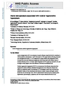
Oct 31, 2017 - and medium caliber, well-focused comma-like vessels were seen (Figure 2). Complete surgical excision of the lesion was performed.
DERMATOLOGY PRACTICAL & CONCEPTUAL www.derm101.com
Nodular lesion with polymorphous vascular pattern Virgínia Coelho de Sousa1, André Oliveira2 1 Department of Dermatology, Hospital de Santo António dos Capuchos, Centro Hospitalar de Lisboa Central, Lisbon, Portugal 2 Centro Académico de Medicina, University of Lisbon, Lisbon, Portugal
Key words: dermatofibroma, dermoscopy, comma-like vessels, dermatopathology Citation: Coelho de Sousa V, Oliveira A. Nodular lesion with polymorphous vascular pattern. Dermatol Pract Concept 2017;7(4):81-83. DOI: https://doi.org/10.5826/dpc.0704a16 Received: July 31, 2017; Accepted: September 8, 2017; Published: October 31, 2017 Copyright: ©2017 Coelho de Sousa et al. This is an open-access article distributed under the terms of the Creative Commons Attribution License, which permits unrestricted use, distribution, and reproduction in any medium, provided the original author and source are credited. Funding: None. Competing interests: The authors have no conflicts of interest to disclose. All authors have contributed significantly to this publication. Corresponding author: Dr. Virgínia Coelho de Sousa, Department of Dermatology and Venereology, Hospital de Santo António dos Capuchos, Alameda de Santo António dos Capuchos, 1169-050 Lisbon, Portugal. Tel. +351 213136300. E-mail: virginiacoelhodesousa@ gmail.com
ABSTRACT
Dermatofibroma (DF) is one of the most common skin neoplasms seen by dermatologists. Out of the various histopathological subtypes of DF, the atrophic variant is considered rare. Clinical and dermoscopic diagnosis of DF is straightforward in most cases. However, deeply atypical clinical and dermoscopic presentations can simulate other benign and malignant tumors. We present a case of atrophic DF, describing its dermoscopic features and the correlation with histopathology.
The Patient
Diagnosis
A 66-year-old female presented to our clinic with a 12-month
Atrophic dermatofibroma.
history of a new, growing, asymptomatic nodule on her right leg. The physical examination revealed a firm, slightly depressible, pink nodule with light brown peripheral pigmentation and superficial visible vessels, measuring 10 mm in maximum diameter (Figure 1). Dermoscopy disclosed a central white structureless area, surrounded by an erythematous halo with areas of light
Clinical Course As it is considered a benign non-melanocytic lesion, a conservative management was proposed. No further unnecessary therapeutic procedures were performed.
brown atypical network. Additionally, fine linear-irregular and medium caliber, well-focused comma-like vessels were
Discussion
seen (Figure 2). Complete surgical excision of the lesion was performed.
Dermatofibroma (DF) is one of the most common skin neo-
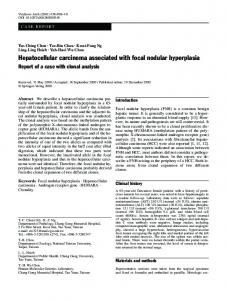
Histopathological examination revealed an intradermic nod-
plasms seen by dermatologists. Out of the various histopatho-
ular lesion with few small fusiform cells, abundant eosino-
logical subtypes of DF, the atrophic variant is considered rare.
philic collagen bundles and capillary vessels (Figure 3). Immu-
Clinical and dermoscopic diagnosis of DF is straightfor-
nohistochemistry was negative to CD34 and S100 protein.
Observation | Dermatol Pract Concept 2017;7(4):16
ward in most cases. However, deeply atypical clinical and
81
Figure 2. Dermoscopic presentation with a central white structureless area, surrounded by areas of light brown atypical network (arrows). Fine linear-irregular (circles) and medium caliber, wellfocused comma-like vessels (arrows) were seen (polarized contact dermoscopy, x10). [Copyright: ©2017 Coelho de Sousa et al.]
Figure 1. Clinical presentation of a nodular lesion located on the leg. [Copyright: ©2017 Coelho de Sousa et al.]
dermoscopic presentations can simulate other benign and malignant tumors [1]. Dermoscopy is a fast, noninvasive technique that increases diagnostic accuracy for both melanocytic and non-melano-
Figure 3. Histopathology showing an intradermic nodular lesion with few small fusiform cells, abundant eosinophilic collagen bundles and capillary vessels (hematoxylin-eosin, x40). [Copyright: ©2017 Coelho de Sousa et al.]
cytic skin tumors, allowing for better differentiation of clinical simulators of melanoma [2]. Common dermoscopic features of DF include pigment
white scar-like patches; peripheral homogeneous area with
network, white scar-like patch and white network. Ten der-
a central white scar-like patch; or peripheral homogeneous
moscopic patterns were described, according to the presence
area with a central white network; and atypical pattern [3].
or absence of peripheral pigment network. DF with peripheral
Vascular structures can be present in 49.5% of DF. The
pigment network are divided in four patterns: total delicate
most common vascular structure is erythema, followed by
pigment network; peripheral delicate
pigment network with
dotted vessels [3]. Vascular structures are one of the criteria
central white scar-like patch; peripheral delicate pigment
used for the dermoscopic diagnosis of melanoma and other
network with a central white network; and peripheral deli-
pigmented and vascular tumoral lesions that may simulate
cate pigment network with a central homogeneous area. DF
melanoma [4].
without peripheral pigment network can present as total
We present a case of a new, growing, nodular lesion pre-
white network; total homogeneous area; total or multiple
senting in an elderly patient. Dermoscopy showed polymor-
82
Observation | Dermatol Pract Concept 2017;7(4):16
phous vascular structures including erythema, linear-irregular
ous tumors. Histopathological examination should always be
and comma-like vessels.
performed in such confounding lesions.
Comma-like vessels are the dermoscopic hallmark of dermal nevi, being rarely described in DF [5]. The presence of comma-like vessels in a regular distribution or as the
References
dominant vascular type is considered a negative predictor for
1. Ferrari A, Argenziano G, Buccini P, et al. Typical and atypical der-
amelanotic melanoma [6]. However, considering the atypi-
moscopic presentations of dermatofibroma. J Eur Acad Dermatol
cal clinical and dermoscopic presentation of polymorphous vascular structures described as a feature of amelanotic melanoma, excision was mandatory to rule out this entity. Histopathology later confirmed the presence of a benign tumor. To our knowledge, this is the first dermoscopic description of the rare atrophic variant of DF. Atrophic DF is identified by dermal atrophy with prominent sclerotic collagen, as well as low cellularity. It has been proposed that dense
Venereol. 2013;27:1375-1380. 2. Argenziano G, Soyer HP, Chimenti S, et al. Dermoscopy of pigmented skin lesions: result of a Consensus Meeting via Internet. J Am Acad Dermatol. 2003;48:679-693. 3. Zaballos P, Puig S, Llambrich A, Malvehy J. Dermoscopy of dermatofibromas: a prospective morphological study of 412 cases. Arch Dermatol. 2008;144:75-83. 4. Argenziano G, Zalaudek I, Corona R, et al. Vascular structures in skin tumors: a dermoscopy study. Arch Dermatol. 2004;140(12):1485-1489.
elastic fibers around the vessels interfere with blood circula-
5. Kilinc Karaarslan I, Gencoglan G, Akalin T, Ozdemir F. Different
tion, causing dermal atrophy, and thus low cellularity [7].
dermoscopic faces of dermatofibromas. J Am Acad Dermatol.
The white strutureless area seen on dermoscopy correlates with the dense collagen fibers found on dermis. Well-focused, medium caliber comma-like vessels represent superficial vessels running above the dermal collagen bundles. DF can thus present with a wide range of dermoscopic patterns, sometimes mimicking melanoma and other cutane-
Observation | Dermatol Pract Concept 2017;7(4):16
2007;57(3):401-406. 6. Menzies SW, Kreusch J, Byth K, et al. Dermoscopic evaluation of amelanotic and hypomelanotic melanoma. Arch Dermatol. 2008;144(9):1120-1127. 7. Alves JV, Matos DM, Barreiros HF, Bártolo EA. Variants of dermatofibroma – a histopathological study. An Bras Dermatol. 2014;89(3):472-427.
83