Noninvasive Fetal ECG Estimation Based on Linear Transformations Mariano Llamedo1,2,3 , Alba Martín-Yebra3,1 , Pablo Laguna1,3 , Juan Pablo Martínez1,3 1
Aragon Institute of Engineering Research, IIS Aragón, University of Zaragoza, Aragon, Spain 2 Universidad Tecnológica Nacional, Buenos Aires, Argentina 3 CIBER of Bioengineering, Biomaterials and Nanomedicine (CIBER-BBN), Spain Abstract
and sophisticated algorithms to separate fetal and mother activity. This year’s PhysioNet challenge consists of locating fetal QRS complexes and estimating FHR and QT interval in mother’s abdomen ECG recordings. The approach presented in this work is based on previously validated algorithms, such as a wavelet based ECG delineator [1], the Woody QRS complexes alignment algorithm [2, 3], and other linear transformation techniques as independent (ICA) [4], periodic (πCA) [5] and principal components analysis (PCA). The strategy consists in performing blind source separation first, followed by simple QRS detection by matching filtering and thresholding. The objective of this work was to develop an algorithm for fetal QRS detection based on the working hypothesis that fetal and maternal sources could be separated by linear transformations.
This year’s PhysioNet challenge consists of locating fetal QRS complexes and estimating QT interval in fetal electrocardiographic (FECG) recordings. The approach presented in this work is based on linear transformations obtained from techniques such as independent (ICA), periodic (πCA) and principal components analysis (PCA). The algorithm consists in a common ECG preprocessing stage, followed by a maternal ECG (MECG) removal, obtaining a preprocessed signal. This preprocessed signal is then rotated by PCA, πCA and ICA , in order to get a more clear representation of the fetal activity. Then QRS complexes are detected in these three signal representations. An statistical model, trained in set A, is applied to the detections obtained from the 12 resulting leads, in order to rank the leads according to their FECG content. With the detections of those leads, an average QRS complex is calculated with the Woody algorithm, and an algorithm for the refinement of the fetal QRS detections (fQRS) is used to end the procedure. An estimation of the SNR gain produced by coherent averaging is used to decide the final validity of the fQRS measurements. For those valid recordings, the QT interval is measured in an average heartbeat, calculated by coherent averaging over all the fQRS. Results in set B were 4714.6 for event 4, and 121.6 for event 5. In conclusion, due to the poor generalization capability of the algorithm, the strategy must be further improved to properly extract FECG.
1.
2.
The data for the challenge consists of three sets of 100 one-minute FECG records. The data were obtained from multiple sources using a variety of instrumentation with differing frequency response, resolution, and configuration, although in all cases they are presented as 1000 samples per signal per second, and 4 leads per recording. The set A includes in addition to the FECG, the reference fetal QRS annotations so that participants can train their algorithms. Set B includes only FECG, remaining the reference annotations hidden to the participants and therefore for evaluation purposes only. Set C is completely hidden and is reserved for open source algorithms, being set B the equivalent for non-open-source entries. The algorithm starts with the preprocessing of the FECG signals, as described in [6], with a low-pass filter (fc = 35 Hz) and baseline wander removal algorithm based on median filtering, as is shown in Figure 1. Then the removal of the maternal ECG activity is performed, the first step consists in detecting maternal QRS activity by applying a conventional QRS detector [1] obtaining the maternal QRS lom m cations q m k . Then with q k a mother template heartbeat tn
Introduction
Fetal Heart rate (FHR) decelerations are known to be associated with fetal distress. The continuous FHR monitoring is accurately performed by direct fetal electrocardiographic, using a fetal scalp electrode. The risk associated with this invasive technique makes it commonly indicated during labor. Noninvasive fetal ECG (FECG) monitoring makes use of electrodes placed on the mother’s abdomen,
ISSN 2325-8861
Materials and methods
285
Computing in Cardiology 2013; 40:285-288.
Woody algorithm
QRS detection
RP
R PCA
abdominal ECG
q fp
RI Preprocessing
Mother cancellation
QRS detection
ICA
Statistical model
SNR Detector
qf
R¼ ¼ CA
Figure 1. Block diagram of the algorithm evaluated in this work. The preprocessing is performed in the blocks of the left, then the linear transformations occur in the middle and the statistical detection of fetal QRS detections in the right blocks. where C is calculated
is calculated using the Woody algorithm [3] , which is then multiplied with a Blackman window in order to smooth the template. Then a mother ECG signal is constructed with the smoothed template repeated for each location in q m k . The FECG estimation F is obtained by subtracting the estimated mother ECG from the preprocessed ECG . Three blind source separation techniques, such as principal (PCA), independent (ICA) and periodic component analysis (πCA) were applied to the fetal signal F in order to enhance the signal-to-noise ratio of fetal ECG components. Using these three methods, a new transformed signal matrix R of N × 4 is defined by the transformation R = F ·P,
C=
Cτ =
(1)
and is the result of computing the eigenvectors of the sample covariance matrix C, calculated as described in [7]. The matrix Q is a diagonal matrix with the eigenvalues of C. This rotation is such that most of the energy of F is projected in the direction of the eigenvector associated with the higher eigenvalues. The πCA technique searches for the optimal linear combination of the available leads in the ECG signal which maximizes the desired periodicity in the transformed lead. In this case, we are interested in combining the leads in such a way that 1-beat maternal periodicity is maximized in the resulting signal Rπ . As it has been shown in [5], the optimal combination is given by solving a generalized eigenvalue problem of the matrix pair (Cτ ,C), where the matrix P is such that simultaneously hold (3)
P−1 .C.P = I,
(4)
1 F(m) .FT (K − 1)N
(6)
In this way, the transformation projects the most periodic component into the first column of Rπ . The last method used was the fast ICA algorithm [4], which identifies the rotation matrix P such that each signals (in R) probability distribution differs from the Gaussian distribution. This is equivalent to find the most independent sources, given they were mixed with P−1 . The next step is to evaluate and find the signal/s where the transformation was able to separate FECG from mother’s residual. For this purpose, we applied the same QRS detector [1] for the three multi-lead signals RP , Rπ and RI , obtaining one fetal QRS location q fp for each of the 12 signals. Also the sequence of RR intervals r fp associated to each q fp were calculated. Then two probabilistic models where estimated in the training set A, the first was a bivariate Gaussian that models the RR interval evolution using r fp and r fp+1 features. The other measurement is the amount of fetal heartbeats detected in one minute, which is modeled with a Weibull distribution, as can be seen in Figure 3. Finally, the likelihood of each sequence q fp of being a true fetal RR series is calculated as the product of both likelihoods. Then the three most likely sequences are selected for calculating an SNR estimate of the gain produced by coherent averaging. The coherent averaging is calculated considering a window of 600 ms centered in each heartbeat detected in q fp . Then the signal energy is calculated in the average pattern. This measurement accounts for the energy which is coherent with respect to
(2)
P−1 .Cτ .P = Q,
(5)
with K the number of beats in the analysis window and N the number of samples of each learning segment. In the same way, we define Cτ , the analogous spatial correlation matrix of the non-periodic components, where F(m) is the equivalent of F after sliding the analysis window m = 1 beats forward.
where P is the 4 × 4 matrix which defines the linear transformation. After the linear transformations were applied we obtained one set of signals for each method used RP , Rπ and RI for PCA, πCA and ICA respectively. For PCA, P is a matrix such that P−1 CP = Q,
1 F.FT (K − 1)N
286
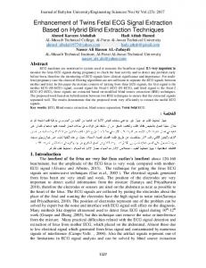
Mother
Fetal
4
3
2
1
Recording a01- 29.5" : 35.5"
29.8" Index #
0.4" 0.4" 2 2
0.8" 3
1.1" 1.2" 4 3
1.6" 5
1.9" 1.9" 6 4
2.3" 7
3.1" 9
2.7" 2.7" 8 5
3.4" 3.5" 10 6
3.8" 11
4.1" 4.2" 12 7
4.6" 13
4.9" 4.9" 14 8
5.3" 15
35.2"
Figure 2. An example of how the linear transformation successfully separates fetal and mother ECGs.
4.
the fetal activity. This energy measurement should be decreased by false detections or missed heartbeats, as long as these errors are not coherent to the fetal activity.
In this work, we presented an algorithm for FECG extraction from abdominal ECG recordings in the context of the PhysioNet/CinC 2013 Challenge. The methodology proposed performed as expected in set A, as can be seen in Figure 2, however the performance achieved in set B shows that the algorithm should be further improved. In dataset A, we found cases where mother and fetal ECG overlapped in the four-dimensional space defined by ECG leads, and therefore could not be linearly separated. Those cases were one of the weaknesses that our strategy presented. Despite this important limitation, the implementation of the algorithm is very simple and could be implemented in real-time once the linear transformation is found. Following this strategy, we could study if mother and fetal ECG activities could be approximately orthogonalized, maybe with a different lead positioning, or by moving the reference of the ECG recorder. In conclusion, due to the poor generalization capability of the algorithm, the strategy must be further improved to properly extract FECG.
On the other hand, the noise is estimated similarly to the signal energy, but considering for the coherent averaging a random sequence of detections q rp of the same length. This sequence is a uniformly distributed random sequence sampled between 1 and the length of the recording. The noise calculation should account for the non-coherent energy with respect to the fetal activity. The ratio of energies, called the signal to noise ratio (SNR), is calculated and a sequence of fetal detections is accepted if the SNR is above 10 db. The QT interval is finally measured on an average heartbeat, calculated by coherent averaging over all the fetal detections, as can be seen in Figure 4. The location of the start of the QRS complex and end of the T wave was also performed using the delineator described in [1].
3.
Discussion and conclusions
Results
Acknowledgments In Figure 2 it is shown the how the algorithm extracts FECG from the abdominal ECG for recording a01. The results achieved in set B were 4714.6 for event 4, and 121.6 for event 5.
This work was supported by projects TEC2010-21703C03-02, TEC2010-19410 from MINECO (Spain) and GTC T-30 from DGA and European Social Fund (EU).
287
QT
#Recordings
10
5
110
120
130
140
150
160
170
#RR intervals in 1 minute
Poincare plot centered
RR(i+1)
0.05
0
Figure 4. The QT interval measured on an average fetal heartbeat calculated from fetal detections. -0.05
-0.04
-0.02
0
0.02
trocardiogram decomposition using periodic component analysis. IEEE transactions on bio medical engineering August 2008;55(8):1935–40. ISSN 1558-2531. URL http://www.ncbi.nlm.nih.gov/pubmed/18632355. [6] Llamedo M, Martínez JP. Heartbeat classification using feature selection driven by database generalization criteria. IEEE Transactions on Biomedical Engineering march 2011; 58(3):616–625. ISSN 0018-9294. [7] Rousseeuw P, Van Driessen K. A fast algorithm for the minimum covariance determinant estimator. Technometrics 1999;41:212–223.
0.04
RR(i)
Figure 3. Models estimated in set A for ranking the quality of the extracted FECG. In the upper panel, the model for the amount of heartbeats in one minute. In the lower panel, the model for the current and next RR interval. The CIBER of Bioengineering, Biomaterials and Nanomedicine is an initiative of ISCIII.
Address for correspondence:
References [1] Martínez JP, Almeida R, Olmos S, Rocha A, Laguna P. A wavelet-based ECG delineator: Evaluation on standard databases. IEEE Transactions on Biomedical Engineering 2004;51:570–581. [2] Sörnmo L, Laguna P. Bioelectrical Signal Processing in Cardiac and Neurological Applications. Elsevier, 2005. ISBN 0-12-437552-9. [3] Harris EK, Woody CD. Use of an adaptive filter to characterize signal-noise relationships. Computers and Biomedical Research 1969;2(3):242–273. [4] Hugo Gävert Jarmo Hurri JS, Hyvärinen A. Fastica - fast independent component analysis. URL http://www.cis.hut.fi/projects/ica/fastica. [5] Sameni R, Jutten C, Shamsollahi MB. Multichannel elec-
288
Mariano Llamedo Soria,
[email protected] I3A - I+D Building, C/ Mariano Esquillor S/N Despacho 4.0.05 – CP: 50018, Zaragoza, España.