After the McNeill Dysphagia Therapy Program. Yue Lan, MD; Mai Ohkubo, PhD; Giédre Berretin-Felix, PhD; Isaac Sia;. Giselle D. Carnaby-Mann, MPH, PhD; ...
Annals of Otology. Rhinohgy & Laryngology 121(8):525-532. © 2012 Annals Publishing Company. All rights reserved.
Normalization of Temporal Aspects of Swallowing Physiology After the McNeill Dysphagia Therapy Program Yue Lan, MD; Mai Ohkubo, PhD; Giédre Berretin-Felix, PhD; Isaac Sia; Giselle D. Carnaby-Mann, MPH, PhD; Michael A. Crary, PhD Objectives: We examined the timing of physiological swallowing events in patients before and after completion of an exercise-based dysphagia intervention (McNeill Dysphagia Therapy Program; MDTP) and compared their performance to that of healthy volunteers. Methods: Eight adults (mean age, 57.5 years) with chronic dysphagia (mean, 45 months) completed 3 weeks of the MDTP. Before and after the MDTP we measured lingual-palatal and pharyngeal manometric pressures during swallows of thin liquid, thick liquid, and pudding material in 5-mL volumes. Using the pressure peak of the pharyngoesophageal segment clearing wave as the anchor point, we measured the relative timing of pressure peaks from the anterior, middle, and posterior parts of the tongue and the manometric peaks from the base of the tongue, the hypopharynx, and the nadir of the pharyngoesophageal segment. We compared these results to identical measures obtained from 34 healthy adults (mean age, 44.0 years). Results: The timing of physiological events before the MDTP was significantly slower than that of the group of healthy volunteers. The timing data from after the MDTP were not significantly different from those of the healthy group. The magnitude change was greatest for thin liquid. Conclusions: Dysphagia therapy with the MDTP improves the timing of physiological events during swallowing. Temporal coordination of swallowing components after therapy approximates that of healthy adults, suggesting a normalization of swallow timing after the MDTP. Key Words: deglutition, dysphagia, manometry, swallowing, temporal coordination, timing.
INTRODUCTION
variables such as slow oral and pharyngeal transit, delayed contraction of pharyngeal musculature, or delay in initiating the swallow.3'4.645,i6 PQJ. example, on the basis of videofluoroscopic analysis, Kendall et al'5 reported that patients with dysphagia after radiotherapy for head and neck cancer demonstrated prolonged pharyngeal bolus transit and delayed laryngeal closure compared to healthy controls. On the basis of manometric analysis, Castell et al'^ reported that patients with dysphagia as a result of oculopharyngeal muscular dystrophy demonstrated prolonged pharyngeal contraction and a slow pharyngeal contraction rate, in addition to temporal incoordination between pharyngoesophageal segment (PES) activity and pharyngeal activity. Furthermore, Olsson et al'° reported significantly shorter PES relaxation duration in patients with dysphagia marked by postswallow residue compared to patients without residue. Collectively, these results suggest that temporal deviations in swallowing reflect some de-
Appropriate timing of physiological events is important for safety and efficiency of swallowing in adults. The temporal aspects of swallowing in both healthy adults and patients with dysphagia have been explored with a variety of methods. Eor example, videofluoroscopy has been used to measure the duration of movement of swallow-related structures'"^ and the timing of bolus movement through the swallow mechanism."^-^ Surface electromyography has been used to measure the total swallowing duration based on neuromotor activation related to movement of swallow structures.^"^ In addition, timing evaluation of manometric events such as lingualpalatal and pharyngeal pressures provides information on the temporal coordination of swallowing in both healthy adults and patients with Temporal incoordination among swallow movements in adults with dysphagia may be reflected in
From the Departments of Speech, Language, and Hearing Sciences (Lan, Ohkubo, Berretin-Felix, Sia, Crai-y) and Behavioral Science and Community Health (Carnaby-Mann), College of Public Health and Health Professions, University of Florida, Gainesville. Florida. Dr Lan is currently in the Department of Rehabilitation Medicine, Third Affiliated Hospital of Sun Yat-Sen University, Guangzhou, China. Correspondence: Michael A. Crary, PhD, Dept of Speech, Language, and Hearing Sciences, College of Public Health and Health Professions, University of Florida, PO Box 100174, Gainesville, FL 32610-0174.
525
526
Lan et al. Normalization of Swallow Timing Afler Therapy
gree of incoordinated movement and may be related to the degree or characteristics of dysphagia in a variety of patient populations. Treatment results in patients with dysphagia have demonstrated variable effects on the timing of swallowing events. Regan et aP observed faster oral and pharyngeal transit times after thermal-tactile stimulation in patients with idiopathic Parkinson's disease. Troche et al'^ reported that after expiratory muscle strength training in patients with Parkinson's disease, hyoid bone displacement was greater, but the duration of hyoid bone excursion did not change. Their results implied that the speed of hyoid bone movement increased after therapy. These studies focused on kinematic aspects of swallowing measured from fluoroscopic evaluation without consideration of the physiological events that contribute to temporal aspects of swallow movements. To our knowledge, no studies have documented changes in the physiological timing of swallow events after successful dysphagia therapy. Evaluation of timing changes in physiological swallow events after dysphagia therapy may provide important information regarding treatment-related changes in temporal coordination in swallow function. In this study, we examined the timing of physiological swallowing events before and after standard administration of an exercise-based dysphagia therapy program. Specifically, we evaluated the timing of lingual-palatal pressure peaks and pharyngeal manometric peaks in patients before and after therapy. On the basis of prior functional and physiological outcomes from this therapy program, we hypothesized that temporal coordination, reflected in increased speed of physiological events, would improve after therapy. We deñned increased speed as reduced time intervals between individual physiological swallow events. To further evaluate this hypothesis, we compared the timing of these physiological swallow events before and after therapy to the same measures obtained from healthy volunteers. METHODS PARTICIPANTS
Patients With Dysphagia. Between November 2006 and September 2009, patients who presented to an outpatient dysphagia clinic housed in a tertiary-care, academic health center and met inclusion criteria were considered for participation in an outpatient dysphagia intervention. Inclusion criteria included clinically significant dysphagia, defined as a score of 5 or less on the Functional Oral Intake Scale'^ and a score of 178 or less on the Mann As-
sessment of Swallowing Ability'^; fluoroscopic evidence of pharyngeal dysphagia, defined as impaired movement of swallow structures (hyolaryngeal excursion, pharyngeal constriction, PES opening) and/or aspiration or postswallow residue; chronic dysphagia, deñned as greater than 12 consecutive months with clinically significant dysphagia symptoms; cognitive abilities sufficient to participate in interactive and intense dysphagia therapy, defined as a score greater than 23 on the Mini Mental Status Examination^"; and willingness to participate in both clinical and physiological evaluation of swallowing performance and to attend 3 consecutive weeks (15 sessions) of therapy. Eight patients participated in the McNeill Dysphagia Therapy Program (MDTP) during the course of the study. Six were male. The average age of all patients was 57.5 years (range, 20 to 70 years). Five of these patients had dysphagia as a result of treatment for oropharyngeal cancer. One patient had dysphagia as a result of a neurologic deficit, and the cause of dysphagia in another 2 patients was a combination of neurologic and cancertreatment deficits. The duration of dysphagia averaged 3.6 years (range, 14 months to 7 years). All patients had received prior swallowing therapy without functional improvement. At the onset of MDTP intervention, 7 ofthe 8 participants were dependent on nonoral tube feeding. The remaining participant was restricted to a single-consistency (puree) diet. Healthy Volunteer Control Subjects. Thirty-four healthy participants (mean age, 44.0 years; SD, 21.12 years; range, 22 to 78 years) served as the control group. The female-to-male ratio in the group was 1:1. The healthy volunteers were recruited from March 2010 to August 2010. None ofthe volunteers had a history of swallowing problems, speech disorders, neurologic illness, or structural disorders of the head and neck, and none were taking medication known to impact swallowing function at the time of their participation in the study. The local Institutional Review Board approved this study, and all participants signed an informed consent form before participation. DYSPHAGIA INTERVENTION
All patients participated in the MDTP, a systematic exercise-based therapy framework for the treatment of dysphagia in adults. The MDTP has been shown to produce functional benefits in adult patients with chronic, treatment-refractory dysphagia.^'-^^ After the MDTP, patients demonstrate increased physiologic effort during swallows as measured by lingualpalatal pressures and surface electromyography.^^ All MDTP sessions followed a previously described standard protocol.^2 In this protocol, a single
Lan et al. Normalization of Swallow Timing After Tiierapy
swallowing strategy was taught to the patient to facilitate swallowing attempts. The criteria and steps for advancement were predetermined, and the program hierarchically incorporated advancing steps of altered bolus volume, bolus consistency, eating rate, and amount of oral intake. Treatment sessions were conducted for I hour a day, 5 days a week, for a maximum of 3 weeks (15 sessions). Participants initiated the MDTP protocol using a bolus consistency and volume that was determined via fluoroscopic examination. The criteria for the starting material and volume were that this material be at least partially swallowed with no evidence of aspiration. The success or failure of individual swallows was based on overt signs of aspiration or expectoration. Once participants "passed" the initial bolus material, they advanced to the next level in the bolus progression. If the initial bolus material was "failed" (signs of aspiration or expectoration), the participant regressed to a lower level on the bolus progression. The bolus progression changed consistency first, and then volume. In later stages of bolus progression, the focus shifted to eating rate and the amount of food liquid consumed. In the MDTP, bolus progression is highly individualized and based on the participant's performance on each bolus material and volume. Based on in-treatment performance, nightly homework was given that consisted of oral ingestion of materials successfully swallowed during therapy. Each participant completed a dietary record of food or liquids consumed each night and noted any difficulties or concerns. The treating clinician reviewed these records during the subsequent therapy session to ascertain compliance with the home activities and identify any difficulties that may have occurred outside the therapy environment. PHYSIOLOGICAL EVALUATION
Physiological measures for this study were obtained by use of the Digital Swallowing Workstation and Swallowing Signals Laboratory (model 7100, Kay PENTAX, Montvale, New Jersey). All data were recorded digitally and analyzed offline at a later time. Lingual-Palatal Pressure. Lingual-palatal pressure during swallowing was measured from 3 airfilled bulbs affixed from front to back on the hard palate with Stomahesive (ConvaTec USA, Skillman, New Jersey). The bulbs were 8 mm apart and mounted on a silica strip. The anterior bulb in this array was placed on the alveolar ridge, just posterior to the maxillary incisors. This placement located the middle bulb near the midpoint of the hard palate, and the posterior bulb anterior to the junction of the hard and soft palates. The disposable bulb array was
527
calibrated before each use to record a range from 0 to 750 mm Hg. Pharyngeal Manometry. Manometric swallow pressures were measured with a lOO-cm solid-state manometric catheter, 2.1 mm in diameter, with three pressure transducers (Gaeltec Devices Ltd, Dun vegan, Scotland). The catheter was calibrated to record a pressure range from -50 to 250 mm Hg before use for each participant. For each subject, the catheter was passed transnasally under videoendoscopic guidance. Once the middle sensor of the catheter was visually within the upper esophageal sphincter, pressure readings from each sensor were monitored. Using a pull-through technique in which the catheter was slowly withdrawn, we noted the increased resting pressure level of the upper esophageal sphincter on the middle sensor and then the distal sensor. At this point, the middle sensor was visible in the inferior hypopharynx. A swallow with the catheter in this position often — but not always — resulted in a characteristic m-wave shape.24 If an m-wave was noted, the catheter was anchored to the exterior part of the nose with tape, and this location was used to obtain manometric data during swallowing. If no m-wave was obtained, the catheter was slowly advanced and withdrawn (under endoscopie guidance), and additional swallows were monitored for the presence of the m-wave. If no mwave was obtained during swallowing, the catheter was anchored to the exterior part of the nose at the location at which increased resting pressure was observed at the distal sensor (upper esophageal sphincter) and the middle sensor was visible in the inferior hypopharynx. DATA COLLECTION After a quiet resting adaptation period (up to 10 minutes), each participant swallowed thin liquid, thick liquid, and pudding materials in 5-mL amounts. All consistencies were prepared 10 minutes before use. Slip-tip syringes were used to measure liquid and pudding volumes to ensure that the bolus quantities were accurate. Gatorade was used as the thin liquid and was combined with Resource Thickenup (Nestle Healthcare Nutrition, Florham Park, New Jersey) to make thick liquid and pudding materials. Both thick liquid and pudding materials were consistently made from a formula. Thick liquid consisted of 4 ounces (118.3 mL) of Gatorade with 1 tablespoon -I- 1 teaspoon -i- 1/8 teaspoon (20.33 mL) of thickener. The mixture was stirred and left to stand for 5 minutes before use. This formula yielded a viscosity of liquid that by consensus judgment of the investigators refiected a nectar consistency. Pudding was made by combining 4 ounces (118.3 mL) of Ga-
528
Lan et al. Normalization of Swallow Timing Afler Therapy
torade with 2 tablespoons -i-1 teaspoon (34.5 mL) of thickener. This mixture was stirred and left to stand for 5 minutes before use. By consensus judgment of the investigators, the viscosity reflected a commercial pudding thickness. The viscosity properties of these materials were evaluated with a viscometer using the Small Sample Adapter (Brookfield Engineering Laboratories, Middleboro, Massachusetts) or Couette geometry. The size of the spindle and the torque range of the viscometer were changed to allow for the different rheological properties of these samples. The true shear rate was determined by the method of Krieger and Elrod.25 These materials do not have a constant viscosity; rather, they are pseudoplastic in nature. Higher shear rates result in decreased viscosity. The thick liquid sample had a K value (consistency index) of nearly 2,300 mPa-s and an n value (pseudoplasticity index) of nearly 0.36. For reference, a constant viscosity sample (Newtonian) would have an n value of 1.0. Smaller numbers indicate increasing pseudoplasticity. The pudding sample had a K value of more than 75,000 mPa-s, which is more than 30 times thicker than the thick liquid. The pudding sample was also more pseudoplastic, with an n value of approximately 0.21. The viscosity of the thin liquid sample was calculated at 1.16 mPa-s at 20°C (just slightly more viscous than water at the same temperature). Once the bolus was placed in the participant's mouth, the participant was instructed to breathe quietly through the nose. Each physiological channel was monitored to ascertain that baseline levels were demonstrated with the bolus being held in the mouth. Once baseline activity levels were demonstrated, the participant was instructed to swallow. All physiological data were recorded on the workstation during the respective examinations. A blinded assessor subsequently measured these examinations. In the case of multiple swallows for a single bolus, only the initial swallow attempt was measured. A total of 6 swallows (3 consistencies at 2 volumes) was measured from each participant at each assessment time point. EVALUATION OF TIMING PARAMETERS
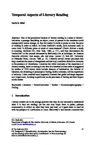
The time points of lingual and pharyngeal pressure peaks were extracted from the pressure waveforms offline and measured in seconds. Using the pressure peak of the PES clearing wave (C-wave) as the anchor point (Fig 1), we calculated the time difference to the peak amplitude point of the respective pressure waveforms. Lingual-palatal pressure peaks included the anterior, middle, and posterior parts of the tongue. Pharyngeal manometric peaks included
Ouration belween anchor point with PES nadir PES C.wave
Fig 1. Graphic depiction of measurement scheme showing duration from anchor point to pharyngoesophageal segment (PES) nadir.
the base of the tongue, the hypopharynx, and the nadir of the PES. The peak pressure for each measure except the PES nadir was defined as the highest value obtained during the positive pressure wave visualized during the primary swallow. The nadir point of the PES during the swallow was defined as the point of lowest pressure during the PES relaxation period. All timing measures were completed by a single investigator who was blinded to the clinical status of the participants. A second blinded judge rated 10% of the total sample to estimate inter-rater reliability. The intraclass correlation values on individual timing measures ranged from 0.88 to 1.00, with an average intraclass correlation across all measures of 0.97. STATISTICAL ANALYSIS
The participant demographics were reviewed with descriptive methods. We compared the means of each timing variable measured before and after MDTP intervention. Because of small numbers and skewed distributions, the treatment effect was analyzed with Wilcoxon signed-rank tests for nonparametric distributions. Next, we compared the timing results from the patient group made before and after the MDTP to those from the group of healthy adult volunteers. An independent-samples ;-test was used to analyze group effects in patients before and after the MDTP and healthy volunteers. An a level of less than 0.05 indicated statistical significance. All analyses were conducted with PASW statistics 18.0. RESULTS FUNCTIONAL OUTCOMES Functional improvement was noted in all participants. The scores on the Mann Assessment of Swallowing Ability improved by an average of 17 points (p < 0.05). The scores on the Functional Oral Intake Scale improved from a baseline mean of 2.11 (SD, 1.27) to a posttreatment mean of 4.56 (SD, 1.74),
Lan et al. Normalization of Swallow Timing After Therapy I Before therapy
D After therapy
529
Control group * *
Fig 2. Mean timing (and standard deviation) of peak pressure points relative to anchor point. Asterisk — p < 0.05. A) 5-mL thin liquid bolus. B) 5-mL thick liquid bolus. C) 5-mL pudding bolus.
and all participants increased functional oral intake (p < 0.05). Finally, at baseline, 7 of 8 participants were dependent on nonoral feeding for nutrition and hydration. After the MDTP, only 3 patients remained partially dependent on nonoral feeding. PHYSIOLOGICAL TIMING
tongue [t = 3.829; df= 39; p < 0.0001]) and for all pharyngeal manometric pressure measures (base of tongue [t = 3.215; df= 38; p = 0.003], hypopharynx [t = 3.725; df= 39; p = 0.001], and PES nadir [t = 3.945; df= 40; p < 0.0001]; Fig 2A). After intervention, the patients demonstrated faster swallow patterns (ie, shorter time intervals) for thin liquids. No significant differences were identified between the results of the patients after MDTP and those of the group of healthy adult volunteers.
Thin-Liquid Consistency. For the thin liquid bolus, the pretreatment timing measures were greater (ie, slower) than the posttreatment measures for all pressure peaks; however, not all pretreatment durations differed significantly from the posttreatment durations. Middle tongue [z = -1.992; p - 0.046], posterior tongue [z = -1.992; p = 0.046], base of tongue [z = -1.992; p = 0.046], and PES nadir [z = -2.240; p - 0.025]) values demonstrated statistically significant differences between the pretherapy and posttherapy assessments (Fig 2A).
Thick-Liquid Consistency. The general timing patterns for thick liquid were similar to those for thin liquid, in that the majority of pretreatment durations were longer than the posttreatment durations. However, no difference between pretreatment and posttreatment durations reached statistical significance (Fig 2B).
Before intervention, the patients demonstrated significantly slower swallow patterns (ie, longer time intervals) than did the healthy control group for all lingual-palatal pressure measures (anterior tongue [/ = 53.773; Í¡Í/= 32; p = 0.001], middle tongue [t = 4.945; df= 33; p < 0.0001], and posterior
Before intervention, the patients demonstrated slower swallow patterns than did the group of healthy volunteers for lingual-palatal pressures and for pharyngeal manometric pressures (Fig 2B). However, the magnitude of the observed mean differences for most measures did not reach statistical significance.
530
Lan et al. Normalization of Swallow Timing Afier Therapy
Only the middle tongue (i = 2.528; df= 35; p = 0.016) and PES nadir (r = 2.351; df ^ 40; p = 0.024) data differed significantly between the patients who had not yet undergone the MDTP and the healthy control subjects. After intervention, no significant differences were identified between the results of the patients and those of the healthy volunteers. Pudding Consistency. The physiological timing results for the pudding bolus were similar to those for thin and thick liquids, in that the pretreatment durations were longer for all pressure point measures. However, the differences between pretreatment and posttreatment measurements failed to reach statistical significance for any measure (Fig 2C). Before intervention, the patients demonstrated slower timing patterns than did the group of healthy volunteers for some, but not all, lingual-palatal and pharyngeal manometric pressures (Fig 2C). The middle tongue (i = 3.664; i//= 34; p = 0.001), hypopharynx (r = 2.053; df= 40; p = 0.047), and PES nadir (t = 2.993; df^ 40; p = 0.005) measures in the patient group were significantly longer (ie, slower) than those in the healthy control group. After intervention, the patient timing patterns reflected faster swallows (shorter intervals), and no timing measures were significantly different between the patients who completed the MDTP and the group of healthy volunteers. DISCUSSION The results of this study demonstrated faster oral and pharyngeal physiological swallowing events after MDTP intervention for severe dysphagia in adults. Furthermore, the timing of the physiological swallowing components after therapy approximated that of healthy adults, suggesting a normalization of swallow timing after the MDTP. These temporal changes were most pronounced with thin liquid. Few clinical treatment studies have described changes in the timing of swallowing events after therapy. Studies that have examined timing changes after therapy have used different timing parameters to indicate improved temporal coordination after treatment.5'26-28 por example, Rosenbek et aP^ demonstrated that both stage transition duration and total swallow duration were reduced after thermal application to the faucial arches. Similarly, Gallas et al2^ found that swallowing coordination improved with a decrease in swallowing reaction time (delay between oral and pharyngeal phases of swallow) for liquids after therapy using transcutaneous electrical stimulation. These investigators suggested that increased speed of pharyngeal swallowing initiation reflected improved coordination and neuromotor
reorganization in swallow control. This argument may be supported by research in limb movement. Limb movement studies in healthy adults have demonstrated that speed of movement is an important factor in coordination; specifically, marked slowing of movement has been linked to reduced ability to coordinate muscle groups effectively.29-3i After exercise training, muscle coordination improves, as reflected by higher-velocity movements .29-31 in this regard, the increased physiological swallow speed in the current study reflects improved temporal coordination among multiple muscle groups related to swallowing. We infer that improved temporal coordination of swallowing muscles may reflect reorganization of neuromotor control of swallowing driven by the exercise-based therapy.^2 Normal swallowing performance involves the rapid and orderly transfer of the bolus from the oral cavity to the esophagus.2'9-33 Rofes et aP^ compared the timing of swallowing in frail elderly patients with oropharyngeal dysphagia to that in healthy volunteers. Healthy volunteers presented a safer and more effective swallow, faster laryngeal closure, and faster upper esophageal sphincter opening than did patients with dysphagia.34 In the present study, we found that the pretreatment timing of individual physiological swallowing events in patients was longer than the timing parameters in healthy control subjects, especially for thin liquids; however, no significant differences were observed in posttreatment timing measures between patients and healthy control subjects. Thus, swallow timing in patients who complete the MDTP approaches normal values. Pretreatment deviance and posttreatment improvement were most pronounced with thin liquid swallows. Similar to our findings, those of Regan et aP indicated that the pharyngeal delay time was reduced in 92% of participants with liquids, compared to 69% of participants with paste, after thermal-tactile stimulation treatment in patients with dysphagia caused by Parkinson's disease. In the present study, the pretreatment timing parameters for thin liquid in the patient group were longer than those for thick liquid or pudding material. This observation is similar to that of Bisch et al,^^ who reported less pharyngeal delay (ie, faster swallow initiation) for paste materials than for liquids in patients with neurogenic dysphagia. One possible explanation for this distinction is that compared to thick liquid or pudding materials, thin liquid demands greater physiological control of swallow components.'^-^-^^ Longer duration of physiological events before therapy may reflect a selective slowing of the impaired swallow to better control thin liquids.
Lan et al. Normalization of Swallow Timing Afler Therapy
Case series are valuable clinical research designs to test initial questions about intervention properties.^'^ Case series with small sample sizes may have increased measurement variability. Yet, with the exception of thin liquid timing performance, variability was not extensive in our patient group. Still, small sample sizes can limit the overall power ofthe analysis and increase the possibility of under-identification of meaningful results (eg, increased type II error or inflated false-negative rate). Given the small sample size in this case series, these results should be viewed as exploratory. Conversely, this study incorporated a control group of healthy participants for comparison with the treatment effects in our patient group. Although our control group was on average slightly younger than our patient group (44 versus 57 years), the age ranges of the two groups overlapped (22 to 78 years and 20 to 70 years, respectively). Thus, as an initial investigation of improved temporal coordination of swallowing after treatment, this study provided control comparison data on timing aspects of swallowing events across different bolus materials. The present results are confined to the oropharyngeal aspects of swallowing and describe changes in peripheral physiological aspects of swallow timing. Future efforts might extend the scope of investigation to include potential effects of this intervention on esophageal aspects of swallowing — more specifically, on pharyngoesophageal interactions before and after successful intervention. Toward this end, the inclusion of high-resolution manometry^^ would be a valuable tool. High-resolution manometry provides the capability to assess swallow physiology
531
from the pharynx through the lower esophageal sphincter and yields the opportunity to simultaneously investigate multiple stages of swallowing and their potential interactions. Further inclusion of simultaneous impedance evaluation-^^ could yield information on bolus flow through the esophagus before and after successful intervention. In addition to peripheral physiological change after successful therapy, future investigations of cortical representations of swallow function may help us interpret both the functional outcomes and the physiological changes. Functional magnetic resonance imaging has been used sparingly to investigate cortical representation of swallow function .'^'^ Future functional magnetic resonance imaging studies of cortical swallow representation before and after successful dysphagia intervention have the potential to disclose important information regarding the collective swallow system. CONCLUSIONS Dysphagia therapy with MDTP improves the temporal coordination of multiple physiological events during swallowing. Most physiological swallow events are faster after MDTP. The improved timing is most pronounced for thin liquids. The temporal coordination of physiological swallowing components after therapy, especially for thin liquids, approached that of healthy adults and reflected a normalization of temporal aspects of swallowing. These results support the positive outcomes reported from prior studies of MDTP in patients with chronic dysh i 2 ' 2 3
REFERENCES 1. dos Santos CM, Cassiani RA, Dantas RO. Videofluoroscopic evaluation of swallowing in Chagas' disease. Dysphagia 2011;26:36l-5. 2. Kendall KA, McKenzie S, Leonard RJ, Leonard RJ, Gonçalves Ml, Walker A. Timing of events in normal swallowing: a videofluoroscopic study. Dysphagia 2000;15:74-83.
examination of psychogenic swallowing disorders. Eur Arch Otorhinolaryngol 2008;265:663-8. 8. Ding R, Logemann JA, Larson CR, Rademaker AW. The effects of taste and consistency on swallow physiology in younger and older healthy individuals: a surface electromyographic study. J Speech Lang Hear Res 2003;46:977-89.
3. Eisbruch A, Lyden T, Bradford CR, et al. Objective assessment of swallowing dysfunction and aspiration after radiation concurrent with chemotherapy for head-and-neck cancer. Int J Radiât Oncol Biol Phys 2002;53:23-8.
9. Hiss SG, Huckabee ML. Timing of pharyngeal and upper esophageal sphincter pressures as a function of normal and effortful swallowing in young healthy adults. Dysphagia 2005;20: 149-56.
4. Troche MS, Sapienza CM, Rosenbek JC. Effects of bolus consistency on timing and safety of swallow in patients with Parkinson's disease. Dysphagia 2008;23:26-32.
10. Olsson R, Castell J, Johnston B, Ekberg O, Castell DO. Combined videomanometric identification of abnormalities related to pharyngeal retention. Acad Radiol 1997;4:349-54. 11. Mielens JD, Hoffman MR, Ciucci MR, Jiang JJ, McCulloch TM. Automated analysis of pharyngeal pressure data obtained with high-resolution manometry. Dysphagia 2011 •263-12.
5. Regan J, Walshe M, Tobin WO. Immediate effects of thermal-tactile stimulation on timing of swallow in idiopathic Parkinson's disease. Dysphagia 2010;25:207-15. 6. Crary MA, Baldwin BO. Surface electromyographic characteristics of swallowing in dysphagia secondary to brainstem stroke. Dysphagia 1997;12:180-7. 7. Vaiman M, Shoval G, Gavriel H. The electrodiagnostic
12. Steele CM, Huckabee ML. The influence of orolingual pressure on the timing of pharyngeal pressure events. Dysphagia 2007;22:30-6. 13. McCuUoch TM, Hoffman MR, Ciucci MR. High-resolu-
532
Lan et al. Normalization of Swallow Timing After Therapy
tion manometry of pharyngeal swallow pressure events associated with head turn and chin tuck. Ann Otol Rhinol Laryngol 2010;119:369-76. 14. Butler SG, Stuart A, Castell D, Russell GB, Koch K. Kemp S. Effects of age, gender, bolus condition, viscosity, and volume on pharyngeal and upper esophageal sphincter pressure and temporal measurements during swallowing. J Speech Lang Hear Res 2009;52:240-53. 15. Kendall KA, McKenzie SW, Leonard RJ, Jones CU. Timing of swallowing events after single-modality treatment of head and neck carcinomas with radiotherapy. Ann Otol Rhinol Laryngol 2000;109:767-75. 16. Castell JA, Castell DO, Duranceau CA, Topart P. Manometric characteristics ofthe pharynx, upper esophageal sphincter, esophagus, and lower esophageal sphincter in patients with oculopharyngeal muscular dystrophy. Dysphagia 1995;10:226. 17. Troche MS, Okun MS, Rosenbek JC, et al. Aspiration and swallowing in Parkinson disease and rehabilitation with EMST: a randomized trial. Neurology 2010;75:1912-9. 18. Crary MA, Mann GD, Groher ME. Initial psychometric assessment of a functional oral intake scale for dysphagia in stroke patients. Arch Phys Med Rehabil 2005;86:1516-20. 19. Mann GD. MASA: Mann Assessment of Swallowing Ability. Clifton Park, NY: Singular, 2002. 20. Folstein MF, Folstein SE, McHugh PR. "Mini-mental state." A practical method for grading the cognitive state of patients for the clinician. J Psychiatr Res 1975; 12:189-98. 21. Crary MA, Carnaby-Mann GD, Lagorio LA, Carvajal PJ. Functional and physiological outcomes from an exercisebased dysphagia therapy: a pilot investigation of the McNeill Dysphagia Therapy Program. Arch Phys Med Rehabil 2012 Feb 24 [Epub ahead of print]. 22. Carnaby-Mann GD. Crai'y MA. Adjunctive neuromuscular electrical stimulation for treatment-refractory dysphagia. Ann Otol Rhinol Laryngol 2008; 117:279-87. 23. Carnaby-Mann GD, Crary MA. McNeill dysphagia therapy program: a case-control study. Arch Phys Med Rehabil 2010;91:743-9. 24. Castell JA, Castell DO. Recent developments in the manometric assessment of upper esophageal sphincter function and dysfunction. Dig Dis 1997;15(suppl l):28-39. 25. Krieger IM. Elrod H. Direct determination of the flow curves of non-Newtonian fluids. II. Shearing rate in the concentric cylinder viscometer. J Appl Phys 1953;24:134-6. 26. Rosenbek JC, Roecker EB, Wood JL, Robbins J. Thermal application reduces the duration of stage transition in dysphagia
after stroke. Dysphagia 1996;11:225-33. 27. Gallas S. Marie JP. Leroi AM, Verin E. Sensory transcutaneous electrical stimulation improves post-stroke dysphagic patients. Dysphagia 2010;25:291-7. 28. Verin E, Leroi AM. Poststroke dysphagia rehabilitation by repetitive transcranial magnetic stimulation: a noncontrolled pilot study. Dysphagia 2009;24:204-10. 29. Seidler RD, Alberts JL. Stelmach GE. Changes in multijoint performance with age. Motor Control 2002;6:19-31. 30. Barry BK, Riek S. Carson RG. Muscle coordination during rapid force production by young and older adults. J Gerontol A Biol Sei Med Sei 2005;60:232-40. 31. Morgan M, Phillips JG, Bradshaw JL, Mattingley JB, Iansek R, Bradshaw JA. Age-related motor slowness: simply strategic? J Gerontol 1994;49:M133-M139. 32. Gabriel DA, Kamen G, Frost G. Neural adaptations to resistive exercise: mechanisms and recommendations for training practices. Sports Med 2006;36:133-49. 33. Groher ME, Crary MA. Dysphagia: clinical management in adults and children. St Louis, Mo: Mosby Elsevier, 2010. 34. Rofes L, Arreóla V, Romea M, et al. Pathophysiology of oropharyngeal dysphagia in the frail elderly. Neurogastroenterol Motil 2010;22:851-8, e230. 35. Bisch EM, Logemann JA, Rademaker AW, Kahrilas PJ, Lazarus CL. Pharyngeal effects of bolus volume, viscosity, and temperature in patients with dysphagia resulting from neurologic impairment and in normal subjects. J Speech Hear Res 1994; 37:1041-59. 36. Clavé P, de Kraa M, Arreóla V, et al. The effect of bolus viscosity on swallowing function in neurogenic dysphagia. Aliment Pharmacol Ther 2006;24:1385-94. 37. Robey RR. A five-phase model for clinical-outcome research . J Commun Disord 2004;37:401-11. 38. Fox M, Hebbard G, Janiak P, et al. High-resolution manometry predicts the success of oesophageal bolus transport and identifies clinically important abnormalities not detected by conventional manometry. Neurogastroenterol Motil 2004; 16: 533-42. 39. Wilson JA, Mainie 1, Tutuian R, Agrawal A, Castell DO. Multichannel intraluminal impedance and esophageal manometry data for unrestricted swallowing: establishing normal values. Dis Esophagus 2008;21:51-6. 40. Li S, Luo C, Yu B. et al. Functional magnetic resonance imaging study on dysphagia after unilateral hemispheric stroke: a preliminary study. J Neurol Neurosurg Psychiatry 2009;80: 1320-9.
Copyright of Annals of Otology, Rhinology & Laryngology is the property of Annals Publishing Company and its content may not be copied or emailed to multiple sites or posted to a listserv without the copyright holder's express written permission. However, users may print, download, or email articles for individual use.