Bio-Medical Materials and Engineering 28 (2017) S107–S111 DOI 10.3233/BME-171631 IOS Press
S107
Numerical simulation and identification of macroscopic vascularised liver behaviour: Case of indentation tests Michaël Kugler a , Alexandre Hostettler b , Luc Soler b , Domenico Borzacchiello c , Francisco Chinesta c , Daniel George a,∗ and Yves Rémond a a
O
PY
Laboratoire des Sciences de l’Ingénieur, de l’Informatique et de l’Imagerie (ICube), Université de Strasbourg, CNRS, 67000 Strasbourg, France b Institut de Recherche contre les Cancers de l’Appareil Digestif (IRCAD), 67000 Strasbourg, France c High Performance Computing Institute (ICI), Ecole Centrale de Nantes, 44300 Nantes, France
R
C
Abstract. Mini-invasive surgery restricts the surgeon information to two-dimensional digital representation without the corresponding physical information obtained in previous open surgery. To overcome these drawbacks, real time augmented reality interfaces including the true mechanical behaviour of organs depending on their internal microstructure need to be developed. For the case of tumour resection, we present here a finite element numerical study of the liver mechanical behaviour including the effects of its own vascularisation through numerical indentation tests in order extract the corresponding macroscopic behaviour. The obtained numerical results show excellent correlation of the corresponding force-displacement curves when compared with macroscopic experimental data available in the literature.
1. Introduction
TH
O
Keywords: Liver, vascularisation, finite element model, numerical simulation, indentation tests, real time
AU
Through mini-invasive techniques, surgery experienced huge progress in terms of patient comfort as well as complication decrease. However, these improvements came with a drastic complexification of the surgeon work. In particular, for liver tumour resection, per-surgery organ visualisation went from a three-dimensional global direct view of the liver to a display of the organ on two-dimensional screens. Up to the surgeon to rebuild, in real time, the third dimension from the different screens. This induced an additional mental strain to the surgeon while simultaneously reducing considerably the quantity of visual and useful information available. The present project led by IRCAD (Institut de Recherche contre les Cancers de l’Appareil Digestif) aims to create optimal surgery conditions for mini-invasive liver surgery by providing an augmented reality deforming in real-time accordingly to the patient organ displacements [1,2]. This image will be enhanced with additional transparency superimposed information such as the vascularisation and tumour positions. The resulting augmented reality 3D image will not only be a huge help for per-surgery actions and decision, but will also allow to build pre-operative specific tools to improve the surgical planning steps [3] or help surgeon students for virtual training. * Corresponding
author. E-mail:
[email protected].
0959-2989/17/$35.00 © 2017 – IOS Press and the authors. All rights reserved
S108
M. Kugler et al. / Numerical simulation and identification of macroscopic vascularised liver behaviour
2. Model development
AU
TH
O
R
C
O
PY
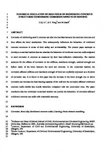
The constitution of a real-time 3D image of the liver in augmented reality requires high precision to help the surgeons. In order to be compliant with an expected medical precision error below 2 mm, we make use of a numerical model, based on finite element (FE) analyses [4] integrating the true mechanical behaviour of the biological tissues (liver and vascularisation) and optimisation procedure. The purpose is to integrate the specific biological mechanical behaviour of the liver as well as its microstructure and determine the way they affect the macroscopic behaviour. Although the integration of the microstructural behaviour into a macroscopic response is sometimes complex [5–12], the use of continuous model [7,8] enable precise determination of the overall mechanical behaviour. To build a compliant mechanical model, the two following steps are applied: first, the geometry of the liver including its vascularisations is extracted from patient imaging data (MRI or CT scan) and integrated within the numerical FE model (see Fig. 1). At the same time, mechanical properties of the different tissue constituting the liver (liver, vessels wall), are extracted from experimental tests from non-invasive techniques [13,14]. From this, a complete geometry, including all mechanical properties for each internal structures, is built. Then, a homogenised FE numerical model [15] is developed for the surgeon, providing precise deformations and displacements. In this study, liver and vessels are modelled using linear elastic material, with a Young modulus of 3 kPa for the liver, 620 kPa for the vessels, and Poisson coefficient of 0.49 [16]. To validate the macroscopic behaviour of the numerical model depending on its integrated vascularisation, numerical indentation tests are simulated and compared to experimental macroscopic information available in the literature [17]. In a second stage, since a homogenized FE numerical model is extremely slow to compute, an optimisation procedure is required. The complete model is processed first through a computational learning method (Locally Liner Embedding – LLE [18–20]). Here, the patient liver and vascularisation specific geometries are treated as parameters and, for each new patient geometry, can be expressed as a combination of all the known geometries in the data-base. Also, since the deformations applied to the liver are bounded to the scope of surgical motions, they can similarly be expressed as parameters. Then,
Fig. 1. FE model of Liver including its vascularisation.
M. Kugler et al. / Numerical simulation and identification of macroscopic vascularised liver behaviour
S109
considering the set of parameters describing the geometries, the mechanical properties and the possible external constrains applied during a surgery, solutions are computed for all given values of the different parameters using a Proper Generalized Decomposition (PGD) Method [21,22]. This allows computing all sets of corresponding internal and external deformed positions of the liver. All these states are then stored in a data-base and will be used to reconstruct, through a real time linear combination, all the local real deformations through the whole organ during the surgery. 3. Results
(a)
AU
TH
O
R
C
O
PY
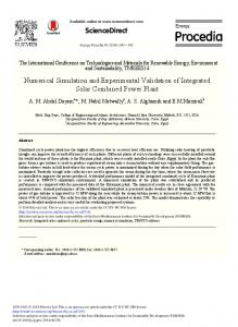
The numerical model validation is completely dependent on each of the patient’s mechanical response for the real time surgery follow up. Hence, to validate the macroscopic behavior as a function of the vascularisation distribution, we present here the comparing results from indentation tests obtained from the numerical model with literature data [17]. The results are presented on Fig. 2. Experimental data [17] were obtained on three pigs’ livers (without knowledge of vascularisation distribution) by force-displacement measurements with an indenter tip of 8 mm radius at different indentation depths and with constant indentation speed of 1 mm/s. In comparison, the numerical results
(b) Fig. 2. Comparison of force–displacement indentation curves. (a) Effects of microstructure and vascularization on the force/displacement curve. (b) Comparison of numerical and experimental results. Symbols represent the mean values and vertical bars correspond to the variations of the different positions of loading. Blue and square data are extracted from Samur [17]. Red and circle data are the numerical results of the current work.
S110
M. Kugler et al. / Numerical simulation and identification of macroscopic vascularised liver behaviour
O
PY
were obtained on a single liver model with identical indenter tip geometry and speed but for different depths and positions of indentation. This has the effect of changing the macroscopic liver rigidity as a function of the vascularisation distribution and can be compared (in range) to Samur’s experimental data. The numerical results are in very good agreement with the experimental data confirming that the mechanical effects due to the differences in mechanical properties and distribution of the vascularisation is well integrated within the numerical model. Experimental results show an increase of the standard deviation measurements with increase of indentation depth. This is to be expected without knowledge of the internal liver microstructures. On the contrary, the standard deviations of the numerical model are shifted towards the higher values (Fig. 2(b)). This shows the influence of the indentation position where harder tissues (blood vessels) are localised (Fig. 2(a)). Since the maximum indentation depth remains reasonably small (10 mm) compared to the liver thickness (between 100 mm and 200 mm), it seems appropriate to think that the measured variabilities are only due to the vascularisation distribution effects rather than the liver geometry. Further work is required to evaluate the impact of indentation speed (viscosity effects), depth and indenter size. More accurate non-linear material behaviour would also be needed for deeper indentation validations (large deformations).
C
4. Conclusion
AU
Conflict of interest
TH
O
R
The construction of a precise augmented reality image of the liver in real time is strongly bounded to the associated numerical model providing the organ internal and external positions. Here we developed a macroscopic liver model integrating vascularisation. Numerical mechanical indentation tests were performed on a vascularised liver and validate the overall macroscopic behaviour compared with literature data. These results will enable the construction of a complete homogenised numerical model to be used as a real help for the surgeons.
The authors have no conflict of interest to report.
References
[1] A. Hostettler, S.A. Nicolau, C. Forest, L. Soler and Y. Remond, Real Time Simulation of Organ Motions Induced by Breathing: First Evaluation on Patient Data, Biomedical Simulation, M. Harders and G. Székely, eds, Springer, Berlin/Heidelberg, 2006, pp. 9–18. [2] A. Hostettler, S.A. Nicolau, L. Soler and Y. Rémond, Toward and accurate real-time simulation of internal organ motions during free breathing from skin motion tracking and an a priori knowledge of the diaphragm motion, Computer Assisted Radiology Surgery 2 (2007), S100–S102. [3] D. Selle, B. Preim, A. Schenk and H.O. Peitgen, Analysis of vasculature for liver surgical planning, IEEE Transaction Medical Imaging 21 (2002), 1344–1357. doi:10.1109/TMI.2002.801166. [4] O. Zienkiewicz and R. Taylor, The Finite Element Method, McGraw-Hill, London, 1977. [5] N. Auffray, F. Dell’Isola, V.A. Eremeyev, A. Madeo and G. Rosi, Analytical continuum mechanics à la Hamilton–Piola least action principle for second gradient continua and capillary fluids, Mathematics and Mechanics of Solids 20 (2015), 375–417. doi:10.1177/1081286513497616.
M. Kugler et al. / Numerical simulation and identification of macroscopic vascularised liver behaviour
S111
AU
TH
O
R
C
O
PY
[6] N. Auffray, F. Dell’Isola, V. Eremeyev, A. Madeo, L. Placidi and G. Rosi, Least action principle for second gradient continua and capillary fluids: A Lagrangian approach following Piola’s point of view, Advanced Structured Materials 38 (2014), 89. [7] A. Madeo, F. Dell’Isola and F. Darve, A continuum model for deformable, second gradient porous media partially saturated with compressible fluids, Journal of the Mechanics and Physics of Solids 61 (2013), 2196–2211. doi:10.1016/j. jmps.2013.06.009. [8] F. Dell’Isola, G. Sciarra and S. Vidoli, Generalized Hooke’s law for isotropic second gradient materials, Proceedings of the Royal Society A: Mathematical, Physical and Engineering Sciences 465 (2009), 2177–2196. doi:10.1098/rspa.2008. 0530. [9] G. Sciarra, F. Dell’Isola, N. Ianiro and A. Madeo, A variational deduction of second gradient poroelasticity, Part I: General theory, Journal of Mechanics of Materials and Structures 3 (2008), 507–526. doi:10.2140/jomms.2008.3.507. [10] A. Madeo, F. Dell’Isola, N. Ianiro and G. Sciarra, A variational deduction of second gradient poroelasticity II: An application to the consolidation problem, Journal of Mechanics of Materials and Structures 3 (2008), 607–625. doi:10.2140/ jomms.2008.3.607. [11] G. Sciarra, F. Dell’Isola and O. Coussy, Second gradient poromechanics, International Journal of Solids and Structures 44 (2007), 6607–6629. doi:10.1016/j.ijsolstr.2007.03.003. [12] F. Dell’Isola, G. Sciarra and R.C. Batra, A second gradient model for deformable porous matrices filled with an inviscid fluid, Solid Mechanics and Its Applications 125 (2005), 221–229. doi:10.1007/1-4020-3865-8_25. [13] A. Hostettler, D. George, Y. Rémond, S.A. Nicolau, L. Soler and J. Marescaux, Bulk modulus and volume variation measurement of the liver and the kidneys in vivo using abdominal kinetics during free breathing, Computer Methods and Programs Biomedicine 100 (2010), 149–157. doi:10.1016/j.cmpb.2010.03.003. [14] M. Nierenberger, D. George, D. Baumgartner, Y. Rémond, S. Ahzi, R. Wolfram, J.L. Kahn and R.A. Rahman, Towards building a multiscale mechanical model for the prediction of acute subdural hematomas, in: ASME 11th Biennial Conference on Engineering Systems Design and Analysis, 2012, pp. 261–266. [15] R.A. Rahman, D. George, D. Baumgartner, M. Nierenberger, Y. Rémond and S. Ahzi, An asymptotic method for the prediction of the anisotropic effective elastic properties of the cortical vein: Superior sagittal sinus junction embedded within a homogenized cell element, Journal Mechanics Materials Structures 7 (2012), 593–611. doi:10.2140/jomms. 2012.7.593. [16] A. Nava, E. Mazza, M. Furrer, P. Villiger and W.H. Reinhart, In vivo mechanical characterization of human liver, Medical Image Analysis 12 (2008), 203–216. doi:10.1016/j.media.2007.10.001. [17] E. Samur, M. Sedef, C. Basdogan, L. Avtan and O. Duzgun, A robotic indenter for minimally invasive characterization of soft tissues, International Congress Series 1281 (2005), 713–718. doi:10.1016/j.ics.2005.03.117. [18] S.T. Roweis and L.K. Saul, Nonlinear dimensionality reduction by locally linear embedding, Science 290 (2000), 2323– 2326. doi:10.1126/science.290.5500.2323. [19] D. Gonzalez, J.V. Aguado, E. Cueto, E. Abisset-Chavanne and F. Chinesta, kPCA-based Parametric Solutions within the PGD Framework, Archives of Computational Methods in Engineering (2016), 1–18. In press. doi:10.1007/s11831-0169173-4. [20] D. Gonzalez, E. Cueto and F. Chinesta, Computational patient avatars for surgery planning, Annals Biomedical Engineering 44 (2016), 35–45. [21] F. Chinesta, R. Keunings and A. Leygue, The Proper Generalized Decomposition for Advanced Numerical Simulations, Springer International Publishing, 2014. [22] S. Niroomandi, D. Gonzalez, I. Alfaro, E. Cueto and F. Chinesta, Model order reduction in hyperelasticity: A proper generalized decomposition approach, International Journal for Numerical Methods in Engineering 96 (2013), 129–149.