Sendai nucleocapsid calculated from an electron micrograph showed that the .... about Io 9 to ~o 1° particles/ml., and very few nucleocapsids could be found in ...
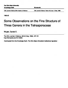
I4I
J. gen. Virol. (I97O), 6, 1 4 i - i 5 o Printed in Great Britain
Observations on the Structure of the Nucleocapsids of some Paramyxoviruses By J. T. F I N C H Medical Research Council Laboratory of Molecular Biology, Hills Road, Cambridge AND A. J. G I B B S
John Curtin School of Medical Research, Australian National University, Canberra (Accepted 9 September I969) SUMMARY
The intact particles and nucteocapsids of mumps, Sendai and measles viruses are of closely similar appearance, size and structure. The intact particles are about 15o nm. in diameter. The filamentous nucleocapsids have a modal length of about ~. I/Am., and are constructed of subunits arranged as a single start helix of pitch 5.0 nm. for Sendai virus, and about 6"o nm. for mumps and measles viruses. A (helical) projection of the structure of the Sendai nucleocapsid calculated from an electron micrograph showed that the structural subunits are hour-glass-shaped and are arranged in the helix with their long axes inclined at an angle of about 6o ° to the long axis of the particle. There are probably J ~ or I3 subunits in each turn of the basic helix. Optical diffraction patterns of electron micrographs of mumps and measles nucleocapsids show that they have closely similar structures.
INTRODUCTION
Electron-microscope investigations (for example Horne, et al. 196o) have shown that the particles of paramyxoviruses are rounded but irregular in shape and mostly about 15° rim. in diameter. They possess an outer envelope containing lipid and covered with spikes, within which is a tightly coiled narrow filament called the nucleocapsid, which contains the ribonucleic acid of the virus particle. The nucleocapsids of paramyxo-viruses, unlike those of the myxoviruses (the influenza viruses), can be obtained relatively easily, either directly from infected cells or by disrupting the virus particles, and they all show a marked periodicity of about 5.0 nm. along their lengths in electron micrographs. The earlier electron-microscope images were interpreted in terms of a double helical structure (Horne & Waterson, 196o) but in later electron micrographs, especially of veiy flexuous nucleocapsids, a single helical arrangement was evident (Choppin & Stoeckenius, 1964). There has been much recent interest in the size and composition of these nucleocapsids. The paramyxoviruses that have been studied are: Mumps virus [cryptogram R/* : */* : S/E :V/O], Newcastle disease virus [cryptogram R/x : (6-6)/0.9: S/E :V/O], Sendai virus (haemagglutinating virus of Japan) [cryptogram R/I : (6-6)/*: S/E :V/O], SV 5 (simian virus 5) [cryptogram R/x : (6.3)/0-9: S/E :V/O]. IO-2
I42
J. T. F I N C H A N D A. J. G I B B S
The nucleocapsids of Sendai virus (Hosaka, Kitano & Ikeguchi, 1966), Newcastle disease virus (Compans & Choppin, ~967a), SV 5 (Compans & Choppin, ~967b) and mumps virus (Hosaka & Shimizu, I968 ) have all been shown to have a length of about r #m. and width of about I8 nm. The electron-microscope study reported here has confirmed these dimensions in the cases of Sendai and mumps viruses and shows that the nucleocapsid of measles virus [cryptogram */*:*/*:S/E:V/O], which is also a paramyxovirus, is of similar size. Optical diffraction from the electron-microscope images confirms that these three nucleocapsids have very similar single helical structures. In the case of a Sendai nucleocapsid, a projection of the structure down the basic helix has been computed from an electron-microscope image using the method of DeRosier & Klug (t968). METHODS
Sendai and mumps virus (Enders strain) were inoculated on to the chorioallantois of 1~- and 7-day-old embryonated hens' eggs and incubated at 35 ° for 2 and 6 days respectively before harvesting the chorioallantoic fluid. Measles virus was grown in BSC-I cells and was kindly supplied by Dr M. F. Warburton of the Commonwealth Serum Laboratories, Melbourne. The virus-containing fluids were clarified by centrifuging at 4ooo g for 5 rain. ; the virus was sedimented by centrifuging at 4o,ooo g for I hr, resuspended in gelatin saline (0"85 °/o (w/v) sodium chloride, 0.05 ~o (w/v) gelatin, o-OzM-boric acid, o-ooaM-magnesium chloride, o-ooo25M-calcium chloride, pH 7.0) clarified at 8,ooo g for 2 rain., sedimented by centrifuging at 75,0oo g for 45 min. and finally resuspended in o.o4M-borate buffer pH 8-o. Nucleocapsids were obtained from the virus particles by treatment with ether (Hoyle, I95z; Sch~ifer & Zillig, 1954; Hoyle, H o m e and Waterson, I96~). A quarter volume of diethyl ether was layered, without stirring, on top of the preparation of purified virus, and the preparation centrifuged at 8,ooo g for 5 rain. to remove the slight precipitate that had formed. For electron microscopy the untreated or ether-treated virus preparations were mixed with an equal volume of neutral 4 % (w/v) sodium phosphotungstate (PTA) negative stain. Droplets of the mixture were pipetted on to carbon films supported on electron-microscope specimen grids, and the excess fluid removed with filter paper. The mounted preparations were examined and 'photographed' in a Siemens Elmiskop I at a magnification of x 2o,ooo or x 4o,ooo. The diameters of intact virus particles and the lengths of the nucleocapsid filaments were measured by comparison with tobacco mosaic virus particles as internal standards of length (300 nm.): these were added to the droplet of stained virus preparation on the carbon film by briefly touching the droplet with the freshly cut edge of a tobacco leaf infected with tobacco mosaic virus. The helical pitches of the nucleocapsids were measured using glutaraldehyde-fixed catalase crystals as an internal standard (Wrigley, I968): these were pipetted on to the carbon film immediately before the droplet of stained virus preparation. Periodicities in the electron-microscope images were analysed and measured by optical diffraction as described by Klug & Berger (1964).
Structure of nucleocapsids of paramyxoviruses
I43
RESULTS
Measles virus and its nucleocapsids were much less easy to obtain in quantity than the other two viruses: concentrated preparations of measles virus contained only about Io 9 to ~o1° particles/ml., and very few nucleocapsids could be found in such preparations after treating with ether. However, sufficient were examined to show that measles is little different from Sendai and mumps viruses. The intact particles of the three viruses were indistinguishable in appearance. All were variable in size and shape, but most particles were approximately circular in outline and about I5 o n m . in diameter. Figure I shows the diameters of particles of mumps and Sendai viruses; the 25 particles of measles virus that were measured also had a mean diameter of about 150 nm. !
!
!
!
M°mPvJ m
--(c) TMV
I O0
200
300
400
rln"~,
Fig. i. Histograms of the diameter of negatively stained particles of (a) Sendai virus, (b) mumps virus, and (c) the lengths of particles of tobacco mosaic virus mounted on the same microscope grids. The vertical scale is in units of 5 particles. Typical electron micrographs of nucleocapsids of the three viruses are shown in Fig. 2. In each case the edges of the images are highly serrated, with a polar appearance as that of a series of interlocking arrow-heads pointing to one end of the nucleocapsid. The nucleocapsid filaments from Sendai virus were fairly straight (Fig. 2a) and of variable length (Fig. 3). There is, however, a maximum in the length distribution close to I. ~ #m. which is interpreted as the length of unbroken filaments and which is close to the value of ~.o/~m. reported by Hosaka et al. 0966). Of the filaments around this maximum (longer than o'9 #m. and less than I-3/~m.) the modal length is I.r I5/~m.
I44
J.T. FINCH
A N D A. J. G I B B S
Fig. 2. Electron micrographs of nucleocapsids negatively stained with sodium phosphotungstate. (a) Sendai (x 165,ooo), (b) mumps ( x 13o,ooo), (c) measles (x 150,000),
Structure of nucleocapsids of paramyxoviruses
]45
The nucleocapsid filaments of mumps are both more flexuous (Fig. zb) and more variable in length (Fig. 3) than those of Sendal virus. Much of the length variation was undoubtedly caused by breakage and re-aggregation of the filaments--there are frequent slight breaks and dislocations which were disregarded when measuring their lengths. Most of the mumps filaments were less than ['5 #m. long, with the most noticeable maxima in the length distribution diagram at about ~'] and o'5 #m, though some filaments were as long as 4/~m. To try to identify the modal length of mumps nucleocapsids, the length-distribution diagram was smoothed by calculating 'running sums'; the sum of the number of filaments in every five adjacent length classes in the diagram was plotted at the central length class of the five. The smoothed lengthdistribution diagram had maxima at o.66 and I"o5/tm. (the latter containing most particles), and smaller maxima at ~.62, 2.o8 and 3"o3/~m. The simplest explanation for I
I
I
,.,l 2.0
I 3.0
- - ( a ) Sendai
40
°I 0
(c) TMV I 1.0
/~m. Fig. 3. Histograms of the lengths of (a) Sendal and (b) mumps nucleocapsids and (c) tobacco mosaic virus particles. these results is that the maximum at [-o5 #m. is the modal length (in agreement with the value of I-o/~m. reported by Hosaka & Shimizu, I968), that at o-66 #m. is of broken nucleocapsids, which can aggregate with those of the modal length to give the maximum at [.62/~m., and the maxima at 2.o8 and 3"o3 #m. are of aggregates of two and three nucleocapsids of modal length. The measles nucleocapsids (Fig. 2 c) were much less flexuous than those of mumps, but not as straight as those of Sendal virus. Of 4[ measles nucleocapsids measured, half were less than 0. 5 #m. long (mostly about o.2 #m.), and the mean length of all longer ones (o. 5 to I'75 #m.) was I.~o #m.
I46
J. T. FINCH AND A. J. GIBBS
The structure of Sendai nucleocapsids The optical diffraction pattern from a length of Sendai nucleocapsid is shown in Fig. 4b. The pattern is typical of that from a regular helical arrangement of subunits. Strong near-meridional intensity occurs along a layer line which corresponds to a helix of pitch close to 5"0 nm. The position of the first intensity maximum shows that this is a single-start helix. Optical transforms from a number of images showed that the pitch of this helix was always within the range 5"o _+o.2 nm. Also visible in the optical transform is near-equatorial intensity occurring along a layer line of spacing 1/22"2 nm. corresponding to near-vertical helices of subunits (or strictly, to helices nearly parallel to the long axis of the nucleocapsid). It has not so far been possible to determine the hand of these helices or that of the basic helix or even to rule out any of the four possible combinations. Nevertheless the data on the layer line corresponding to the basic helix are sufficient to enable a projection of the structure down this helix to be calculated. The procedure used was that described by DeRosier & Klug 0968). The image shown in Fig. 4a was converted, using a microdensitometer, to a set of numbers representing the optical density at regular points on a grid across the image. These numbers were transformed by computation into a set of Fourier amplitudes and phases. In particular, the amplitudes and phases of the Fourier components arising from the helical structure of the filament (i.e. those corresponding to the layer lines already noted in the optical transforms) were determined. Considering firstly the ~/5.o nm. layer line, the phases of corresponding maxima on opposite sides of the meridian differ by ~8o°, as would be expected since they arise from opposite sides of a single-start basic helix. The first and second maxima are comparable in amplitude and arise from features in the original particle at radii about 8.o and 3"o nm. The phases of these two maxima differ by about 18o °. The amplitudes and phases of the Fourier components along the layer line of spacing 1/2"5 nm. are in agreement with it being the second order of that at i/5.o nm. From the data on these two layer lines and the equator, the projection of the structure along the basic helix on to a plane through the particle axis was calculated by Fourier-Bessel transformation (Klug, Crick & Wyckoff, 2958), and is shown in Fig. 4c. Contours have been drawn at regularly spaced density levels. The thick contour is one which preserves both the continuity of the structure between inner and outer radii and the discrete protuberances on the outside of the particle and is taken to indicate the boundary of the projected structure. The nucleocapsid has a central hole of diameter about 5 nm. and the outermost limit of the structure has a diameter of about 2o nm.--rather bigger than the value obtained by directly measuring the width of the particle image. However, this larger value is perhaps more likely to be correct since it is obtained using information from the centre of the image of the nucleocapsid, whereas it is difficult to measure accurately the width of a filament of this type as the positions of the extreme edges of the filament are difficult to determine in relatively thick stain. However, the radial density distribution which is included in the helical projection (as the contribution from the equatorial layer line) must be regarded as somewhat tentative since the Fourier-Bessel transformation of the equator is based on the assumption that the distribution of stain around the particle axis is cylindrically symmetric which, because of the interaction with the substrate, it is clearly not. Nevertheless, apart from this possibility of radial distortion, the broad
Structure of nucleocapsids of paramyxoviruses
~47
1 2.5 nm.
1 5"0 nrn. 1 22.2 rim.
~ "~
I 2"0
1 4"0
I ., I F 6-0 8"0 1 0 " 0
Radius (nm.)
Fig. 4. (a) Electron-microscope image of a length of Sendai nucleocapsid negatively stained with sodium phosphotungstate ( x 4oo,ooo). (b) Optical diffraction pattern from (a). (c) Computed projection of the structure of (a) down the basic helix on to a plane through the axis of the nucIeocapsid. The shaded area indicates the probable extent of the projection of a single protein subunit. (Since the computer output is not square, the two scales shown are not identical.)
I4 8
J. T. F I N C H
A N D A. J. G I B B S
features of Fig. 4 c are correct and very similar density distributions have been obtained by similarly processing other images. The helical projection has density maxima at radii of about 3"0 and 8.o nm. Since both of these positions appear very exposed, it is unlikely that either of them corresponds to the position of the RNA, but to the protein fraction of the nucleocapsid. The projection of the protein structural subunit of the helical arrangement, which as discussed below is probably one protein molecule, thus includes one inner and one outer density maximum in Fig. 4c, and to be consistent with the observed convexity and concavity of the ends of the nucleocapsids, the general direction of the subunits must be as indicated by the shading in the Plate. The long axes of the subunits thus make an angle of about 60 ° to the long axis of the nucleocapsid. Between the subunits there is a density shoulder at a radius of about 5 nm. which could be the location of the RNA: this radius is rather smaller than the value of 8 nm. suggested by Hosaka & Shimizu 0968) for the radius of the R N A in Newcastle disease virus, based on composition of the nucleocapsids and assuming the same weight per length as the R N A in tobacco mosaic virus. The corresponding maxima on opposite sides of the meridian on the layer line of spacing 1/22"2 nm., differ in phase by close to I8O°, showing that there is an odd number of near vertical helices (this is the integral number closest to the number of subunits per turn of the basic helix). The first intensity maximum occurs at a reciprocal spacing of o.oo28 nm. -1, which would be due to features in the structure at a radius of 6q, 7"3, 8"5 or 9"7 nm. if the number of helices were 9, t I, I3 or ~5 respectively. It is possible to see these helices running right across the particle image when it is viewed at a glancing angle in the direction of the particle axis. One would therefore expect the same outer radius to be contributing here as in the layer line of spacing I/5-o nm., namely about 8.o nm., and on this basis the number of near-vertical helices is either 11 or I3. An independent estimation of this number can be made from the electron-microscope images of short lengths of Sendai nucleocapsids which occur occasionally, especially in old preparations. When seen end-on the appearance is disc-like with a number of protuberances, presumably arising from the projections of the near-vertical helices. Of I2 discs examined, the number of protuberances counted was within the range 9 to I3 and mostly I I.
Mumps and measles nucleocapsids The optical transforms from images of straight lengths of these nucleocapsids showed features very similar to those of Sendal. While the pitch of the basic helix for both these viruses is somewhat larger (6.o + o'3 nm.), the intensity distributions along the layer lines corresponding to this helix are in all three cases very much alike, indicating that they are structurally very similar, i.e. they are built of structure units arranged at an angle to the radial direction of the particle.
DISCUSSION
Our observations on the particles of these three paramyxoviruses confirm and extend the observations of other workers, and show that except in two trivial aspects the three viruses are morphologically indistinguishable; they differ in the flexuousness, and in the pitch of the basic helix, of the nucleocapsids. Sendai virus has straight nucleo-
Structure of nucleocapsids of paramyxoviruses
I49
capsids, measles virus has slightly more flexuous nucteocapsids resembling those of SV 5 (Choppin & Stoeckenius, I964; Compans & Choppin, I967b), and mumps has very flexuous fragile nucleocapsids like those of Newcastle disease virus (Kingsbury & Darlington, 1968 ). The difference in pitch of the basic helix of the nucleocapsids of the three viruses is less than the variation in pitch reported for a single paramyxovirus by other workers (Choppin & Stoeckenius, I964; Kingsbury & Darlington, I968). These differences may indicate how tightly adjacent turns of the helix are bound together, and seem to be correlated with differences in the fragility of the nucleocapsids of the different viruses. The nucleocapsids of all paramyxoviruses show a similar herring-bone like structure which, as has been shown above, is because the structure units are not set perpendicular to the long axis of the particle. Of viruses other than the paramyxoviruses, only the nucleocapsids of the myxoviruses seem to have a similarly angled arrangement of subunits; the subunits of the helically constructed particles of some plant and bacterial viruses seem to have their subunits arranged with their long axes more nearly perpendicular to the long axis of the particle, though the subunits of some, for example, tobacco rattle virus [cryptogram R/I:2"3/5:E/E:S/Ne], may be slightly angled (Harrison & Woods, I966). This difference may be because the nucleocapsids of myxoviruses and paramyxoviruses have to be flexible enough to be tightly coiled within the virus particles, whereas the others do not. The contact surface between adjacent turns of the basic helix of the nucleocapsids of the paramyxoviruses is approximately conical, and thus contact would be maintained between adjacent turns of helix and the RNA presumably remain protected, even when the nucleocapsid was sharply curved. The angled arrangement of the subunits also explains the observation of Choppin & Stoeckenius (I964) that in the case of SV 5 the length of the short axis of the subunits was less than the pitch of the helix. The angling of the subunits and their hour-glass-like shape gives rise to quite complex superposition patterns in the electron-microscope images of negatively stained particles, and it is the interpretation of these in terms of structural detail from a single radius that has given rise to reports of two-start helical structures ( H o r n e & Waterson, I96o; Hosaka, Nishi & Fukai, I96I). Similar reports of two-start helices in myxovirus nucleocapsids could have the same origin. We have found the number of near-vertical helices (i.e. subunits per turn) in the nucleocapsid of Sendai virus to be 11 or i3, whereas Hosaka (1968) deduced that the rotational symmetry of end-on views of short lengths of the nucleocapsid was 15. Hosaka deduced this number however by rotationally superposing images, a method that is unreliable unless the image being rotated can be accurately centred, and, more important still, unless one already knows the substructure, for otherwise one cannot decide which superposition pattern represents it best. The results given above show that there are between 2400 and 2800 structure units in the Sendal nucleocapsid. Most recent reports agree that the paramyxoviruses have a nucleic acid with a molecular weight near 6"5 x lO6 (Compans & Choppin, I967b, ~968; Klenk & Choppin, I969) and that this constitutes about 4"5 % of the nucleocapsid. These values when combined with our observations suggest that the structure units of the paramyxoviruses have a molecular weight between 5"2 x lo 4 and 6 x Io 4, which agrees well with the estimates of 5"4 x lo 4 and 6.2 x Io 4 for the molecular weight of the nucleocapsid protein of Newcastle disease virus obtained by acrylamide
I50
J. T. F I N C H
AND
A. J. G I B B S
gel electrophoresis studies by Haslam, Cheyne & White, (I969) and Evans & Kingsbury (I969) respectively. The structure unit of the nucleocapsid thus contains one chemical protein subunit. The computations described above were carried out using a programme system of Dr D. J. DeRosier and we thank him and Dr A. Klug for helpful discussions. We also thank Miss Marion Holder for much technical assistance. REFERENCES CHOPPIN, P. W. & STOECKENIUS, W. (I964)- T h e m o r p h o l o g y of SV 5 virus. Virology 23, 195. COMPANS, R. W. & CHOPPIN, P. W. (I967a). T h e length of the nucleocapsid of Newcastle disease virus. Virology 33, 344. COMPANS, R. W. & CHOPPIN, P. W. (1967b). Isolation a n d properties o f the helical nucleocapsid o f parainfluenza SV 5. Proceedings of the National Academy of Sciences of the United States of America 57, 949COMPANS, R. W. & CHOPI"IN,1'. W. (1968). T h e nucleic acid of parainfluenza virus SV 5. Virology 35, 289. DEROSlER, D.J. & KLU6, A. (1968). R e c o n s t r u c t i o n of three-dimensional structures f r o m electron micrographs. Nature, London 2x7, I3O. EVANS, M. J. & KINGSBURY, D. W. (1969). Separation of Newcastle disease virus proteins by polyacryla m i d e gel electrophoresis. Virology 37, 597. HARRISON, B. D. & WOODS, R. D. (1966). Serotypes a n d particle dimensions of tobacco rattle viruses from E u r o p e a n d America. Virology 28, 61o. HASLAM, E. A., CHEYNE, I. M. & WHITE, D. O. (1969). T h e structural proteins of Newcastle disease virus. Virology 39, I I8. HORNE, R. W. & WATERSON, A. a. (1960). A helical structure in m u m p s , Newcastle disease a n d Sendal viruses. Journal of Molecular Biology 2, 75. I-IORNE, R. W., WATERSON, A. P., WILDY, P. & FARNHAM, A. E. (1960). T h e structure a n d c o m p o s i t i o n o f the myxoviruses. I. Electron microscope studies of the structure of m y x o v i r u s particles by negative staining techniques. Virology xi, 79. HOSAKA, Y. (i968). Isolation a n d structure of the nucleocapsid of HVJ. Virology 35, 445. HOSAKA, Y. & SHIMIZU, K. (1968). L e n g t h s of the nucleocapsids of Newcastle disease a n d m u m p s viruses. Journal of Molecular Biology 35, 369. HOSAKA~ Y., KITANO, H. & IKEGUCHI, S. (1966). Studies on the p l e o m o r p h i s m of H V J virions. Virology 29, 205. HOSAKA, Y., NISHI, Y. & FUKAI, K. (1961). T h e structure of HVJ. II. T h e fine structure of the subunits. Bikens Journal 2, 243. HOYLE, L. (1952). Structure of the influenza virus. T h e relation between biological activity and the chemical structure of virus fractions. Journal of Hygiene 5o, 229. HOYLE, L., HORNE, R. W. & WATERSON, A. V. (I96I). T h e structure a n d c o m p o s i t i o n of the myxoviruses. i I . C o m p o n e n t s released f r o m the influenza particle by ether. Virology I3, 448. KINGSBURY, D. W. & DARLINGTON, R. W. (1968). Isolation and properties of Newcastle disease virus nucleocapsid. Journal of Virology 2, 248. KLENK, H. D. & CHOPPIN, P. W. (I969). Chemical c o m p o s i t i o n of the parainfluenza virus SV 5. Virology 37, t55. KLUG, A. & BERGER, J. E. (I964). A n optical m e t h o d for the analysis of periodicities in electron micrographs, a n d s o m e observations on the m e c h a n i s m of negative staining. Journal of Molecular Biology io, 565. KLUG, A., CRICK, F. H. C. & WYCKOFF, H. w. (1958). Diffraction by helical structures. Aeta Crystallographiea i i , 199. SCH;4FER, W. & ZlLLIO, W. (I954). U b e r den A u f b a u des Virus-Elementarteilchens der klassischen Gefliigelpest. I. G e w i n n u n g , physikalischchemische u n d biologische Eigenschaften einiger Spaltprodukte. Zeitschrift fiir Naturforsehung 9b, 779-788. WRIOLEY, N. ~. (1968). T h e lattice spacing of crystalline catalase as a n internal s t a n d a r d o f length in electron microscopy. Journal of Ultrastrueture Research 24, 454.
(Received 3I July 1969)