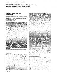
Sep 6, 1989 - c-myc has been found in the genome of the human (Bernard et al. ,1983), the cat ..... human c-myc protein (Dang and Lee, 1988), and 5' to this.
committed cells (Metcalfe, 1987), and final differentiation of cells within a ..... by Drs Jane Dodd and Thomas Jessell (Columbia), antibodies against the glial ...
Sep 10, 1983 - ZE'EV LEV,'* NOEMI LEIBOVITZ,1 ORIT SEGEV,1 AND BEN-ZION SHILO2. Department ofBiology, Technion-Israel Institute of Technology, ...
Sep 10, 1983 - agarose gel, transferred to nitrocellulose, and hybridized with the genomic probe. A single transcript, 6.2 kilobases. (kb) long, reacted with the ...
of novel microsatellite markers for the Common. Pheasant (Phasianus colchicus) using RAD-seq. Biao Wang, Xuan Xie, Simin Liu, Xuejing Wang, Hong Pang ...
Stem cells possess two main attributes, namely, a self-renewal capacity and .... The Sox2 protein contains one HMG box, as well as a transactivation domain in ...
caused by exposure to nonionizing radiation and may play a mechanistic role in ... induction by ionizing radiation, we found concurrent activation of K-ras and ...
al., 1987; Ben-Bassat et al., 1997; Kersemaekers et al., 1999). Moreover, HeLa ..... Azizkhan JC, Jensen DE, Pierce AJ and Wade M. (1993). Crit. Rev. Eukaryot.
David Polsky*,1 and Carlos Cordon-Cardo2. 1Oncology Section of the Skin and .... heterodimer. Oncogenes in melanoma. D Polsky and C Cordon-Cardo. 3088.
ago, with the characterization of the ipt, iaaM and iaaH oncogenes. However, the simplistic .... tumefaciens oncogene-mediated IAA biosynthesis pathways.
Colour genes, oncogenes and melanocyte differentiation. DOROTHY C. BENNETT. St George's Hospital Medical School, Cranmer Terrace, London SW17 ORE, ...
The heterogeneous biology of cancer cells or the adjustments ... identification of the key genes regulating cellular response to therapy with ... With an in vivo half-life range of several minutes and size of. < 50 KDa and 6nm, ... paclitaxel treatme
Jul 8, 2014 - at early stages, but the female phallus later regresses. Then, we compared male and female genital development be- tween ducks and ...
Apr 1, 1993 - bling Menetrier's disease in transgenic mice overexpressing transforming growth factor a in the stomach. J. Clin. Invest., 90: 1161-1167. 1992.
27 Oct 2005 - How did the researchers know ... they analyzed 10 separate 500-kb ... that affords a fascinating glimpse ... of mechanisms of gene regulation .... enzyme and its partner Loqacious (Loqs), a double-stranded RNA-bindingâ ...
Jun 25, 2007 - Publisher: Taylor & Francis. Informa Ltd Registered in ... Taylor and Francis 2007 ...... Cook, J.K.A., Kinloch, S. & Ellis, M.M. (1993b). In vitro and ...
Received December 29, 1986; accepted February 12,1987. cellular genes. Thus, each viral oncogene possesses a cellu- lar counterpart. (proto-oncogene) that ...
pleiotropic cellular effects. Since Ras is ... oncogenes and tumor suppressors may affect glutamine ... ing the Warburg effect: the metabolic requirements of cell.
Sep 30, 2010 - transcribed as primary miRNA precursors (pri-miRNAs), which are converted into ,100 ... the functions of p- oncomirs and p-mirsupps can be thought of as representing two ... miRNA family data in miRBase [30] using the method of Zhang e
Oct 22, 2009 - evolution and development of dermal elements in the pectoral apparatus, .... with the addition of Bismarck brown Y, and Verhoeff elastin stain.
layer of the lateral plate mesoderm. Controversy surrounds the origin of the scapula, a major component of the shoulder girdle, with both somitic and lateral plate ...
interspecific brood parasitism will not be favored in precocial birds. ... this difference between altricial and precocial species: (1) altricial birds have relatively ...
The genera Leucocytozoon, Haemoproteus and. Hepatocystis. In: Kreier JP, Baker JR, editors. Parasitic protozoa. 2nd ed. Cambridge: Academic Press; 1993. p.
&p.1:Abstract The musculature of the vertebrate tongue is composed of cells recruited from the somites. In this pa- per we have investigated the migration and ...
the cell membrane and cytoplasm to the genome are obviously implicated inthis .... v-myc + II-mil v-myb. II-myb + v-ers. lI-erbA + v-erbB. Fig. 1. c-myc expression in the early chick ... cells. c-myc expression was clearly linked to a process of cell.
Int..I.
Dc\'. Riolo J-I: 61-6X (11)1)0,
hi
Oncogenes and aVian development FRANCOISE DIETERLEN-LiEVRE'*, THIERRY JAFFREDO', NAIMA BACHNOU', ALA EDDIN AL MOUSTAFA' and SIMON SAULE' Ilnstitut
d'Embryologie
Cellula ire et Moleculaire du CNRS et du College de France, Nogent JUnite d'Onc%gie Moleculaire, Instit(Jt Pasteur, Lille, France
sur Marne
and
ABSTRACT In order to detect signs of oncogene activity and elucidate their possible role in avian ontogeny we implemented two different strategies. One was to detect either the protein product or messenger RNA in situ at various stages of development. The other was to try and disturb development with retroviruses carrying one or several oncogenes in their activated forms. Time- and tissue-specific expression of c-mye was apparently not related to particular phases of cell evolution, such as population amplification. Rather the presence of c-myc immunoreactive product at particular stages appeared to depend on cell types. cmyb and c-ets messenger RNAs were found expressed preferentially in the blood system, respectively in hemopoietic and differentiating endothelial cells. The developing embryo heart was found to be uniquely sensitive to the effect of retroviruses provided that two conditions were respected. The first was the injection of the virus or construct prior to E3.5. The second was the presence of the v-myc gene. whether alone or associated with one or several other v-one. In such cases a large proportion (70%) of chick and all quail embryos developed multiple heart rhabdomyosarcomas within 10 days. In chickens the association of a second v-one or of two others induced the formation of secondary tumors. whose type was determined by the nature of the other oncogene(s). v-myc + v-mil induced excessive proliferation of endothelial cells; v-myc + v-myb, v-mye + v-ets, or v-myc + v-myb + v-ets induced solid hemopoietic cell tumors; finally the association ofv-mycwith v-erbA induced the differentiation ofectopic cartilage nodules within the heart rhabdomyosarcomas. When the viruses were injected in E5 embryos, none of these cardiac pathologies were observed. KEY WORDS:
m'iall
/'mhl}'o,
II/(('(mr
()//(og(,1/l's. dl'l'f'(o/J/III'llln(
Introduction Cell multiplication and differentiation are regulated during embryonic development according to a finely adjusted balance. Growth signals and the multiple steps of their transduction through the cell membrane and cytoplasm to the genome are obviously implicated in this regulation. Many investigators are now trying to pinpoint the actions of oncogenes that code for proteins mediating these steps during embryonic development. With that aim. the expression of many of some 50 oncogenes identified to date has been analyzed in Drosophila. Xenopus. chicken or mouse ovogenesis or embryogenesis (Adamson, 1987; Ohlsson and Pfeifer-Ohlsson. 1987). The developmental significance of the patterns of expression thus uncovered is apparent only in a minority of instances. in which expression is restricted to a cell type and/or to a particular stage of development. Another popular approach has consisted in introducing protooncogenes under the control of ubiquitous ortissue-specific promoters into transgenic mice (review in Cory and Adams. 1988). This strategy has yielded information on -Address sur Marne
for reprints: Institut Cede)(, France
O:! 14-62X2(1)O ~LlKPrC\, Pr",t,'J",Spain
d'Embryotogie
Cettutaire
et Moleculaire
lUll/on
the oncogenic potency of various oncogenes. In avian embryos, we have adopted two tactics. One is a cataloguing process that others have also carried out on embryos of various species. The other consists in subjecting the developing embryotothe effects of oncogene-