interest for detection (without producing false signal and results); and (4) ...... [88] Draper HH, Hadley M. Malondialdehyde determination as index of lipid peroxida- ...... [44] Schanen BC, Karakoti AS, Seal S, Drake III DR, Warren WL, Self WT.
OXIDATIVE STRESS AND BIOMATERIALS
This page intentionally left blank
OXIDATIVE STRESS AND BIOMATERIALS
Edited by
THOMAS DZIUBLA Department of Chemical and Materials Engineering, University of Kentucky, Lexington, KY, United States
D. ALLAN BUTTERFIELD Department of Chemistry, Markey Cancer Center, Spinal Cord and Brain Injury Research Center, and Sanders-Brown Center on Aging, University of Kentucky, Lexington, KY, United States
AMSTERDAM • BOSTON • HEIDELBERG • LONDON NEW YORK • OXFORD • PARIS • SAN DIEGO SAN FRANCISCO • SINGAPORE • SYDNEY • TOKYO Academic Press is an imprint of Elsevier
Academic Press is an imprint of Elsevier 125 London Wall, London EC2Y 5AS, UK 525 B Street, Suite 1800, San Diego, CA 92101-4495, USA 50 Hampshire Street, 5th Floor, Cambridge, MA 02139, USA The Boulevard, Langford Lane, Kidlington, Oxford OX5 1GB, UK Copyright © 2016 Elsevier Inc. All rights reserved. No part of this publication may be reproduced or transmitted in any form or by any means, electronic or mechanical, including photocopying, recording, or any information storage and retrieval system, without permission in writing from the publisher. Details on how to seek permission, further information about the Publisher’s permissions policies and our arrangements with organizations such as the Copyright Clearance Center and the Copyright Licensing Agency, can be found at our website: www.elsevier.com/permissions. This book and the individual contributions contained in it are protected under copyright by the Publisher (other than as may be noted herein). Notices Knowledge and best practice in this field are constantly changing. As new research and experience broaden our understanding, changes in research methods, professional practices, or medical treatment may become necessary. Practitioners and researchers must always rely on their own experience and knowledge in evaluating and using any information, methods, compounds, or experiments described herein. In using such information or methods they should be mindful of their own safety and the safety of others, including parties for whom they have a professional responsibility. To the fullest extent of the law, neither the Publisher nor the authors, contributors, or editors, assume any liability for any injury and/or damage to persons or property as a matter of products liability, negligence or otherwise, or from any use or operation of any methods, products, instructions, or ideas contained in the material herein. British Library Cataloguing-in-Publication Data A catalogue record for this book is available from the British Library. Library of Congress Cataloging-in-Publication Data A catalog record for this book is available from the Library of Congress. ISBN: 978-0-12-803269-5 For Information on all Academic Press publications visit our website at http://www.elsevier.com/
Publisher: Joe Hayton Acquisition Editor: Cari Owen Editorial Project Manager: Lucy Beg Production Project Manager: Sruthi Satheesh Designer: Mathew Limbert Typeset by MPS Limited, Chennai, India
CONTENTS List of Contributors ix Preface xiii
1. A Free Radical Primer
1
Prachi Gupta, Andrew Lakes and Thomas Dziubla 1.1 Free Radical Biology—Importance 1 1.2 RED/Ox Chemistry 3 1.3 Biological Oxidation Events 17 1.4 Conclusion and Final Thoughts 27 References 27
2. Oxidative Stress, Inflammation, and Disease
35
Shampa Chatterjee 2.1 Introduction 35 2.2 ROS and Oxidative Stress: A Major Activator of Inflammatory Pathways 37 2.3 Inflammation: A Major Cause of Oxidative Stress 40 2.4 Oxidant Stress and Inflammation in Cellular Transformation, Apoptosis, and Necrosis 42 2.5 Exploring the Link Between Oxidative Stress and Inflammation and the Onset of Various Diseases 43 2.6 Antioxidants and Anti-Inflammatory Agents: Perspectives in Therapeutics 47 2.7 Conclusions and Perspectives 51 Abbreviations 51 References 52
3. Oxidative Stress, Inflammation, and the Corrosion of Metallic Biomaterials: Corrosion Causes Biology and Biology Causes Corrosion 59 Jeremy L. Gilbert and Gregory W. Kubacki 3.1 Introduction 59 3.2 Oxidation, Reduction, and Tribocorrosion at Metallic Biomaterial Surfaces 61 3.3 Immune Cells, Inflammation, and ROS 67 3.4 Metal Ions and Wear Debris Effects on Local Tissues 73 3.5 Reduction Reactions and Cellular Viability 76 3.6 ICIC of CoCrMo and Ti Alloys: ROS Effects on Corrosion and Wear 79 3.7 Summary and Conclusions 83 Acknowledgments 84 References 84 v
vi
Contents
4. Oxidative Stress and Biomaterials: The Inflammatory Link
89
Isaac M. Adjei, Glendon Plumton and Blanka Sharma 4.1 Introduction 89 4.2 FBR to Biomaterials 90 4.3 Effect of Physicochemical Properties of Biomaterial on Inflammation 95 4.4 Relationship Between Inflammation and Oxidative Stress 98 4.5 Oxidative Stress as By-Product of Inflammatory Response to Biomaterial 102 4.6 Impact of Oxidative Stress on Implanted Cells and Induction of Inflammation 107 4.7 Conclusion 109 References 109
5. Nanoparticle Toxicity and Environmental Impact
117
Yiqun Mo, Aihua Gu, David J. Tollerud and Qunwei Zhang 5.1 Introduction 117 5.2 Nanotoxicology 118 5.3 Free Radicals, Reactive Oxygen Species, and Oxidative Stress 119 5.4 Nanoparticle-Induced ROS Generation and Oxidative Stress 120 5.5 Inflammation and Nanoparticles 123 5.6 Systemic Toxicity 125 5.7 Mechanisms of Nanoparticle Toxicity 125 5.8 Genotoxic Effects of Nanoparticles 128 5.9 Ecotoxicity of Nanoparticles and Its Environmental Impact 129 5.10 Conclusion 133 Acknowledgments 134 References 134
6. In Vitro Cellular Assays for Oxidative Stress and Biomaterial Response
145
Mihail I. Mitov, Vinod S. Patil, Michael C. Alstott, Thomas Dziubla and D. Allan Butterfield 6.1 Introduction to the In Vitro Cellular Assays 145 6.2 Choice of Cell Lines and Animal Models 146 6.3 “Real-Time” Cellular Assays for Detection of Oxidative Stress 149 6.4 Fluorescent Probes and Dyes Based Detections 150 6.5 Seahorse FX Technology Based Assays 157 6.6 EPR Methods 166 6.7 “Static” Assays for Detection of Oxidative Stress 167 6.8 Conclusion 175 Abbreviations 176 References 179
Contents
vii
7. Redox Interactions Between Nanomaterials and Biological Systems 187 Devrah Arndt and Jason Unrine 7.1 Introduction 187 7.2 Oxidative Stress by Inorganic Nanomaterials 189 7.3 Oxidative Stress by Organic Nanomaterials 192 7.4 Oxidative Stress and Nanomaterial Surface Chemistry 194 7.5 Techniques for Evaluating Oxidative Stress Due to Nanomaterial Exposure 196 Acknowledgments 200 References 200
8. Hydrocyanines: A Versatile Family of Probes for Imaging Radical Oxidants In Vitro and In Vivo 207 Kousik Kundu and Niren Murthy 8.1 Introduction 207 8.2 The Hydrocyanines: A New Family of Fluorescent ROS Probes 208 References 221
9. Oxidation State as a Bioresponsive Trigger
225
John R. Martin and Craig L. Duvall 9.1 Introduction 225 9.2 Oxidation-Responsive Polymer Systems: Phase Transition Versus Polymer Degradation 225 9.3 Utilizing Oxidation-Responsive Polymers in Drug Delivery 227 9.4 Utilizing Oxidation-Responsive Polymers in Biodegradable Tissue Engineering Scaffolds 240 9.5 Conclusion 244 References 246
10. Antioxidant Polymers as Biomaterial
251
Robert van Lith and Guillermo A. Ameer 10.1 Introduction 251 10.2 Passive Delivery of Antioxidant Molecules by Polymers 255 10.3 Intrinsically Antioxidant Polymers: Nonenzymatic Antioxidants 263 10.4 Intrinsically Antioxidant Polymers: Enzymatic Antioxidants 274 10.5 Intrinsically Antioxidant Polymers: Metal-Chelating Polymers 278 10.6 In Vivo Oxidative Stress Modulation With Antioxidant Polymers 281 10.7 Conclusions and Perspectives 286 References 289
viii
Contents
11. Oxidation of Total Joint Implants and Antioxidant Strategies: Designing Implants for Oxidative Stress Resistance
297
Ebru Oral 11.1 A Brief History of Total Joint Implants 297 11.2 Oxidation Mechanisms of Total Joint Implants 299 11.3 In Vitro Simulation of Oxidation and Characterization 303 11.4 The Introduction of Antioxidants into Medical Grade UHMWPE 309 11.5 Conclusion 315 References 315
12. Targeted Antioxidant Interventions for Vascular Pathologies
323
Elizabeth D. Hood, Vladimir V. Shuvaev and Vladimir R. Muzykantov 12.1 Introduction 323 12.2 Vascular Oxidative Stress and Inflammation in Dangerous Acute Conditions 324 12.3 Markers of Oxidative Stress and Inflammation 326 12.4 Antioxidant Interventions and Untargeted Delivery Systems 329 12.5 Targeted Delivery of AOEs 334 12.6 Conclusion: Challenges and Perspectives 341 References 342
13. Oral Mucositis as a Target for Antioxidant Biomaterial Therapy
351
Nihar M. Shah 13.1 Introduction 351 13.2 Pathophysiology of OM 352 13.3 Management and Treatment of OM 353 13.4 Oxidative Stress Management and Antioxidant Therapy for OM 354 13.5 A Case for Curcumin as an OM Therapeutic 356 13.6 Challenges With Curcumin Delivery 357 13.7 Advances in Curcumin Delivery Technologies 357 13.8 Curcumin Delivery From Poly(Beta-Amino Ester) Polymers 361 13.9 Conclusion 367 References 368 Index 373
LIST OF CONTRIBUTORS Isaac M. Adjei J. Crayton Pruitt Family Department of Biomedical Engineering, University of Florida, Gainesville, FL, United States Michael C. Alstott Markey Cancer Center, University of Kentucky, Lexington, KY, United States Guillermo A. Ameer Biomedical Engineering Department, Northwestern University, Evanston, IL, United States; Department of Surgery, Feinberg School of Medicine, Chicago, IL, United States; Chemistry of Life Processes Institute, Northwestern University, Evanston, IL, United States; Simpson Querrey Institute for BioNanotechnology in Medicine, Northwestern University, Chicago, IL, United States Devrah Arndt Department of Plant and Soil Sciences, University of Kentucky, Lexington, KY, United States D. Allan Butterfield Department of Chemistry, Markey Cancer Center, Spinal Cord and Brain Injury Research Center, and Sanders-Brown Center on Aging, University of Kentucky, Lexington, KY, United States Shampa Chatterjee Institute for Environmental Medicine, University of Pennsylvania Medical Center, Philadelphia, PA, United States Craig L. Duvall Department of Biomedical Engineering,Vanderbilt University, Nashville, TN, United States Thomas Dziubla Department of Chemical and Materials Engineering, University of Kentucky, Lexington, KY, United States Jeremy L. Gilbert Syracuse Biomaterials Institute, Syracuse University, Syracuse, NY, United States; Department of Biomedical and Chemical Engineering, Syracuse University, Syracuse, NY, United States; Institute of Medical and Biological Engineering, University of Leeds, Leeds, United Kingdom Aihua Gu State Key Laboratory of Reproductive Medicine, Institute of Toxicology, Nanjing Medical University, Nanjing, Jiangsu, People’s Republic of China Prachi Gupta Chemical and Materials Engineering Department, University of Kentucky, Lexington, KY, United States
ix
x
List of Contributors
Elizabeth D. Hood Department of Systems Pharmacology and Translational Therapeutics, Center for Translational Targeted Therapeutics and Nanomedicine of the Institute for Translational Medicine and Therapeutics, Perelman School of Medicine, University of Pennsylvania, Philadelphia, PA, United States Gregory W. Kubacki Syracuse Biomaterials Institute, Syracuse University, Syracuse, NY, United States; Department of Biomedical and Chemical Engineering, Syracuse University, Syracuse, NY, United States Kousik Kundu Nalco-Champion, An ECOLAB Company, Houston, TX, United States Andrew Lakes Chemical and Materials Engineering Department, University of Kentucky, Lexington, KY, United States John R. Martin Department of Biomedical Engineering,Vanderbilt University, Nashville, TN, United States Mihail I. Mitov Markey Cancer Center, University of Kentucky, Lexington, KY, United States Yiqun Mo Department of Environmental and Occupational Health Sciences, School of Public Health and Information Sciences, University of Louisville, Louisville, KY, United States Niren Murthy Department of Bioengineering, University of California, Berkeley, CA, United States Vladimir R. Muzykantov Department of Systems Pharmacology and Translational Therapeutics, Center for Translational Targeted Therapeutics and Nanomedicine of the Institute for Translational Medicine and Therapeutics, Perelman School of Medicine, University of Pennsylvania, Philadelphia, PA, United States Ebru Oral Orthopaedic Surgery, Harvard Medical School; Harris Orthopaedic Laboratory, Massachusetts General Hospital, Boston, MA, United States Vinod S. Patil Chemical and Materials Engineering Department, University of Kentucky, Lexington, KY, United States Glendon Plumton J. Crayton Pruitt Family Department of Biomedical Engineering, University of Florida, Gainesville, FL, United States Nihar M. Shah Bluegrass Advanced Materials, LLC, A268 ASTeCC, Lexington, KY, United States Blanka Sharma J. Crayton Pruitt Family Department of Biomedical Engineering, University of Florida, Gainesville, FL, United States
List of Contributors
xi
Vladimir V. Shuvaev Department of Systems Pharmacology and Translational Therapeutics, Center for Translational Targeted Therapeutics and Nanomedicine of the Institute for Translational Medicine and Therapeutics, Perelman School of Medicine, University of Pennsylvania, Philadelphia, PA, United States David J. Tollerud Department of Environmental and Occupational Health Sciences, School of Public Health and Information Sciences, University of Louisville, Louisville, KY, United States Jason Unrine Department of Plant and Soil Sciences, University of Kentucky, Lexington, KY, United States Robert van Lith Biomedical Engineering Department, Northwestern University, Evanston, IL, United States Qunwei Zhang Department of Environmental and Occupational Health Sciences, School of Public Health and Information Sciences, University of Louisville, Louisville, KY, United States
This page intentionally left blank
PREFACE After decades of free radical biology research and development, the conclusions are clear; there are no simple solutions to oxidative stress relateddisorders. Dietary antioxidants are not enough to stave off the countless diseases that involve excess production of reactive oxygen species. The failed clinical trials have shown that there is no panacea to use for all conditions. Much like the combustive processes involved in oxidation, such disappointments result in scorched earth that turns researchers off from continued development in the area. However, out of the wreckage of these shattered therapeutic dreams emerges a deeper understanding of the way the body deals with oxidation events, inflammation and attempts at reverting to homeostasis. As the entire field of biomaterials explores more deeply the ability to control the inflammatory response, improve material biocompatibility and even delve into the field of regenerative medicine, this understanding provides fertile ground for exploring new ways to improve the materials and devices we create for biomedical applications. With the above paragraph in mind, this book, we believe, serves as a starting point to introduce investigators to the importance of oxidative stress to biomaterials and broaden the work being done in the area. To facilitate this objective, we separated the chapters into three main sections: (1) a background summary of oxidative biology and its links to inflammation and biomaterial biocompatibility; (2) an overview of analytical approaches currently available to characterize oxidative stress responses; and (3) a series of examples of in which researchers have taken advantage of oxidative stress biology to improve biomaterial function. We hope as you read this book you will agree with us that the future of biomaterials must include on some level, a mechanism by which such substances interact with cellular redox systems. We look forward to seeing what new, phoenix-like devices are created out these antioxidant ashes. Sincerely, Thomas Dziubla, D. Allan Butterfield
xiii
This page intentionally left blank
CHAPTER ONE
A Free Radical Primer Prachi Gupta1, Andrew Lakes1 and Thomas Dziubla2 1
Chemical and Materials Engineering Department, University of Kentucky, Lexington, KY, United States Department of Chemical and Materials Engineering, University of Kentucky, Lexington, KY, United States
2
1.1 FREE RADICAL BIOLOGY—IMPORTANCE Free radicals, represented with a superscript “dot” (A • ), are defined as any atom or molecule containing an unpaired electron, which has a strong tendency to gain another electron to achieve a nonradical state [1]. These molecules are considered to be highly reactive and are capable of reacting with a nonradical molecule in their quest for self-stabilization. Radical species can be formed through a variety of mechanisms, one of which is abstraction of an electron from an atom or a molecule. Radicals can also be generated by the splitting of a molecule at a very high-energy state. A classic example would be radiation-induced homolysis of a water molecule into a hydroxyl radical and a hydrogen atom [2] (Eq. (1.i)) H 2O + eV → H• + OH• (1.i) As these radical ions exist in a high and unstable potential energy state, they can react in variety of ways. For example, two radical species can react with each other to form a nonradical molecule or one radical can donate an electron to another yielding two stable compounds [3]. As oxygen serves as the primary player in most biological free radical reactions, free radicals are also commonly known as reactive oxygen species (ROS). In a similar way, nitrogen base free radicals are called reactive nitrogen species (RNS). Table 1.1 lists some of the most common free radicals/oxidants/ROS important in biology. Most of the essential cellular matrix components (protein, cellular membrane, DNA, lipids, PUFAs (polyunsaturated fatty acids), etc.) in physiological systems are stable nonradical entities and perform their regular function of energy production in the form of ATP and maintain the cellular redox balance. But, the presence of more than the basal level of free radical molecules/ROS can lead to reactions of ROS with alternative cellular components in a quest to stabilizing themselves, damaging the chemical integrity of cellular Oxidative Stress and Biomaterials. Doi: http://dx.doi.org/10.1016/B978-0-12-803269-5.00001-2
© 2015 2016 Elsevier Inc. All rights reserved.
1
2
Prachi Gupta, Andrew Lakes and Thomas Dziubla
Table 1.1 Potential Free Radical Species ROS/RNS Symbol
ROS/RNS
Symbol
Superoxide Hydroperoxyl Nitric oxide
O2− • OOH NO•
Hydroxyl Peroxyl Nitrogen oxide
•
Peroxynitrite Hypochlorous acid
ONOO− HOCl
Singlet oxygen
1
LH+OH•
OH ROO• NO•2 O2
L•+H2O O2 LOO•
LOOH LH
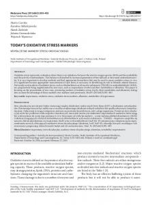
Figure 1.1 Lipid peroxidation cycle in presence of excess free radicals, specifically hydroxyl radical ( OH • ).
biomolecules [4,5]. One well-known example is lipid membrane damage via lipid peroxidation, where the hydroxyls radical react with PUFAs of the cellular membrane, extracting an electron and yielding a lipid free radical. If this reaction phenomena is not controlled in a timely manner, it can initiate a chain reaction of free radical molecules with intact lipids resulting in overall cellular membrane damage [6] (Fig. 1.1). Although most of the research concerning the role of ROS/RNS has been done towards the potential cellular damage and subsequent pathological events, they do serve beneficial role to the body under certain conditions. When present at optimum concentrations, they help maintain the redox homeostasis in the cellular environment that regulates cell functioning and cell signaling and responds to endogenous and exogenous stimuli [7]. Under normal physiological conditions, a balance between generation and elimination of ROS/RNS via endogenous antioxidant enzymes/ molecules helps the redox-sensitive signaling proteins to function properly [8]. Endogenous ROS generating enzymes like myeloperoxidase and NADPH oxidase (NOX), also known as phagocyte oxidase, actually help the neutrophils perform their phagocytic function against microbial intrusion. In a process known as the respiratory burst, NOX-catalyzed superoxide production further forms hydrogen peroxide with the help of superoxide
3
A Free Radical Primer
dismutase (SOD), which in turn converts to hypochlorus acid (HOCl), which has strong bactericidal properties [9,10]. oxidase SOD • NADPH + O2 NADPH → O− → 2
Myloperoxidase → HOCl H 2O 2 (1.ii)
Indeed, the dual nature of biological oxidation and reduction processes is the very reason for its importance in biomaterial/tissue interaction. Through a proper understanding of the underlying chemistry, it is possible to design next generation materials which can harness this intrinsic biological signaling mechanism. This chapter will cover the key points in free radical chemistry, the various pathways and vocabulary and finally, the way these processes occur in a biological setting.
1.2 RED/OX CHEMISTRY 1.2.1 Oxidation/Reduction Reactions and Voltage Potentials Originally, the term “oxidation” was described as a process of any element, primarily metals, to combine with oxygen to form metal oxides and “reduction” defined as a process that will convert the metal oxide back to pure metal. For example, conversion of magnesium (Mg) to magnesium oxide is oxidation, while smelting of magnesium oxide to magnesium at high temperature in presence of carbon is reduction. Later, the discovery of electrons changed the definition of oxidation–reduction to the transfer of electrons from one species to another. As per the law of conservation of mass applied to electrons, oxidation and reduction are always linked to one another. Meaning, if one species is oxidized, the counter reactant species will be reduced [11,12]. 2Mg + O2 → 2MgO Oxidation (1.iii) MgO + C → Mg + CO Reduction (1.iv)
By definition, oxidation of any given element or molecule will involve a loss of electron and the element/molecule will be known as a reducing
4
Prachi Gupta, Andrew Lakes and Thomas Dziubla
agent while the reduction of any molecule will be a gain of electron and that molecule will be known as an oxidizing agent [13]. In reference to free radicals or ROS they are commonly known to be strong oxidizing agents and have a tendency to get reduced [14,15]. This is because most radicals have one unpaired electron, and the addition of another electron (a.k.a. getting reduced) with the opposite electron spin would help in stabilizing the electron pair, taking them to a more inert state. However, this is not always true. Oxidizing tendency of any free radical will depend on its affinity to gain an electron or its own reduction potential against the affinity or potential of the coreactant. It is possible that free radical “A” gets reduced in presence of “B” but might get oxidized itself by losing another electron in presence of “C” because the reduction potential or electron gaining affinity of “C” is greater than “A.” 1.2.1.1 Types of Redox Reactions 1.2.1.1.1 Corrosion and Rusting
Iron has served as a common example of corrosion, which is the electrochemical oxidation of metals in presence of oxygen to form respective oxides. In reference to iron, it is specifically termed as formation of “rust” (Eq. (1.v)) [16]. 4Fe + 3O2 → 2Fe2O3 Oxidation of Fe to Fe3 + (1.v) In presence of an acid, iron (II) is oxidized to iron (III) by reaction with hydrogen peroxide, which acts as an oxidizing agent [17], although iron (III) can be reduced back to iron (II) in the presence of stronger reducing agents or free radicals, such as superoxide anion [18]. This oxidation–reduction chemistry of iron with hydrogen peroxide is popularly known as the Fenton and Haber–Weiss reaction mechanisms (see Section 2.4) and are critical in maintaining the redox state of the cell. 2Fe2 + + H 2O2 + 2H+ → 2Fe3 + (oxidized ) + 2H 2O(reduced)Oxidation of Fe 2 + toFe3 + (1.vi) 1.2.1.1.2 Nitrification and Denitrification
Nitrification often occurs naturally and is a biologically oxidative process where ammonia is oxidized to nitrite followed by formation of nitrate by nitrifying bacteria. On the other hand, reduction of nitrate to nitrogen in the presence of an acid is termed as denitrification and is often used as water purification process [19]. These nitrates have the ability to diffuse
A Free Radical Primer
5
through the cellular membrane and play a significant role in the production of RNS in a cellular environment. + Nitrification : NH3 + O2 → NO− 2 ( oxidized ) + 3H (1.vii) + 2e−( ammonia to nitrite) − + − (1.viii) NO− 2 + H 2O → NO3 ( oxidized ) + 2H + 2e ( nitrite to nitrate )
− + Denitrification : NO− 3 + 10e + 12H → N 2 ( reduced ) (1.ix) + 6H 2O (oxidized)
1.2.1.1.3 Dismutation Reaction
Dismutation or disproportionation is a specific kind of redox reaction, where both oxidized and reduced forms of a chemical species are produced. For example, superoxide free radicals produced in mitochondria dismutate to hydrogen peroxide and oxygen (Eq. (1.x)) [20] or ascorbyl radical to ascorbate (vitamin C) and dehydroascorbate (DHA) (Eq. (1.xi)) in order to maintain intracellular nutrient requirements for the cells [21,22]. + 2O− → H 2O2 ( reduced ) + O2 (oxidized ) (1.x) 2 + 2H
(1.xi) 2 ascorbyl • + H+ → ascorbate(oxidized) + DHA(reduced) 1.2.1.1.4 Cellular Respiration
Oxidation of glucose to carbon dioxide with simultaneous reduction of oxygen to water is another example of natural oxidation–reduction reaction, and is required for energy production in living organisms [23]. C6H12O6 + 6O2 → 6CO2 + 6H 2O (1.xii) All the reactions shown (Eqs. (1.v)–(1.xii)) and their tendency to either undergo oxidation or reduction are controlled by their reduction potential. Reduction potential (Eo) is defined as a tendency of a chemical species to be reduced by gaining an electron and is defined with electrochemical reference of hydrogen, which is globally given the reduction potential of zero [24]. As this is an electric potential, it is measured in volts and each chemical species has its own intrinsic reduction potential. Numerically, the more positive the potential, the stronger the affinity of the species is to acquire an electron and get reduced.
6
Prachi Gupta, Andrew Lakes and Thomas Dziubla
1.2.1.2 Redox Potential A normal redox reaction example could be as given below (Eq. (1.xiii)): (1.xiii) Fe3 + + Cu+ → Fe2 + + Cu2 + and this can be broken down into two parts: (1.xiv) Fe3 + + e− → Fe2 + Reduction (1.xv) Cu+ → Cu2 + + e− Oxidation For a combined redox reaction, overall redox potential is estimated by o o ∆E = E acceptor − Edonor (1.xvi)
Electrochemical potential or reduction potential stated above is directly related to the Gibbs free energy (∆G ) of the reaction: (1.xvii) (∆G ) = −nF ∆E n, number of electrons associated with the reaction F, Faraday’s constant. For a reaction to proceed, total Gibb’s free energy (∆G ) must be negative or ∆E should be positive. In Eq. (1.xv), Cu is the electron donor with redox potential of Cu2+ to Cu+ is + 0.16 V and that of Fe+ to Fe2+ is 0.77 V. Therefore, overall redox potential becomes + 0.61 V. To state this simply, for a system to undergo a redox reaction, the redox potential of a species to be reduced should be higher than the species to be oxidized. Table 1.2 lists some of the common redox potential of certain half-cell redox couples and of some important biomolecules important to physiological redox environment.
1.2.2 Thermodynamic Treatment (Ellingham Diagram) Voltage potential is not the only way to determine the direction of reaction. As stated earlier, for a reaction to proceed, ∆G should be negative which requires ∆E to be positive, but ∆G is also a function of temperature (Eq. (1.xviii)). An alternative method to determine the direction of a reaction can be obtained by a simplified thermodynamic analysis of the reaction equilibrium. (1.xviii) ∆G = ∆H − T ∆S where T, reaction temperature
7
A Free Radical Primer
Table 1.2 Half-Cell Reduction Potentials of Biologically Significant Molecules Redox Couple Eo (V)
+0.816
O2 + 4H+ + 4e− → 2H 2O −
NO2 + e
→
NO− 3
+
+1.04
NO− 2 −
+ 4H + 3e +
→ NO + H 2O
+0.96
→ 2H 2O
+0.82
−
H2O2 + 2H + 2e
Fe3 + + e− → Fe2 +
+0.77
+
+0.68
−
O2 + 2H + 2e
→ H2O2
2I2 + 2e− → 2I−
+0.54
2H2O + O2 + 4e− → 4OH−
+0.40
2Cu
2+
−
+ 2e
2 cytochromec
+0.34
→ Cu 3+
2 cytochrome b
3+
−
+ 2e
2H + 2e +
+0.070
→ 2 cytochrome b
−
→ succinate
Fumarate + 2H + 2e −
+0.254
2+
−
+ 2e
+
+
→ 2 cytochromec
2+
+0.031 0.00
→ H2 +
−
FAD + 2H + 2e
→ FADH 2 ( free coenzyme )
−0.22
FAD+ + 2H+ + 2e− → FADH 2 ( in flavoprotein ) −0.166
Oxaloacetate + 2H+ + 2e− → malate +
−
Pyruvate + 2H + 2e +
+
−
NAD + 2H + 2e +
+
→ NADH + H −
NADP + 2H + 2e −
O2 + e
→
−0.185
→ lactate +
−0.320
+
→ NADPH + H
+
−0.324 −0.33
O− 2 +
−
Succinate + CO2 + 2H + 2e Na + + e− → Na
→ α − ketoglutarate + H2O
−0.324 −2.71
ΔS, entropy change ΔH, enthalpy change. The temperature dependence of a reaction using Eq. (1.xviii) is also utilized in redox chemistry to drive the direction of reactions of metal oxides and sulfides to pure metal. This phenomenon is usually illustrated in the form of Ellingham’s diagram, which represents the stability of a metal oxide as a function of temperature in reference to ΔG (Figure 1.2). Free energy versus temperature plot for a particular metal shows the free energy values
8
Prachi Gupta, Andrew Lakes and Thomas Dziubla
for its metal oxide formation where slope depicts the ΔS values while the y-intercept gives ΔH. The position of the line of a metal with reference to another helps in depicting the metal oxide reduction ability in presence of another metal. To illustrate, the lower the position of metal ( Mg + O → MgO) is on Elligham’s diagram (Fig. 1.2), the more stable its oxide is at a particular temperature than the one that lies above it ((4 / 3)Al + O2 → ( 2 / 3)Al2 O3 ). Therefore, magnesium can reduce aluminum oxide to metallic aluminum. Looking at the diagram, all the metal H2/H2O ratio
–8
10–4
10–2
–2
10
2 +O O4 e3 M 4F
100
200 4Cu
+ O2
U O = 2C 2
1
=6
1
M
–2
10
O
M
NiO
O2
i+
2N
300
2Co
+ O2
o = 2C
1
M
=2
–4
10
M
2Fe +
O2
eO = 2F
2
10
–6
10 2
10
400 O
Zn
500
O2
+ Zn
=2
2
M
C
OK
10 10–6
O3 Fe 2
2
Standard free energies of formation of oxides (–∆Gº = RTIn pO ) kJmol–1 O2
H
pO2
10–4
–6
10 10–8
CO/CO2 ratio
600
O n+ 2
Si +
/3
–8
10
M
4
10
M M
nO
= 2M
2M
700
O3 Cr 2
=2
O2
r+
C 4/3
M M MnO
B
10–10
B
iO 2
O2
=S
–12
iO 2
O2 Ti +
10
6
=T
10
800
l 2O 3 /3 A
l 3A
4/
900
=2
O
Mg
M + Mg
1000
+ O2
O2
2
M
=2
O2 a+
6
10
B
8
B
O
Ca
=2
10
10–14
–16
10
2C
Change of state
M
Element
Melting Point Boiling Point
1100
Oxide
8
10
M B
M B
–18
10
10
10 1200
200
0
400
600
800
1000 1200 1400 1600 Temperature (ºC)
1800
–200
14
–100
10
–70
10
–60
10
–50
10
–42
10
–38
10
12
10
–34
10
–20
10
2200 2400
10
CO/CO2 ratio H2/H2O ratio 10
2000
–30
10
–28
10
10
10
–26
10
–22
10
–24
10
Figure 1.2 Ellingham’s diagram. The figure is reproduced with permission from the University of Cambridge Dissemination of IT for the Promotion of Materials Science (DoITPoMS) website. (www.doitpoms.ac.uk).
A Free Radical Primer
9
oxide formations have a positive slope while carbon oxidation to carbon monoxide has a negative slope and it cuts across many of the metals at particular temperatures. Therefore, carbon becomes a very useful reducing agent for metal oxides at higher temperatures. For example, carbon can reduce manganese oxide (MnO) to its metallic form Mn once the reaction temperature goes above 1400°C while it will be able to reduce TiO2 to Ti above 1600°C [25–27]. As highlighted here, while the reduction potential of two reacting molecules decides their affinity to get reduced or oxidized, it is important to remember that the change in other system properties (eg, temperature) can alter the fate of one species getting reduced in presence of another.
1.2.3 Combustion Sequences and/or Metal Oxides Combustion is a classic example of free radical reactions and generation. It is a high temperature exothermic process involving multiple redox reactions of a fuel (hydrocarbons) with an oxidant mostly oxygen resulting in oxidized products primarily carbon dioxide and water, to generate heat and light. The overall combustion process of oils is described by the reaction: n + 1 Cn H 2( n +1) + 4n + O2 → nCO2 + (n + 1)H 2O (1.xix) 2 While this equation is a useful simplification of the combustion process, the exact chemistry of combustion is highly complex, multifaceted, and not easy to describe. Molecular oxygen in its ground state is a very stable molecule and unreactive to hydrocarbons until a catalyst is introduced. However, at elevated temperatures as high as 2200°C, oxygen converts into highly reactive singlet oxygen (O21) [28] and is known to have high oxidizing power. Singlet oxygen can react with and break carbon–carbon or carbon–hydrogen bonds of large hydrocarbons into smaller molecules and, subsequently, hydrogen and water. In the process, it is also capable of initiating numerous radical chain reactions via reaction with molecular hydrogen (H2) resulting in hydroxyl radical (OH• ) and proton (H+). Combination of many such reactions results in the generation and simultaneous consumption of hydroperoxyl (HCOO• ), formyl (HCO• ) radicals, and carbon monoxide [29,30]. As for hydrocarbon pyrolysis, the process involves generation of various aliphatic and aromatic radicals.
10
Prachi Gupta, Andrew Lakes and Thomas Dziubla
Below are some of the common series of radical reactions that can occur during combustion process: O2 + H+ → O•− + OH• (1.xx) O12 + H 2 → H+ + OH• (1.xxi) C3H8 + M → C2H5• + CH•3 + M (1.xxii) C2H•5 + M → C2H4 + H+ + M (1.xxiii) C3H8 + OH• → C3H•7 + H 2O (1.xxiv) C3H8 + H+ → C3H•7 + H 2 (1.xxv) C3H8 + O•− → C3H•7 + OH• (1.xxvi)
C3H•7 → C3H6 + H+ (1.xxvii)
Decay tounsaturated hydrocarbons These radicals react with oxygen radical to produce formyl radical and formaldehyde and CH•3, CH2, H2CO finally oxidize to CO2 and H2O. C3H6 + O•− → C2H5 + HCO (1.xxviii) C2H4 + O2 → 2CO + H 2 (1.xxix) CO + (1 / 2)O2 → CO2 (1.xxx) Looking at all the reactions that are involved in the process of producing heat energy via oxidation of long/short chain hydrocarbon fuels, it is clear that a milieu of free radicals are generated. Yet, a spatial relationship also exists in radical production, which is described by the “zone theory.” This concept divides the combustion process into four zones: zone 1-free flame zone (fuel zone), zone 2-high temperature flame zone (>1200°C), zone 3-postflame thermal zone (600–1200°C), and zone 4-gas quench cool and surface catalysis zone (1200°C Hydrocarbon + O2 = heat energy
Free radicals
Incomplete reaction Postflame zone 600° > T >1200°C partial combustion NO, semiquinone, phenoxyl radicals
Oxidative phosphorylation
Partial oxygen reduction/ proton leak
Carbohydrates+ O2 = ATP (cellular energy)
(O2+e– = O2–) Oxygen reduction to superoxide
Cool zone T < 600°C Fe2+/ Cu2+
EPFRs
ROS/RNS production 2+ H2O2 Fe / Cu2+
.
OH– + OH
Figure 1.3 Analogous comparison of various stages of combustion zone theory with cellular respiration during energy production along with free radical generation [27].
12
Prachi Gupta, Andrew Lakes and Thomas Dziubla
formation. In both cases, the role of radicals produced at this stage are boosted by the presence of transition metal ions. Transition metal ion combination with radicals makes more environmentally stable EPFRs and are reactive to other biological species. During cellular metabolism, transition metal ions act as a catalyst towards production of more reactive and damaging oxidants namely hydroxyl radical (OH• ).
1.2.4 Fenton/Haber–Weiss Chemistry Both the Fenton and Haber–Weiss reactions are associated with iron and the production of hydroxyl radicals. The Fenton reaction describes the • formation of hydroxide (OH−) and hydroxyl (OH ) radical by a reaction between Iron (II) (Fe2+) and hydrogen peroxide (H2O2) [33], Haber– Weiss reaction is where hydroxyl and hydroxide ions are generated from •− the reaction of H2O2 and superoxide ion (O2 ) catalyzed by iron [34]. Fe2 + + H 2O2 → Fe3 + + OH− + OH• O•2− + H 2O2 → OH− + OH• + O2
Fenton reaction (1.xxxi)
Haber - Weiss reaction (1.xxxii)
The Haber–Weiss cycle is actually a two-step reaction, where the ferric ion reduces to ferrous ion via reaction with superoxide, which, in turn, reacts with H2O2 to form OH− and OH• ions, converting ferrous back to ferric ion. 3+ O•− → Fe2 + + O2 (1.xxxiii) 2 + Fe
Fe2 + + H 2O2 → Fe3 + + OH− + OH• (1.xxxiv) Henry J.H. Fenton reported for the first time the oxidation power of H2O2 and Fe2+ towards tartaric acid in 1876 but never mentioned the existence of the hydroxyl radical intermediate in the oxidation process, • although the reaction was named after him [35]. The existence of OH was proposed by two German chemists, Fritz Haber and Joseph Joshua Weiss in 1934 via reaction of hydrogen peroxide and superoxide in presence of iron as a catalyst [36]. Formation of this hydroxyl radical via transition metal catalyzed reaction has gained plenty of attention over the years and is considered to be one of the key processes for the production of the highly reactive OH• in the cellular redox chemistry. Not only hydroxyl radicals but also Fenton/Haber–Weiss chemistry gives rise to several other intermediates including hydro peroxides (HOO• ), superoxide (O2−), etc. during various chain initiation and
A Free Radical Primer
13
propagation reactions shown in Eqs. (1.xxxv)–(1.xliii). OH• acts as the chain carrier with the ability to react with Fe2+, H2O2, or any organic species to propagate the reaction. But in some instances, chain termination also comes into effect via combination of two radicals (OH• / OH• or OOH• / OH•) producing just hydrogen peroxide, water, and oxygen as shown in Eqs. (1.xlii) and (1.xliii) [37]. The existence and propagation of any of these reactions highly depends on the density of iron in its required oxidation state as well as the rate constant. An important condition for iron-induced reduction of hydrogen peroxide is the low pH requirement between 3 and 6. Fe2 + + H 2O2 → Fe3 + + OH− + OH• (1.xxxv) K = 5.7 × 10² M−1 s−1 Fe3 + + H 2O2 → Fe2 + + H+ + OOH• / O− 2 −3 −1 − 1 (1.xxxvi) K = 2.6 × 10 M s OH• + H 2O2 → OOH• / O− 2 + H 2O 7 −1 − 1 (1.xxxvii) K = 3.3 × 10 M s 2+ Fe3 + + OOH• / O− + O2 + H + 2 → Fe (1.xxxviii) K = 3.1 × 10⁵ M−1 s−1
Fe2 + + OH• → Fe3 + + OH− (1.xxxix) K = 3.2 × 10⁸ M−1 s−1 3+ Fe2 + + OOH• / O− + H 2 O2 2 → Fe 6 −1 − 1 (1.xl) K = 6.6 × 10 M s − • OOH• / O− 2 + OOH / O2 → H 2O2 (1.xli) K = 2.3 × 10⁶ M−1 s−1 • OOH• / O− 2 + OH → H 2O + O2 (1.xlii) K = 8.9 × 10⁹ M−1 s−1
OH• + OH• → H 2O2 (1.xliii) K = 5.2 × 10⁹ M−1 s−1
14
Prachi Gupta, Andrew Lakes and Thomas Dziubla
Chain propagation and termination reactions associated with iron and hydrogen peroxide with rate constant measured at pH = 5 [38–40]. Fenton chemistry is commercially utilized to treat water pollution, contaminated soils, sludge, etc. by oxidizing the pollutants such as benzene, formaldehyde, rubber chemicals, and pesticides. The rate constant for the initial reaction between Fe2+ and H2O2 is generally observed to be around 102 M−1 s−1 but in biological systems, this rate of reaction with free Fe2+ is not enough for the oxidation to occur. However, when bound to ADP, ATP, or citrate, the oxidation rate increases by at least 2 orders of magnitude, resulting in a reaction rate fast enough for the iron-catalyzed redox process to occur [41].
1.2.5 Thiol Chemistry (–SH;–SS–) Thiol–disulfide reactions are one of the most important biological redox reactions. In biology, oxidation/reduction governs the metabolic redox state of a cell. Thiol–disulfide interchange/exchange reactions are significant in itself where a thiol (RSH) reacts with another disulfide containing molecule (R′SSR″) to give a new oxidized disulfide (R′SSR) and the corresponding reduced thiol (R″SH) [42]. The reaction is base catalyzed and is proposed to proceed through SN2, 2 step reactions as follows [43,44]: RSH � RS− + H+ (1.xliv) RS− + R′SSR″ � R′SSR + R″S− (1.xlv) H+ + R″S− � R″SH (1.xlvi) d[ R″RS− ] = kRS− [ RS− ][ R′SSR″] (1.xlvii) dt d[R″S− ] = kobs [ RSH ]total [ R′SSR′ ] (1.xlviii) dt kRS− = kobs (1 + 10 pK a − pH ) (1.xlix) where R, attacking group; R′, central group; R″, leaving group
A Free Radical Primer
15
kRS− , calculated rate constant dependent on thiolate concentration but independent of pH kobs, observed rate constant dependent on total thiol concentration and pH. In this reaction mechanism, the thiolate anion (RS−) acts as an active nucleophile or reactive species to propagate the reaction. In the cytosol, glutathione plays a key role in the thiol redox chemistry as it is the most abundant small molecule cellular antioxidant (see Section 1.3.2 for an in-depth discussion on glutathione redox reactions). One of the important factors that contribute towards the occurrence of this thiol exchange reaction is the pKa of the corresponding thiol and the pH of the reaction environment [45]. For example, a thiol with pKa value of 10, 0.1% of the total thiol will form thiolate at pH 7 while at pH 8, 10 times higher thiolate ions would be present in comparison [43]. In other words, we can say that thiol–disulfide interchange is most favorable at pH near to the pKa values of the thiol. Oxidation of glutathione (GSH) (pKa = 9.33) to its oxidized form (GSSG) is faster at higher pH of 9.43 with rate constant k = 45 L/mol s as compared to at pH of 8.46 with k = 9.1 L/mol s [46]. As the pKa is a function of the structure of thiol molecule, chemical properties of thiol molecules involved in the reaction will dictate the extent of reaction and their existence in reduced or oxidized form. For instance, the reaction of 2-mercaptoethanol (pKa = 10.14) with Ellman’s disulfide (5-(3-carboxy-4-nitrophenyl)disulfanyl-2-nitrobenzoic acid) (pKa = 4.5) is faster than mercaptoethanol with oxidized glutathione (GSSG) (pKa = 9.33) by the order of 104 in water [47,48]. Also, the exchange reaction is faster when the pKa of a nucleophilic thiol (RSH) is as high as possible and that of the corresponding disulfide molecule (R′SH/R″SH) is as low as possible. Stearic interference is another factor that determines the rate constants of the thiol–disulfide exchange reactions. It is most pronounced when there is any carbon substitution at α-position to sulfur. For instance, reaction of bis (t-butyl) disulfide with 1-butylthiolate (k = 0.26 M−1 s−1) is 106 times faster than that with t-butylthiolate (k = 10−7 M−1 s−1). Similarly, charge on the thiolate anion also affects the rate constants [49]. The effect is higher when the charged entity is near to the sulfur group [50]. The rate of reaction is often correlated with the Brønsted plot, which displays the relationship between pKa of the reacting thiols/ disulfide and the rate constant. The slope of the plot is defined by the
16
Prachi Gupta, Andrew Lakes and Thomas Dziubla
13. Thiol-disulfide interchange 1.0
4
3
0.6 2 0.4
log KRS–
θ = [RS–]/([RS–]+[RSH])
0.8
1
0.2
0
0.0 5
6
7 K p a
8
9
Figure 1.4 Correlation between pKa of reacting thiol and rate constant (kRS−)/degree of dissociation (θ) where kRS− was calculated using Eq. (1.l), considering pKa of R′SH and R″SH groups as 8.5. This figure is reproduced with permission from [43].
Brønsted coefficient (β), which normally lies between 0.4 and 0.5 for different thiol–disulfide exchange reactions. Eq. (1.l) is generally recommended for calculating kRS− values for the thiolate content [51] and Fig. 1.4 shows the graphical correlation between pKa of thiol versus log(kRS− ) at pH 7 for a thiol–disulfide exchange reaction. Fig. 1.4 also shows the degree of dissociation (θ) of the thiol and observed rate constant for the thiol/disulfide exchange reaction are predicted by Eq. (1.li). Therefore, the highest observed rate constant for an exchange reaction is RSH observed at a pK a that is equivalent to the pH of the solution, pH = 7 in the figure.
log(kRS−) = 6.3 + 0.59pK a RSH − 0.40 pK a R′SH − 0.59pK a R″SH (1.l) kobs = θkRS− (1.li)
A Free Radical Primer
17
1.3 BIOLOGICAL OXIDATION EVENTS 1.3.1 Oxygen and Nitrogen Currency Oxygen and nitrogen are the most abundant diatomic gaseous molecules in the atmosphere and play a significant role in regulating both, human and plant metabolism. Nitrogen is known to be highly inert and even oxygen (O2) in its ground state is a stable molecule. But O2 at a higher energy state form such as singlet oxygen (O21) [52] or in the reduced form of superoxide (O•− 2 ), it can become an active source of free radical generation leading to disruption of the cellular redox state [53], while oxide forms of nitrogen (NO, NO2) are generally produced enzymatically and can readily diffuse through lipid membranes and lead to formation of intracellular free radicals or RNS. Indeed, these processes has led to the Superoxide Theory of Oxygen Toxicity which states that the partially reduced form of oxygen, ie, superoxide via enzyme, auto-oxidation, mitochondrial electron transport chain, heme proteins, etc. is a primary cause of cellular toxicity [54]. Oxygen, upon its one electron reduction to superoxide, form can further yield hydrogen peroxide via the dismutation process [55]. Hydrogen peroxide can reduce to hydroxyl radicals in the presence of transition metal ions like Fe2+/Cu2+, a highly reactive radical in cellular biology capable of reacting with DNA, lipid membranes, proteins, etc. [1]. Reactivity and reduction potential at various stages of oxygen radical production chain are different and are often enzyme catalyzed. For example, dismutation of superoxide to hydrogen peroxide mainly occurs via SOD-catalyzed reaction [56,57], but a part of hydrogen peroxide production also takes place via a two-step process of oxygen conversion to peroxyl radicals followed by further reduction to H2O2. In the oxygen-derived free radical production chain, H2O2 is not considered a highly reactive molecular species but serves as an intermediate to the production of hydroxyl radical (OH• ) [58] via Fenton reaction, which is highly reactive towards all the cellular components and can lead to complete cell damage. Similarly, superoxide ion by itself is not very reactive towards nonradical species but it can react very quickly with NO• or phenoxyl radicals [59]. This radical has a unique selectivity towards its oxidizing properties. For example, it does not readily react with free NADH/NAD+ but can easily oxidize enzyme-bound (lactate dehydrogenase) NADH to NAD+ [60]. Apart from being an oxidizing agent (oxidation of ascorbate to ascorbyl radical), superoxide can also serve as a reducing agent where it can reduce cytochrome c or Fe3+ to Fe2+ which is one of the steps in cellular redox cycling discussed later [61,62].
18
Prachi Gupta, Andrew Lakes and Thomas Dziubla
Singlet oxygen 1
O2
hv E°= –0.95V
Dioxygen O2
Superoxide radical e– E°= –0.33V
O2•–
Peroxide radical
e–
O22– K = 4.5×10⁵ M–1S–1 E°= +0.94V
H+
E°= –0.46V
HO2•
O2•– +H+ K = 8×10⁷ M–1S–1 E°= +1.06V
Peroxyl radical
2H+
H2O2
Fe2+ K ⇒ 2×10⁹ M–1S–1 E°= +0.32
Hydrogen peroxide
OH•
Hydroxyl radical
Figure 1.5 Free radical production/reactive oxygen species with oxygen as a precursor [1].
In reference to biological free radical production, oxygen nucleated free radicals are produced from all the oxygen that is inhaled or consumed by organisms and mitochondria is considered to be the main source of superoxide radical production due to oxygen leakage in electron transport chain during ATP production and is reported to be produced by complexes I and III [63–65]. Reported in vitro studies show that about 4% of the oxygen consumed is converted into superoxide, though in vivo studies have reported about 10-fold lower production, which is still a significant amount of ROS and translates to about a 10-µM intracellular concentration of superoxide [66]. This charged radical readily diffuses through mitochondrial membranes and can react with biomolecules like proteins, PUFAs, or produce other reactive species (H2O2, OH•) (Fig. 1.5). Although gaseous nitrogen is an extremely stable and inert molecule, nitrogen in its oxide forms, such as nitric oxide (NO), acts as a precursor to a variety of free radicals collectively known as reactive nitrogen species, which are equally capable of carrying out cell damage if produced in excess. Nitric oxide ( NO• ) due to its lipophilic property readily diffuses through cell membranes from the atmosphere where it is a byproduct of various combustion processes [67]. More importantly, in the cellular environment, it is produced as a result of the reaction of l-arginine and oxygen with NADPH in the presence of nitric oxide synthase (NOS) to give nitric oxide, l-citrulline, and NADP+ [68]. Nitric oxide by itself is not very reactive towards nonradical species similar to superoxide anion but can generate highly reactive free radicals such as nitrogen dioxide (NO•2 ) via a slow reaction with oxygen (O2) [69]. NO•2 with the Eo value of 1.04 V is a potent oxidant in metabolic redox system and in RNS chain, it further reacts with NO• leading to production of N2O3, which eventually decomposes to give another reactive nitrite (NO2−) free radical [70]. In the presence of superoxide, NO• reacts to form peroxynitrite (ONOO−),
19
A Free Radical Primer
O2
NO
NO
2NO + O2 L-arginine
+ NADPH + O2
2NO 2
N2O3 RSH/RNH2
O2– NO + NO2– ONOO–
NO2–
OONOH
NO2
NO2
ONOO–
OH
Thiol nitrosation (RSNO, RSOH) Nitrosation of amine RSH/RNH2
Figure 1.6 Reactive nitrogen species production.
which in itself is also a powerful oxidizing molecule [71,72]. It is present in the form of acidified peroxynitrous acid (ONOOH) or its activated form ( • NO2 … OH• ), both forms are capable of oxidizing most biological molecules such as DNA, proteins, and lipids. NO• also takes part in the modification of some biological molecules with thiols or amine groups during the process called nitrosation via direct reaction with thiols or via a metal bound NO• reaction with thiols/amines, resulting in formation of nitrosothiols (RSNO) or nitrosamine (RN2O), which are also regarded as nonradical RNS [73,74] (Fig. 1.6).
1.3.2 Cellular Redox Chemistry Within the cellular environment, there exists an equilibrium between oxidants and antioxidants. Major players of these processes are defined in Fig. 1.7. Of particular note is the small molecule thiolated antioxidant, glutathione (GSH), which is the most abundant small molecule in eukaryotic cells, roughly found intracellulary from 1 to 10 mM and about 1 to 10 µM extracellular [75–78]. Synthesized within the cell from the three amino acids, glutamate, glycine, and cysteine, GSH production is rate limited by cysteine content, a semi-essential amino acid in biology. Cysteine transported from extracellular space in the disulfide form, cystine [79].
20
Prachi Gupta, Andrew Lakes and Thomas Dziubla
G6PD HNO3
NADP+
NADPH
AO/GSH Glu, Cys, Gly ONOO– NOS Arg O2
NO XO COX
NADPHOX
GSS
LPO, PCO
GSSG
2GSH
AO/ GSH O2– OH Fe2+ Cu2+ SOD
GST GSH GS-LPO GS-PCO
GR
GCL
H2O
L, P CAT
GPx ROOH, RH, H2O, O2 H2O2 Cl–
H2O + O2
MPO HOCl
GSH/AO
GSA, HCl, H2O
Abbreviations: AO, antioxidant CAT, catalase G6PD, glucose-6-phosphate dehydrogenase GCL, glutamate cysteine ligase GPX, glutathione peroxidase GR, glutathione reductase GSA, glutathione sulfonamide GSH, glutathione GSS, glutathione synthetase GSSG, glutathione disculfide GST, Glutathione S-transferase L, lipid LPO, lipid peroxide MPO, myeloperoxidase NOS, nitrous oxide synthase P, protein PCO, protein carbonyl SOD, superoxide dismutase
Figure 1.7 Significant cellular redox molecule interactions (green, glutathione processes; red, oxidant species reduced by glutathione processes). (For interpretation of the references to color in this figure legend, see the color plate.)
Reaction of cysteine with glutamic acid in presence of glutamate– cysteine–ligase (GCL) followed by the reaction with glycine via glutathione synthetase (GSS) results in GSH production [79]. GSH may act in several mechanisms as an antioxidant, whether directly through electron donation to ROS/RNS in the intracellular or extracellular space, or via enzymatic routes such as with glutathione S-transferase (GST) to irreversibly reduce toxic species like lipid peroxides and protein carbonyls [80]. In addition to direct chemical reaction with oxidative species, GSH participates in several mechanisms to regenerate other important antioxidant enzymes such as glutathione peroxidase (GPx), glutaredoxin (GRX), and peroxiredoxins nonspecifically. Oxidized glutathione (GSSG) may be reduced back to GSH via glutathione reductase (GR), which is driven by NADPH and the glucose-6-phosphate dehydrogenase (G6PD) cycle via oxidation of NADPH to NADP+. There are also other thiol-based oxidoreductases such as thioredoxin, which utilizes cysteine thiol–disulfide exchange (instead of GSH) and is regenerated with thioredoxin reductase, similarly utilizing NADPH for regeneration akin to GSSG with GR. Depending on the cellular compartment, redox equilibrium may vary. For instance, the cytosol (GSH:GSSG 100:1) and nucleus (GSH:GSSG > 100:1) maintain a reducing environment [76,81], whereas there are high concentrations of oxidants in the endoplasmic reticulum (GSH:GSSG of 1–3:1 [82], where disulfide bonds fortify the
A Free Radical Primer
21
protein structures), mitochondria (GSH:GSSG of 20–40:1 [81]), secretory pathways, and extracellular space (GSH:GSSG 20:1 [78]). It is important to note that extracellularly, while the GSH:GSSG ratio is around 10–20:1, cysteine and cystine are in greater concentration by about an order of magnitude, and maintain the oxidative environment around cysteine:cystine of 0.2:1 [78]. In a similar manner, NADPH is a reducing coenzyme involved in processes like fatty acid synthesis and is required to drive various redox reactions in the metabolic pathway. Therefore, NADPH:NADP+ (200:1) ratios are found to be in the same order of magnitude as GSH/GSSG redox equilibrium to drive the reaction forward. On the other hand, another coenzyme NADH plays an essential part in both reduction and oxidation in general, hence a significant concentration of both oxidized and reduced forms is maintained in the cell with NAD+ at higher concentrations. Cytoplasmic NADH, which is produced by oxidation of cytoplasmic NAD+, acts as an electron donor and is transported to the mitochondrion to reduce mitochondrial NAD+ to NADH, which in turn is oxidized again during oxidative phosphorylation process to generate ATP. It is postulated via several experimental studies that NAD+/NADH ratio in cytoplasm is around 700:1, though the overall cellular ratio varies between 0.5:1 and 4:1. This ratio is involved in regulation of several metabolic enzymes such as glyceraldehyde 3-phosphate dehydrogenase and pyruvate dehydrogenase used in conversion of pyruvate to acetyl-CoA.
1.3.3 Radical Generation in Metabolism and Role of Enzymes in Redox Cycle ROS/RNS production takes place in a cellular environment via multiple metabolic pathways with the aid of specific enzymes at various sites in a cellular matrix and its organelles. In that, mitochondria are one of the most important cellular components with the greatest potential to produce free radicals ranging from hydroxyl to peroxynitrite to hypochloride radicals. Figure 1.8 depicts the role of mitochondrion in the redox of cycle of a cell. Mitochondria are known to be the energy producer or ATP production center of the cell which takes place via tetravalent reduction of oxygen (O2) in the presence of mitochondrial cytochrome c oxidase (COX) also called complex IV [83]. About 2–4% of this oxygen taken by mitochondria undergoes univalent reduction to superoxide ion (O2.−) via complex I enzyme (NADH-ubiquinone oxidoreductase) to a concentration of about 10 µM [66,84]. Oxidation of NADPH to NADP+
22
Prachi Gupta, Andrew Lakes and Thomas Dziubla
by O2 in the presence of NOX is another source of O•− 2 which occurs as an important inflammatory response in phagocytes against bacterial infection and this process is often termed as oxidative burst [85–87]. NOX also facilitates O•− 2 production in nonphagocytic systems like fibroblasts, endothelial cells, and smooth muscle cells, although rate of O•− 2 production here is one-third than that in neutrophils [7,88,89]. This superoxide anion is further converted to hydrogen peroxide by mitochondrial manganese superoxide dismutase (Mn-SOD) [90,91]. Apart from controlling the O•− 2 concentrations inside the mitochondrion, cytosolic CuZn-SOD also catalyzes the superoxide dismutation to H2O2, 104 times faster than an uncatalyzed reaction (k = 2 × 10⁹ M−1 s−1) [92]. Hydrogen peroxide is also known to be formed by two electron reduction of O2 by cytochrome P-450 or acetyl coenzyme A oxidase [93–95]. As described earlier, hydrogen peroxide is relatively stable and membrane permeable nonradical ROS. In the cellular system, it can be converted to water and oxygen via the enzymes like catalase, glutathione peroxidase (GPx), etc. [96,97], or it can be further reduced to a highly reactive hydroxyl radical under transition metal ion catalytic conditions (oxidation of Fe2+ or Cu2+ to Fe2+ or Cu3+) (Fig. 1.9) [98] based upon Fenton/Haber– Weiss style reactions. In this pathway, oxidized iron or copper (Fe/Cu3+) is reduced back to ferrous or cuprous ions (Fe2+/Cu2+) by superoxide ion present in the vicinity, which in turn produces more hydroxyl radicals. The concentrations of superoxide anion, transition metal ions significantly define the free radical production rates [99,100]. Other active routes of OH• production are through disproportionation of peroxynitrous acid intermediate (HO…NO2) to hydroxyl and nitrous oxide (NO2.) radical [101]. Due to the extremely high reactivity of the hydroxyl radical, it is very difficult to monitor its real-time concentration in vivo (Fig. 1.8). Peroxynitrite is a product of reaction of superoxide with endogenously generated nitric oxide ( NO• ) radical [102,103]. NO is also generated in the cellular matrix via reduction of L-arginine in presence of nitric oxide synthase (NOS). NOS has three isoforms namely: (1) NOS1 (NOSI) found in neural tissues, (2) NOS II or iNOS, inducible NOS, which is found in various cell types stimulated upon inflammatory response, and (3) eNOS found in endothelium [104]. Mitochondrial NOS (mtNOS) is another isoform, which facilitates the formation of NO• inside the mitochondria. This NO formation plays an important role in regulating the mitochondrial respiration and has the potential to reversibly inhibit cytochrome oxidase activity, which is responsible for superoxide production [105]. Reaction of superoxide with nitric oxide under physiological
23
A Free Radical Primer
H2O2
GSSG
NADP+
H2O2 (10–8M)
HO
B BH2
ROOH (10–6M/s)
H2O + ROH
NADPH + H+
GSSG
ONOO–
DH2
NADP+
2GSH
GPer
NO
Peroxisomes Cat . H2O2
Cat RH
O2
DH2
D
H2O2
SOD
2GSH
NADPH + H+
A
O2 (10–11M)
SOD 2H2O
AH2
Cytosolic Endopiasmic enzymes reticulum
O2
Mitocondria
H2O2
O2
UQH
O.2–
ecSOD
H2O2
D
Fe
.
Lipid radicals
OH
EXTRACELLULAR ENDOTHELIUM
Xanthine oxidase
eNOS
PLASMA MEMBRANE
nNOS NO
O2. –
PM oxidoreductase/ NAD(P)H oxidase SR/T-Tubule NAD(P)H oxidases
ONOO–
CYTOSOL PLA2-dependent processes
O.2–
CuZnSOD
H2O2
GPx CAT
H2O
VDAC? Electron transport chain eNOS
MnSOD GPx H2O2 H2O O.2– NO
ONOO– Mitochondrion
Figure 1.8 Examples of various routes of metabolic free radical production and detoxification pathways.
24
Prachi Gupta, Andrew Lakes and Thomas Dziubla
conditions to produce peroxynitrite is found to be three times faster than hydrogen peroxide production and again is also a highly reactive RNS just like OH.− [106]. Hydrogen peroxide also contributes to the production of hypochlorite radical upon reaction with chloride ion (Cl−) in activated leukocytes by the action of myeloperoxidase (neutrophils and monocytes) and eosinophil peroxidase (eosinophils) [107,108]. Their ideal function is to act as antimicrobial agents, but excess or unregulated production can result in fragmentation and aggregation of proteins [108,109]. As for the production of singlet oxygen, it is known to be generated at the catalytic sites of multiple enzymes in the cellular matrix or via dismutation of unstable oxidation products like superoxide both spontaneous and enzyme catalyzed. It is also known to be produced as a byproduct of Table 1.3 Role of Enzymes in Carrying Out Various Redox Reaction and Maintaining the Cell Homeostasis Enzyme Function Refs.
SOD (CuZn-SOD, Mn-SOD, Fe-SOD) NOS (eNOS, iNOS, NOSI) NADPH oxidase GPx Myeloperoxidase (abundant in neutrophils) Catalase (in peroxisomes) 5-Lipoxygenase Cyclooxygenase Xanthine oxidase
Monoamine oxidase (outer mitochondrial membrane)
Catalyzes dismutation of superoxide to hydrogen peroxide Oxidation of l-arginine with production of nitric oxide (NO) Superoxide production from dioxygen molecule Reduction of GSH to GSSG with simultaneous conversion of hydrogen peroxide to water Produces HOCl from H2O2 and Cl− ion, oxidation of tyrosine to tyrosyl radical in presence of H2O2 Decomposition of hydrogen peroxide to water and oxygen Inducible source of ROS production in lymphocytes ROS generation in TNF-α stimulated cells Source of ROS in diabetes mellitus catalyzes oxidation of hypoxanthine to xanthine with simultaneous production of H2O2, xanthine further catalyzes to uric acid and H2O2, sometimes can also produce superoxide radical Catalyzes oxidative deamination of biogenic amines, large source of H2O2 and OH• radical as well
[112,113] [68,114] [68,115] [116,117] [118] [119,120] [121,122] [123,124] [125]
[126]
25
A Free Radical Primer
O2
O2
–
Fe3+
Fe2+
– OH + OH
H2O2
GR
GPx H2O + O2
NADP+ + H+
GSH
GSSG
NADPH
Figure 1.9 Concept of redox cycle in maintaining the healthy redox state of a cell.
peroxyl radical reaction between hydrogen peroxide and hypochlorite via the Russell mechanism, found to occur in stimulated neutrophils mostly during the respiratory burst phenomena (antimicrobial cell lysis process) [110,111] (Table 1.3; Fig. 1.9).
1.3.4 Known ROS Targets and Concerns Proteins, PUFAs, and carbohydrates are very common targets to ROS. These molecules constitute very important cell organelles including lipid membranes, mitochondria, DNA, and other nucleic acids. Reaction with these biological components starts with the excess production of ROS and insufficient endogenous antioxidants available to stabilize them, resulting in disruption of the cellular redox balance. Extent or affinity of ROS/ RNS to react with the cellular components varies with the type of ROS depending on its reduction potential. For instance, hydroxyl and peroxynitrite radicals discussed in Section 3.1 have relatively higher reduction potential amongst other oxidants (Table 1.2) and are considered to be the most reactive towards lipids, DNA, etc. It is almost difficult to detect or measure their quantity in vivo as their rate constants in the range of 107– 1010 M−1 s−1 almost match their diffusion rates [127,128]. In theory, oxidation of proteins, peptides, and amino acids by these free radicals can take place via several routes including hydrogen abstraction (favorable with aliphatic amino acids), electron transfer, dimerization, disproportionation, rearrangement, and many more [129,130]. Protein and amino acids have multiple sites of attack on their backbone and their reactivity depends on stability of the resulting radical and degree of carbon hydrogenation [131]. Aromatic amino acids have a tendency to undergo addition reaction with free radicals rather than hydrogen abstraction due to their stabilizing resonance property forming hydroxylated/quinone molecules after reaction with hydroxyl radical [132,133].
26
Prachi Gupta, Andrew Lakes and Thomas Dziubla
One of the most sensitive target sites to free radicals are mitochondria and their DNA components (mtDNA) [134,135]. This is because, mitochondria is the key production site of ROS/RNS where superoxide anion concentration is found to be 5–10 times higher than in the cytosol, with mtDNA to the closest proximity in comparison to nuclear DNA to the cytosolic ROS [136,137]. There are numerous amount of studies demonstrating that the mitochondrial dysfunction due to excess ROS/RNS production is a kick-off event towards the final stage of cell apoptosis or necrosis [127]. The mitochondrial dysfunction is thought to start via mtDNA damage and protein deactivation. This leads to the incapability of the mitochondria to maintain its membrane potential, resulting in the net loss of ATP production, destructuring of the mitomembrane (inner and outer both), resulting in release of excess calcium ion (Ca2+), cytochrome c, and lysosomes. These apoptogenic proteins/ enzymes finally activate the apoptotic pathway leading to cell death [138]. Another potential target to ROS like singlet oxygen, peroxynitrite, or hydroxyl radical is the cellular lipid membrane, which is made of unsaturated fatty acids. It is thought that the central carbon of the unsaturated fatty acids (L) is attacked by ROS/RNS to produce lipid free radical ( L• ), which oxidizes further to its alkoxyl ( LO• ) and peroxyl ( LOO• ) radical forms [139]. These radicals, considered to be important intermediates in lipid oxidation process, again react with another fatty acid chain (L) to produce more L• radical and the chain reaction continues. In this propagation event, radicals like NO• can act as antioxidant towards termination of the chain by forming stable alkyl nitrites (LONO2) or as pro-oxidant to form more LO• and NO2 through the peroxynitrite pathway [140]. Due to this process, lipid fatty acid chains are often considered as secondary free radicals as they form reactive intermediates that propagates the lipid peroxidation chain reaction resulting in membrane degeneration and membrane protein dysfunction [106]. This degeneration leads to loss of membrane integrity making membrane bound protein even more susceptible to free radical attack. Alkyl peroxyl radicals (another form of secondary radicals) generated by decomposition of alkyl hydroperoxide in presence of transition metal ion also contribute in the propagation of lipid peroxidation [141–144]. (1.lii) LOO• + NO• → LOONO LOONO → LO• + NO2 Chain propagation(14% ) (1.liii) LOONO → LONO2 Chain termination( 86% ) (1.liv)
A Free Radical Primer
27
1.4 CONCLUSION AND FINAL THOUGHTS As seen, the exact mechanisms by which free radical chemistry progresses are extremely complex, with multiple interacting pathways. Yet, even an incomplete understanding has led to major impacts into the way we approach fields as diverse as metallurgy, combustion, and most relevant here, biology. As we gain new knowledge in the field of free radical biology, the opportunities to improve our biomedical treatments and therapies also grow. Researchers have already started harnessing these opportunities to develop new and exciting biomaterials that no longer remain “inert,” but rather actively respond and control design a more nuanced level of biomaterial/host interaction.
REFERENCES [1] Gutowski M, Kowalczyk S A study of free radical chemistry: their role and pathophysiological significance. Acta Biochim Pol 2013;60(1):1–16. [2] Cameron MD, Poyer JF, Aust SD. Identification of free radicals produced during phacoemulsification. J Cataract Refract Surg 2001;27(3):463–70. [3] Neuman RC Jr. Free radical addition and substritution reactions. Organic Chemistry; 1992–2013. [4] Halliwell B. Oxygen and nitrogen are pro-carcinogens. Damage to DNA by reactive oxygen, chlorine and nitrogen species: measurement, mechanism and the effects of nutrition. Mutat Res 1999;443(1–2):37–52. [5] Halliwell B, Gutteridge JM. Oxygen toxicity, oxygen radicals, transition metals and disease. Biochem J 1984;219(1):1–14. [6] Mylonas C, Kouretas D. Lipid peroxidation and tissue damage. In Vivo 1999;13(3):295–309. [7] Dröge W. Free radicals in the physiological control of cell function. Physiol Rev 2002;82:47–95. [8] Trachootham D, et al. Redox regulation of cell survival. Antioxid Redox Signal 2008;10(8):1343–74. [9] Aratani Y, et al. Candida albicans and Aspergillus fumigatus. Med Mycol 2002;40(6):557–63. [10] Babior BM. NADPH oxidase. Curr Opin Immunol 2004;16(1):42–7. [11] Jacobs JJ, Gilbert JL, Urban RM. Current concepts review—corrosion of metal orthopaedic implants*. 1998;80:268–82. [12] Bodner G. Oxidation and reduction. Available from: [cited 11.05.15]. [13] Rademann SBAJ. The power of functional resins in organic synthesis. J.W.a. Sons, Hoboken, New Jersey; 2008. [14] Cadet J, Wagner JR. DNA base damage by reactive oxygen species, oxidizing agents, and UV radiation. Cold Spring Harb Perspect Biol 2013;5(2). [15] Thannickal VJ, Fanburg BL. American Journal of Physiology, Lung Cell and Molecular Physiology. 2000;279:L1005–28. [16] Hœrlé S, et al. Advances in understanding atmospheric corrosion of iron. II. Mechanistic modelling of wet–dry cycles. Corrosion Sci 2004;46(6):1431–65.
28
Prachi Gupta, Andrew Lakes and Thomas Dziubla
[17] Rao JR, Nayak R. Ferrous-ferric oxidation in pickle liquor. J Sci Ind Res (India) 2003;62:1094–7. [18] Rose AL, Waite TD. Reduction of organically complexed ferric iron by superoxide in a simulated natural water. Environ Sci Technol 2005;39(8):2645–50. [19] Wastewater Management Fact Sheet. Denitrifying filters. U.S.E.P. Agency. [20] Chin DH, et al. Proton-induced disproportionation of superoxide ion in aprotic media. J Am Chem Soc 1982;104(5):1296–9. [21] Buetyner GR, Bors W. The vitamin C radical and its reactions Fuchs LPAJ, editor. Vitamin C in helath and diseases. USA: Marcel Dekker, Inc. 1997. [22] May JM, et al. Reduction of the ascorbyl free radical to ascorbate by thioredoxin reductase. J Biol Chem 1998;273(36):23039–45. [23] Lodish HBA, Zipursky SL. Oxidation of glucose and fatty acids Molecular cell biology. New York: W.H. Freeman; 2000. [24] Wardman P. Reduction potentials of one-electron couples involving free radicals in aqueous solution. J Phys Chem Reference Data 1989;18(4):1637–755. [25] Yang JJ, et al. Metal/TiO2 interfaces for memristive switches. Appl Phys A 2011;102(4): 785–9. [26] Desmoulins-Krawiec S, et al. Synthesis of nanostructured materials in supercritical ammonia: nitrides, metals and oxides. J Mater Chem 2004;14(2):228–32. [27] https://tuebingen.mpg.de/startseite/detail/geschickt-gebaut-struktur-einer-zellulaerenkraftstoffpipeline-entschluesselt.html. [28] Starik AM, Kozlov VE, Titova NS. On the influence of singlet oxygen molecules on characteristics of HCCI combustion: a numerical study. Combust Theory Model 2013;17(4):579–609. [29] Wilson Jr, WmE. A critical review of the gas-phase reaction kinetics of the hydroxyl radicals. J Phys Chem 1972;1(2). [30] Pollutant formation and control combustion, In: Air pollution. [31] Vejerno EWP. Formation and stabilization of combustion-generated environmentally persistent free radicals on transition metal oxides supported on silica Department of chemistry. Baton Rouge, LA: Louisiana State University; 2011. [32] Vejerano E, Lomnicki S, Dellinger B. Lifetime of combustion-generated environmentally persistent free radicals on Zn(II)O and other transition metal oxides. J Environ Monit 2012;14(10):2803–6. [33] Barbusiński K. Toxicity of industrial wastewater treated by fenton’s reagent. Pol J Environ Stud 2004;14(1):11–16. [34] Kehrer JP. The Haber–Weiss reaction and mechanisms of toxicity. Toxicology 2000;149(1):43–50. [35] Barbusiński K. Henry John Horstman Fenton—Short biography and brief history of fenton reagent discovery. Chemistry-Teaching-Ecology-Metrology 2009:101–5. [36] Haber F, Weiss J. The catalytic decomposition of hydrogen peroxide by iron salts. 1934;147:332–51. [37] Bielski BHJ, et al. Reactivity of HO2/O2− radicals in aqueous solution. J Phys Chem Ref Data 1985;14(4):1041–100. [38] Kremer ML. Mechanism of the Fenton reaction. Evidence for a new intermediate. Phys Chem Chem Phys 1999;1(15):3595–605. [39] Friedrich LC, et al. Mechanistic implications of zinc(II) ions on the degradation of phenol by the Fenton reaction. J Brazil Chem Soc 2012;23:1372–7. [40] Benatti CT, Tavares CRG. Fenton’s process for the treatment of mixed waste chemicals. In: Organic pollutants ten years after the Stockholm convention—Environmental and analytical update; 2012. [41] Vile GF, Winterbourn CC, Sutton HC. Radical-driven Fenton reactions: studies with paraquat, adriamycin, and anthraquinone 6-sulfonate and citrate, ATP, ADP, and pyrophosphate iron chelates. Arch Biochem Biophys 1987;259(2):616–26.
A Free Radical Primer
29
[42] Fernandes PA, Ramos MJ. Theoretical insights into the mechanism for thiol/disulfide exchange. Chemistry 2004;10(1):257–66. [43] Singh R, Whitesides GM. Thiol–disulfide interchange Sulphur-containing functional groups (1993). John Wiley & Sons, Inc; 2010.633.58. [44] Gilbert HF. Thiol/disulfide exchange equilibria and disulfide bond stability. Methods Enzymol 1995;251:8–28. [45] Chandrasekhar S, et al. Thiol–disulfide exchange in peptides derived from human growth hormone. J Pharm Sci 2014;103(4):1032–42. [46] Guo W, Pleasants J, Rabenstein DL. Nuclear magnetic resonance studies of thiol/disulfide chemistry. 2. Kinetics of symmetrical thiol/disulfide interchange reactions. J Org Chem 1990;55(1):373–6. [47] Szajewski RP, Whitesides GM. Rate constants and equilibrium constants for thiol– disulfide interchange reactions involving oxidized gluthathione. J Am Chem Soc 1980;102:2011–26. [48] Whitesides GM, Lilburn JE, Szajewski RP. Rates of thiol–disulfide interchange reactions between mono- and dithiols and Ellman’s reagent. J Org Chem 1977;42:332–8. [49] Fava A, Iliceto A, Camera E. Kinetics of the thiol–disulfide exchange. J Am Chem Soc 1957;79(4):833–8. [50] Hupe DJ, Wu D. Effect of charged substituents on rates of the thiol–disulfide interchange reaction. J Org Chem 1980;45(15):3100–3. [51] Nagy P. Kinetics and mechanisms of thiol–disulfide exchange covering direct substitution and thiol oxidation-mediated pathways. Antioxid Redox Signal 2013;18(13):1623–41. [52] Weishaupt KR, Gomer CJ, Dougherty TJ. Identification of singlet oxygen as the cytotoxic agent in photo-inactivation of a murine tumor. Cancer Res 1976;36(7 Part 1):2326–9. [53] McCord JM. Oxygen-derived free radicals in postischemic tissue injury. N Engl J Med 1985;312(3):159–63. [54] Gerschman R, et al. Oxygen poisoning and x-irradiation: a mechanism in common. Science 1954;119(3097):623–6. [55] Freeman BA, Crapo JD. Biology of disease: free radicals and tissue injury. Lab Invest 1982;47(5):412–26. [56] McCord JM, Fridovich I. Superoxide dismutase: an enzymic function for erythrocuprein (hemocuprein). J Biol Chem 1969;244(22):6049–55. [57] Klug D, Rabani J, Fridovich I. A direct demonstration of the catalytic action of superoxide dismutase through the use of pulse radiolysis. J Biol Chem 1972;247(15): 4839–42. [58] Chen SX, Schopfer P. Hydroxyl-radical production in physiological reactions. A novel function of peroxidase. Eur J Biochem 1999;260(3):726–35. [59] Nagy P, Kettle AJ, Winterbourn CC. Superoxide-mediated formation of tyrosine hydroperoxides and methionine sulfoxide in peptides through radical addition and intramolecular oxygen transfer. J Biol Chem 2009;284(22):14723–33. [60] Petrat F, et al. Initiation of a superoxide-dependent chain oxidation of lactate dehydrogenase-bound NADH by oxidants of low and high reactivity. Free Radic Res 2005;39(10):1043–57. [61] Harel S, Salan MA, Kanner J. Iron release from metmyoglobin, methaemoglobin and cytochrome c by a system generating hydrogen peroxide. Free Radic Res Commun 1988;5(1):11–19. [62] Fessenden RW, Verma NC. A time-resolved electron spin resonance study of the oxidation of ascorbic acid by hydroxyl radical. Biophys J 1978;24(1):93–101. [63] Wang W, et al. Superoxide flashes in single mitochondria. Cell 2008;134(2):279–90. [64] Muller FL, Liu Y, Van Remmen H. Complex III releases superoxide to both sides of the inner mitochondrial membrane. J Biol Chem 2004;279(47):49064–73.
30
Prachi Gupta, Andrew Lakes and Thomas Dziubla
[65] Barja G. Mitochondrial oxygen radical generation and leak: sites of production in states 4 and 3, organ specificity, and relation to aging and longevity. J Bioenerg Biomembr 1999;31(4):347–66. [66] Boh EE, et al. Mitochondrial chemiluminescence elicited by acetaldehyde. J Bioenerg Biomembr 1982;14(2):115–33. [67] Chiueh CC. Neuroprotective properties of nitric oxide. Ann N Y Acad Sci 1999;890:301–11. [68] Ghafourifar P, Cadenas E. Mitochondrial nitric oxide synthase. Trends Pharmacol Sci 2005;26(4):190–5. [69] Ignarro LJ, et al. Oxidation of nitric oxide in aqueous solution to nitrite but not nitrate: comparison with enzymatically formed nitric oxide from l-arginine. Proc Natl Acad Sci USA 1993;90(17):8103–7. [70] Kissner R, et al. Formation and properties of peroxynitrite as studied by laser flash photolysis, high-pressure stopped-flow technique, and pulse radiolysis. Chem Res Toxicol 1997;10(11):1285–92. [71] Hsiai TK, et al. Hemodynamics influences vascular peroxynitrite formation: implication for low-density lipoprotein apo-B-100 nitration. Free Radic Biol Med 2007;42(4):519–29. [72] Pacher P, Beckman JS, Liaudet L. Nitric oxide and peroxynitrite in health and disease. Physiol Rev 2007;87(1):315–424. [73] Gow AJ, Buerk DG, Ischiropoulos H. A novel reaction mechanism for the formation of S-nitrosothiol in vivo. J Biol Chem 1997;272(5):2841–5. [74] Ford PC. Probing fundamental mechanisms of nitric oxide reactions with metal centers. Pure Appl Chem 2004;76(2):335–50. [75] Ballatori N, et al. Molecular mechanisms of reduced glutathione transport: role of the MRP/CFTR/ABCC and OATP/SLC21A families of membrane proteins. Toxicol Appl Pharmacol 2005;204(3):238–55. [76] Saito G, Swanson JA, Lee KD. Drug delivery strategy utilizing conjugation via reversible disulfide linkages: role and site of cellular reducing activities. Adv Drug Deliv Rev 2003;55(2):199–215. [77] Ottaviano FG, Handy DE, Loscalzo J. Redox regulation in the extracellular environment. Circ J 2008;72(1):1–16. [78] Banerjee R. Redox outside the box: linking extracellular redox remodeling with intracellular redox metabolism. J Biol Chem 2012;287(7):4397–402. [79] Lu SC. Glutathione synthesis. Biochim Biophys Acta 2013;1830(5):3143–53. [80] Eaton DL, Bammler TK. Concise review of the glutathione S-transferases and their significance to toxicology. Toxicol Sci 1999;49(2):156–64. [81] Hu J, Dong L, Outten CE. The redox environment in the mitochondrial intermembrane space is maintained separately from the cytosol and matrix. J Biol Chem 2008;283(43):29126–34. [82] Hwang C, Sinskey AJ, Lodish HF. Oxidized redox state of glutathione in the endoplasmic reticulum. Science 1992;257(5076):1496–502. [83] Nohl H. Is redox-cycling ubiquinone involved in mitochondrial oxygen activation? Free Radic Res Commun 1990;8(4–6):307–15. [84] Cuzzocrea S, et al. Antioxidant therapy: a new pharmacological approach in shock, inflammation, and ischemia/reperfusion injury. Pharmacol Rev 2001;53(1):135–59. [85] Jones OT. The regulation of superoxide production by the NADPH oxidase of neutrophils and other mammalian cells. Bioessays 1994;16(12):919–23. [86] Babior BM. NADPH oxidase: an update. 1999;93:1464–76. [87] Paravicini TM, Touyz RM. NADPH oxidases, reactive oxygen species, and hypertension: clinical implications and therapeutic possibilities. Diabetes Care 2008;31(Suppl. 2):S170–80.
A Free Radical Primer
31
[88] Bae YS, et al. Epidermal growth factor (EGF)-induced generation of hydrogen peroxide: role in EGF receptor-mediated tyrosine phosphorylation. J Biol Chem 1997;272(1):217–21. [89] Griendling KK, et al. Angiotensin II stimulates NADH and NADPH oxidase activity in cultured vascular smooth muscle cells. Circ Res 1994;74(6):1141–8. [90] MacMillan-Crow LA, Crow JP. Does more MnSOD mean more hydrogen peroxide? Anticancer Agents Med Chem 2011;11(2):178–80. [91] Miwa S, et al. Superoxide and hydrogen peroxide production by Drosophila mitochondria. Free Radic Biol Med 2003;35(8):938–48. [92] Goto JJ, Gralla EB, Valentine JS, Cabelli DE. Reactions of hydrogen peroxide with familial amyotrophic lateral sclerosis mutant human copper-zinc superoxide dismutases studied by pulse radiolysis. J Biol Chem 1998;273(46):30104–9. [93] Haberland A, et al. Comparison of malondialdehyde and hydrogen peroxide modified CuZnSOD by EPR spectroscopy. Agents Actions 1993;40(3–4):166–70. [94] Goeptar AR, Scheerens H, Vermeulen NP. Oxygen and xenobiotic reductase activities of cytochrome P450. Crit Rev Toxicol 1995;25(1):25–65. [95] Li Y, Tharappel JC, Cooper S, Glenn M, Glauert HP, Spear BT. Expression of the hydrogen peroxide-generating enzyme fatty acyl CoA oxidase activates NF-kB. DNA Cell Biol 2000;19(2):113–20. [96] Apel K, Hirt H. Reactive oxygen species: metabolism, oxidative stress, and signal transduction. Annu Rev Plant Biol 2004;55(1):373–99. [97] Doroshow JH, Locker GY, Myers CE. Enzymatic defenses of the mouse heart against reactive oxygen metabolites: alterations produced by doxorubicin. J Clin Invest 1980;65(1):128–35. [98] Puppo A, Halliwell B. Formation of hydroxyl radicals from hydrogen peroxide in the presence of iron. Is haemoglobin a biological Fenton reagent? Biochem J 1988;249(1):185–90. [99] Kanner J, Harel S, Hazan B. Muscle membranal lipid peroxidation by an “iron redox cycle” system: initiation by oxy radicals and site-specific mechanism. J Agric Food Chem 1986;34(3):506–10. [100] Deguillaume L, Leriche M, Chaumerliac N. Impact of radical versus non-radical pathway in the Fenton chemistry on the iron redox cycle in clouds. Chemosphere 2005;60(5):718–24. [101] Coddington JW, Hurst JK, Lymar SV. Hydroxyl radical formation during peroxynitrous acid decomposition. J Am Chem Soc 1999;121(11):2438–43. [102] Koppenol WH, et al. Peroxynitrite, a cloaked oxidant formed by nitric oxide and superoxide. Chem Res Toxicol 1992;5(6):834–42. [103] Ignarro LJ. Nitric oxide as a unique signaling molecule in the vascular system: a historical overview. J Physiol Pharmacol 2002;53(4 Pt 1):503–14. [104] Guzik TJ, Korbut R, Adamek-Guzik T. Nitric oxide and superoxide in inflammation and immune regulation. J Physiol Pharmacol 2003;54(4):469–87. [105] Carreras MC, et al. The biological significance of mtNOS modulation. Front Biosci 2007;12:1041–8. [106] Powers SK, Jackson MJ. Exercise-induced oxidative stress: cellular mechanisms and impact on muscle force production. 2008;88:1243–76. [107] Hawkins CL, Brown BE, Davies MJ. Hypochlorite- and hypobromite-mediated radical formation and its role in cell lysis. Arch Biochem Biophys 2001;395(2): 137–45. [108] Hawkins CL, Davies MJ. Hypochlorite-induced damage to proteins: formation of nitrogen-centred radicals from lysine residues and their role in protein fragmentation. Biochem J 1998;332(Pt 3):617–25.
32
Prachi Gupta, Andrew Lakes and Thomas Dziubla
[109] Chen YR, et al. Involvement of protein radical, protein aggregation, and effects on NO metabolism in the hypochlorite-mediated oxidation of mitochondrial cytochrome c. Free Radic Biol Med 2004;37(10):1591–603. [110] Kamat JP, Devasagayam TP. Biological significance of singlet oxygen. Indian J Exp Biol 2002;40:680–92. [111] Miyamoto S, et al. Biological hydroperoxides and singlet molecular oxygen generation. IUBMB Life 2007;59(4–5):322–31. [112] Beauchamp C, Fridovich I. Superoxide dismutase: improved assays and an assay applicable to acrylamide gels. Anal Biochem 1971;44(1):276–87. [113] Grankvist K, Marklund SL, Taljedal IB. CuZn-superoxide dismutase, Mn-superoxide dismutase, catalase and glutathione peroxidase in pancreatic islets and other tissues in the mouse. Biochem J 1981;199(2):393–8. [114] Bredt DS, et al. Cloned and expressed nitric oxide synthase structurally resembles cytochrome P-450 reductase. Nature 1991;351(6329):714–8. [115] Bedard K, Krause KH. The NOX family of ROS-generating NADPH oxidases: physiology and pathophysiology. Physiol Rev 2007;87(1):245–313. [116] Sies H, et al. Glutathione peroxidase protects against peroxynitrite-mediated oxidations: a new function for selenoproteins as peroxynitrite reductase. J Biol Chem 1997;272(44):27812–7. [117] Rotruck JT, et al. Selenium: biochemical role as a component of glutathione peroxidase. Science 1973;179(4073):588–90. [118] Hampton MB, Kettle AJ, Winterbourn CC. Inside the neutrophil phagosome: oxidants, myeloperoxidase, and bacterial killing. 1998;92:3007–17. [119] Scibior D, Czeczot H. [Catalase: structure, properties, functions]. Postepy Hig Med Dosw (Online) 2006;60:170–80. [120] Góth L. A simple method for determination of serum catalase activity and revision of reference range. Clin Chim Acta 1991;196(2–3):143–51. [121] Rådmark O, Samuelsson B. 5-Lipoxygenase: mechanisms of regulation. J Lipid Res 2009;50(Suppl.):S40–5. [122] Cook-Moreau JM, et al. Expression of 5-lipoxygenase (5-LOX) in T lymphocytes. Immunology 2007;122(2):157–66. [123] Dubois RN, et al. Cyclooxygenase in biology and disease. FASEB J 1998;12(12):1063–73. [124] Feng L, et al. Involvement of reactive oxygen intermediates in cyclooxygenase-2 expression induced by interleukin-1, tumor necrosis factor-alpha, and lipopolysaccharide. J Clin Invest 1995;95(4):1669–75. [125] Pacher P, Nivorozhkin A, SzabÓ C. Therapeutic effects of xanthine oxidase inhibitors: renaissance half a century after the discovery of allopurinol. Pharmacol Rev 2006;58(1):87–114. [126] Richardson JS. On the functions of monoamine oxidase, the emotions, and adaptation to stress. Int J Neurosci 1993;70(1–2):75–84. [127] Valdez LB, Lores Arnaiz S, Bustamante J, Alvarez S, Costa LE, Boveris A. Free radical chemistry in biological system. Biol Res 2000;33(2):65–70. [128] Yan EB, et al. Novel method for in vivo hydroxyl radical measurement by microdialysis in fetal sheep brain in utero. 2005;98:2304–10. [129] Davies KJ. Protein damage and degradation by oxygen radicals. I. General aspects. J Biol Chem 1987;262(20):9895–901. [130] Dean RT, et al. Biochemistry and pathology of radical-mediated protein oxidation. Biochem J 1997;324(Pt 1):1–18. [131] Hawkins CL, Davies MJ. Generation and propagation of radical reactions on proteins. Biochim Biophys Acta (BBA) Bioenerg 2001;1504(2–3):196–219.
A Free Radical Primer
33
[132] Davies M. Free radicals, oxidants and protein damage. Sydney, Australia: Australian Society for Biochemistry and Molecular Biology; 2012. [133] Goeschen C, Wille U. Damage of polyesters by the atmospheric free radical oxidant NO(3) (•): a product study involving model systems. Beilstein J Org Chem 2013;9:1907–16. [134] Green K, Brand MD, Murphy MP. Prevention of mitochondrial oxidative damage as a therapeutic strategy in diabetes. Diabetes 2004;53(Suppl. 1):S110–8. [135] Yakes FM, Van Houten B. Mitochondrial DNA damage is more extensive and persists longer than nuclear DNA damage in human cells following oxidative stress. Proc Natl Acad Sci USA 1997;94(2):514–9. [136] Gutierrez J, et al. Free radicals, mitochondria, and oxidized lipids: the emerging role in signal transduction in vascular cells. Circ Res 2006;99(9):924–32. [137] Cadenas E, Davies KJA. Mitochondrial free radical generation, oxidative stress, and aging. Free Radic Biol Med 2000;29(3–4):222–30. [138] Crow MT, et al. The mitochondrial death pathway and cardiac myocyte apoptosis. Circ Res 2004;95(10):957–70. [139] Ayala A, et al. Lipid peroxidation: production, metabolism, and signaling mechanisms of malondialdehyde and 4-hydroxy-2-nonenal. Oxid Med Cell Longevity 2014;2014:31. [140] Hogg N, Kalyanaraman B. Nitric oxide and lipid peroxidation. Biochim Biophys Acta (BBA) Bioenerg 1999;1411(2–3):378–84. [141] Goldstein S, Lind J, Merenyi G. Reaction of organic peroxyl radicals with • NO2 and • NO in aqueous solution: intermediacy of organic peroxynitrate and peroxynitrite species. J Phys Chem A 2004;108(10):1719–25. [142] O’Donnell VB, et al. Nitric oxide inhibition of lipid peroxidation: kinetics of reaction with lipid peroxyl radicals and comparison with alpha-tocopherol. Biochemistry 1997;36(49):15216–23. [143] O’Donnell VB, Freeman BA. Interactions between nitric oxide and lipid oxidation pathways: implications for vascular disease. Circ Res 2001;88(1):12–21. [144] Abuja PM, Albertini R, Esterbauer H. Simulation of the induction of oxidation of low-density lipoprotein by high copper concentrations: evidence for a nonconstant rate of initiation. Chem Res Toxicol 1997;10(6):644–51.
This page intentionally left blank
CHAPTER TWO
Oxidative Stress, Inflammation, and Disease Shampa Chatterjee Institute for Environmental Medicine, University of Pennsylvania Medical Center, Philadelphia, PA, United States
2.1 INTRODUCTION Oxidative stress is a term coined to denote the state of imbalance between the generation of oxidants (ie, free radicals and ions collectively called reactive oxygen species (ROS) and the availability of endogenous antioxidants to scavenge this ROS resulting in an excess production of oxidants [1–3]. As this ROS produced in biological systems can readily react with lipids, proteins, and DNA, an excessive production of ROS can be detrimental. Indeed studies have shown that ROS either through direct damage to biomolecules or modifications in proteins and genes are pivotal in triggering signaling cascades that lead to the onset of numerous pathologies and ultimately cell injury and death [4–7]. It is now well established that ROS and intermediate free radicals are the primary source of oxidative damage. Paradoxically, there is also extensive evidence to suggest that ROS serve as critical signaling molecules in maintenance of physiological function such as cell growth, proliferation, and survival [8–10]. ROS produced in vivo by cells, such as endothelial, inflammatory, and immune cells, has two faces; first is its participation in redox signaling and the second is its role in oxidative stress or injury. Redox signaling occurs when low levels of ROS produced induce activation of signaling pathways to initiate biological processes while oxidative stress defines high levels of ROS production that damages biomolecules. Although work over the last few decades implicates oxidative stress in the pathophysiology of many diseases, it seems increasingly evident that both redox signaling and oxidative stress underlie conditions ranging from cardiovascular to neurodegenerative diseases [11]. Both ROS-induced modifications of proteins and DNA as ROS-induced oxidative damage can lead to the onset of signaling that seems to be at the heart of inflammatory Oxidative Stress and Biomaterials. Doi: http://dx.doi.org/10.1016/B978-0-12-803269-5.00002-4
© 2015 2016 Elsevier Inc. All rights reserved.
35
36
Shampa Chatterjee
disorders. Inflammation is a “host defense” mechanism against pathogens which involves an enhanced or exaggerated ROS generation by activated inflammatory and immune cells. ROS that is produced as part of the inflammatory response facilitates clearance of tissue invasive bacteria, but when produced for prolonged periods can promote oxidative stress and chronic inflammation associated disorders. Additionally although inflammation induces oxidant injury, the reverse sequence of events is also true. Thus inflammation and oxidative stress are inextricably interrelated. While most of these modifications lead to irreparable damage, some modifications are more subtle and fully reversible. The reversible modifications can initiate signaling cascades known as “redox signaling.” This chapter reviews the role of oxidative stress and inflammation in the onset of signaling that underlies the pathology of several diseases. This link between oxidative stress and inflammation is highlighted in Figs. 2.1 and 2.2. The factors that cause inflammation and its amplification, leading to oxidative stress and the reverse sequence of events (oxidative stress induced inflammation), and the intersection of these are also reviewed. Finally, the antioxidant regulatory mechanisms that modulate the balance between host defense, inflammation, and oxidative stress are discussed.
Environmental agents
Cellular oxidants
Low ROS generation Signaling pathways
High ROS generation
Oxidative stress
Lipids Proteins DNA
Mutation
Pro-inflammatory mediators
Inflammation
Cell proliferation
Cell survival
Cellular transformation Carcinogenesis
Cell invasion/angiogenesis
Cell death Cell growth and tissue remodeling
Figure 2.1 Interrelationship between ROS, oxidative stress, inflammation, and cellular physiology and pathology.
37
Oxidative Stress, Inflammation, and Disease
Transcription factors NF-κB, AP-1, HIF-1α, Nrf-2
ICAM/VCAM, MCP-1, TNF-α, TGF-β
Oxidative stress
Inflammation
MAP Kinase (ERK, JNK, p38Kinase) PI3-Kinase Growth and remodeling
Figure 2.2 ROS and redox-dependent signaling pathways in oxidative stress and inflammation. ROS is reported to activate transcription factors NF-κB, AP-1, and HIF-1α that drive expression of pro-inflammatory genes leading to induction of proteins such as CAMs, MCP-1, TNF-α, IL-1, and transforming growth factor (TGF-β). ROS can also modify tyrosine kinases, such as Src, Ras, PI3K, EGFR, MAPK, that is, p38MAPK, JNK, and ERK. Activation of these redox-sensitive pathways results in numerous cellular responses.
2.2 ROS AND OXIDATIVE STRESS: A MAJOR ACTIVATOR OF INFLAMMATORY PATHWAYS ROS is a term that encompasses a wide variety of free radicals, ions, and reactive species that are generated by enzymatic and nonenzymatic sources in various cell types. For a summary of these players see chapter “A Free Radical Primer.” Briefly, the major ROS players in biological systems are superoxide anion (O2−), hydrogen peroxide (H2O2), the highly reactive hydroxyl radical (HO·), singlet oxygen (1O2), and ozone (O3). Besides ROS, the other physiologically important reactive species are the reactive nitrogen species comprising of nitric oxide (NO·) and the reactive radical, peroxynitrite anion (ONOO−, produced via interaction of O2−· and NO·). Nitric oxide can be converted into peroxynitrous acid and ultimately into hydroxyl radical and nitrite anion (NO2−) [12]. ROS are produced by biological systems through normal metabolic pathways. Cells also possess endogenous enzymes such as superoxide dismutases (SOD) which protect against ROS-induced damage by scavenging these free radicals [13–15]. Enzymatic sources of ROS are xanthine oxidase (XO), cyclooxygenases (COX), lipooxygenases, myeloperoxidases
38
Shampa Chatterjee
(MPO), cytochrome P450 monooxygenase, uncoupled nitric oxide synthase (NOS), peroxidases, and NADPH oxidase (NOX) while nonenzymatic sources are leakage from electron transport chain, free iron, xenobiotic, etc. [16–18]. High levels of ROS from external sources (such as environmental pollutants) or produced endogenously (via activation of XO, NOX, or mitochondrial pathways) can result in direct or indirect damage or both. Direct damage to proteins, DNA, and lipids alters these moieties, affecting their normal cellular functions. Indirect damage can occur via modification of proteins (by oxidation of amino acid residue side chains) or DNA base pairs [19,20]. As several of these proteins act as regulatory and transcription factors for cellular processes, modification of their structure activates signaling cascades that lead to altered cellular function. The biological consequences include a loss of transforming activity as well as mutagenicity and genotoxicity. Redox-sensitive (or ROS-regulated) transcription factors such as nuclear factor kappa B (NF-κB) are, when in an inactive form, localized in the cytoplasm of cells and bound to cytosolic proteins like IκB. Oxidants or ROS drive modification of IκB proteins, such as phosphorylation by a serine kinase, inhibitor of NF-κB kinase (IKK), which leads to its degradation, thus freeing NF-κB that can then translocate to the nucleus where, either alone or in combination with other transcription factors, it induces expression of several genes that express inflammatory proteins. ROS from various sources, external or endogenous (XO or NOX derived or mitochondrial ROS), have been demonstrated to be needed for NF-κB activation [21–24]. Besides NF-κB, other transcription factors, such as activator protein-1 (AP-1), the hypoxia-inducible factor (HIF-1α), peroxisome proliferator activator receptor gamma (PPAR-γ), β-catenin/Wnt, and nuclear factor like 2 (Nrf-2), have also been observed to be ROS regulated [25–27]. The activation primarily occurs via their release from a complex and subsequent translocation into the nucleus where it can “turn on” the expression of specific genes that have DNA-binding sites for the transcription factor in its promoter region. Transcription factors, such as the ones mentioned above, can alter hundreds of genes that induce expression of cellular adhesion molecules (CAMs), growth factors (such as the vascular endothelial growth factor, VEGF), cytokines, and chemokines. Cytokines are mediators of inflammation signaling; they bind to their respective receptors and either initiate ROS production or activate kinases or transcription factors that lead
39
Oxidative Stress, Inflammation, and Disease
Table 2.1 Redox Regulated Transcription Factors, Chemokines, and Cytokines
Transcription NF-κB factors AP-1 HIF-1α Nrf-2 p53 PPAR-γ Cytokines
TNF-α and -β (tumor necrosis factor) IL-1 to -15 (Interleukins)
Immune response, inflammation, proliferation Cell differentiation, survival, apoptosis, cytokine synthesis Vascularization, angiogenesis, cell migration, tumor invasion Antioxidant enzymes, inhibits inflammation Apoptosis, anti-proliferative, DNA repair Lipid handling, adipose regulation, inflammation Recruits neutrophils, macrophages Pro-inflammatory, mobilizes neutrophils, promotes angiogenesis Immune-regulatory; inhibits growth and activation Monocyte migration
TGF-α and -β (transforming growth factor) Chemokines MCP-1 and -3 (Monocyte Chemoattractant Protein) MIP-1α (Macrophage Macrophage activation, allergy Inflammatory Protein-1 RANTES (regulated on Cell migration activation, normal T-expressed and secreted, CCL5) IL-8 Neutrophil activation
to induction of other inflammatory signals. Chemokines are cytokines of low molecular weight (7–15 kDa) that bind to their respective receptors; some of these are released at the site of infection and drive inflammatory responses. Overall cytokines and chemokines induce recruitment of well-defined subsets of immune cells (leukocytes, polymorphonuclear neutrophils (PMN), macrophages, dendritic cells, etc.) [28–31]. Table 2.1 summarizes the redox or ROS-regulated players (transcription factors, cytokines, and chemokines) that are involved in various aspects of cellular and tissue inflammatory response. A pivotal event in the inflammatory response is the recruitment and adherence of phagocytes or immune cells (PMN, macrophages, monocytes) to the blood vessel wall. This is followed by extravasation of the adherent immune cells into the vascular wall. Once inside the tissue, these cells release ROS required for clearance of tissue invasive bacteria [32,33]. Excessive ROS produced during the inflammatory response can, in turn, cause further
40
Shampa Chatterjee
damage and exacerbate the oxidative stress-inflammation cycle. In general, the longer the inflammation persists, the higher the risk of pathologies such as atherosclerosis, cancer, and neurodegenerative diseases (Fig. 2.1). Adhesion molecules, cytokines, chemokines, and growth factors released by ROS-induced signals promote inflammation; conversely inflammatory processes by releasing ROS and other oxidants also promote oxidative stress and injury. Excessive ROS from external or other sources can, by generating lipid peroxidation products and other adducts, lead to oxidative damage. This also causes the onset of inflammation as peroxidized adducts trigger immune cell recruitment to “phagocytose” cell debris, etc. This is discussed in Section 2.4.
2.3 INFLAMMATION: A MAJOR CAUSE OF OXIDATIVE STRESS Inflammation is a pathological condition characterized by immune cell infiltration (monocytes, macrophages, lymphocytes, PMN, and plasma cells) into the vascular wall, extravasation of the immune cells into the tissue and release of ROS by these cells leading to tissue injury. PMN generate ROS predominantly via the enzyme NADPH oxidase 2, the activity of which is mediated through the assembly of catalytic subunit gp91phox (NOX2). The O2− produced spontaneously or enzymatically (SOD) dismutates to H2O2. H2O2 also gives rise to other highly reactive radicals, such as hydroxyl radical (·OH) or hypochlorous acid (HOCl) when catalyzed by the enzyme, MPO. Both these radicals can cause tissue injury besides oxidizing a variety of proteins, etc. ROS generated by inflammatory cells (besides causing direct oxidative stress to remove pathogens) also stimulates pathways that lead to amplification of inflammation. ROS-induced activation of kinases (or enzymes that facilitate phosphorylation), such as protein kinase C (PKC), c-Jun-N-terminal kinase (JNK), and p38 mitogen-activated protein kinase (MAPK), leads to the activation of transcription factors, which in turn triggers the generation of pro-inflammatory cytokines and chemokines. Once triggered, these cytokines and chemokines bind to their respective receptors (such as plateletderived growth factor receptor,VEGF receptor, and epidermal growth factor receptor (EGFR) that are well established to generate ROS [34,35]. ROS is thus both upstream and downstream of the inflammation cycle (Fig. 2.2). Besides facilitating the production of cytokines such as tumor necrosis factor-alpha (TNF-α), interleukins (IL-1 and -6), and chemokines such
Oxidative Stress, Inflammation, and Disease
41
as monocyte chemoattractant protein-1 and 2 (MCP-1/2), macrophage inflammatory protein (MIP-1/2) and RANTES (regulated on activation, normal T cell expressed and secreted), ROS induces (via transcription factor NF-κB) the expression of plasminogen activator inhibitor-1 (PAI-1), macrophage migration inhibition factor (MMIF), inducible NOS (iNOS), matrix metalloproteinases (MMP) 9, and MMP-2 [36]; all of which are important in recruitment and adherence of various subsets of immune cells. Recruitment and adherence are complex processes that involve cytokine-chemokine gradient to control influx and CAMs to regulate immune cell-vessel wall interaction. This is followed by PAI, MMIF, and MMPs induced disruption of adherens junctions that facilitate migration of immune cells into tissue. Postactivation, immune cells release oxidants and several proteases. Neutrophils produce neutrophil elastase, a serine protease which has direct antimicrobial activity. Besides ROS, other free radicals like HOCl are also produced when H2O2 is catalyzed by MPO, an enzyme which is highly abundant in inflammatory and immune cells. The highly reactive HOCl can oxidize a variety of molecules, including proteins, to cause tissue injury and dysfunction. ROS generation and oxidative stress can continue the cycle of inflammation, thus contributing to a chronic state which drives several inflammatory pathologies [37,38]. Recent advances in understanding the complexities of inflammatory signaling point to discrete pathways that are activated with recognition of molecular patterns derived from either pathogens or oxidative damage. Thus immune receptors can sense pathogen-associated molecular patterns (PAMPs), derived from invading pathogens or danger-associated molecular patterns (DAMPs) as a result of endogenous stress and trigger downstream signaling cascades. The multiprotein complex that assemble upon sensing PAMPs or DAMPs are termed as inflammasomes; these initiate a caspase signaling pathway that leads to cell injury and death [39–41]. Inflammasome induced cell injury and apoptosis contributes to and amplifies the pathology of inflammation diseases. Indeed, inflammasomes have been linked to a variety of inflammatory diseases from neurodegenerative (multiple sclerosis (MS), Alzheimer’s disease (AD), and Parkinson’s disease (PD)) to metabolic (atherosclerosis, type 2 diabetes, and obesity) [42]. Although the canonical pathway for inflammasome activation is via the PAMP-DAMP “sensing,” there are several reports that ROS and other oxidants activate the inflammasome complex. This would point to a “ROS induced inflammation induced oxidative stress” process [43,44]. As shown in Fig. 2.3, ROS are essential secondary messengers in
42
Shampa Chatterjee
Non pathogen mediated
Pathogen mediated
UV radiation
DAMPs
TLR2
PAMPs, DAMPs (bacterial toxins)
ATP Fatty acids Uric acid
LPS
ROS
NF-κB, AP-1
NLRP3 Inflammasome
ASC
Caspase-1 IFN-, TNF Pro-IL-1β, Pro-IL-18
IL-1β, IL-18
Inflammation Figure 2.3 Activation of the inflammasome. PAMPs and DAMPs arising from bacterial or viral ligands (such as LPS) or some environmental stress factors (asbestos, silica, alum) or endogenous agents of cellular stress (ATP, uric acid). In response to DAMPS and PAMPs, NLRP3 inflammasomes are activated (by among other agents, ROS) and recruit adaptor protein ASC and pro-caspase-1, which is cleaved to caspase-1. Caspase-1 facilitates the cleaving of Pro-IL-1β, Pro-IL-18 (which is expressed via transcription factors NF-κB and AP-1) to IL-18 and IL-1β, potent mediators of inflammation.
DAMP-PAMP–induced inflammasome activation and subsequent inflammatory signaling.
2.4 OXIDANT STRESS AND INFLAMMATION IN CELLULAR TRANSFORMATION, APOPTOSIS, AND NECROSIS Alterations or damage that arise from oxidative stress or from inflammation can cause cellular changes that may lead to cell death by necrosis, apoptosis, aging or to cell survival mechanisms such as autophagy where oxidized or defective proteins are degraded. ROS-induced cell death occurs either directly by damaging cell membrane/cellular structures or by activating caspases (enzymes that drive cell death) via lipid peroxidation products such as 4-hydroxy-2-nonenal
Oxidative Stress, Inflammation, and Disease
43
(HNE). HNE, when accumulated in cells, can induce apoptosis by activation of the “death receptor” or the “mitochondrial” pathway. The death receptor pathway is activated when lipid peroxides induced cytokines activate “initiator” caspases (8 and 10) which cleave “effector or executioner” caspases that in turn cleave intracellular proteins to trigger apoptosis. The mitochondrial pathway is activated when ROS and products of lipid peroxidation and damage lead to mitochondrial outer membrane permeabilization, which results in both caspase-dependent and caspase-independent apoptosis. To make matters worse, ROS and HNE can also activate transcription factors NF-κB and AP-1, propagating the pro-inflammatory signaling cascades [45,46]. ROS and redox signals participate in cell protection mechanisms such as autophagy. Complexes and adducts of peroxidized proteins, such as HNE-protein Michael-adducts, are degraded by the autophagy mechanism. Autophagy, which involves the clearance of (defective or mutated) proteins and organelles, is characterized by the formation of an autophagosome or vacuole by the fusion of membrane edges. Once generated, this vacuole eliminates its contents through the canonical lysosomal degradation pathway. Reports show that ROS (superoxide anion and H2O2) are crucial in autophagy as treatment with antioxidants partially or completely reverses the process. ROS probably act via autophagosome activation that degrades defective and damaged proteins or autophagy execution, that is, activation of lysosomal cysteine proteases that are crucial for protein degradation [47–49]. Low ROS levels reportedly act as secondary messengers of signal transduction pathways involved in cell growth, transformation, and apoptosis [3,37,38,50]. These occur via activation of signaling pathways that result in the induction of either growth stimulatory genes like c-fos, c-jun, and c-myc or cell death stimulating enzymes (caspases, etc.) [51–53].
2.5 EXPLORING THE LINK BETWEEN OXIDATIVE STRESS AND INFLAMMATION AND THE ONSET OF VARIOUS DISEASES Both oxidative stress and inflammation can cause injury to cells [54]. Arguably, one cell type that is uniquely sensitive to this injury is the vascular endothelium that is the site for inflammation. Besides serving as a conduit to transport nutrients to organs and acting as a barrier between blood flow and tissue, the endothelium is also a site for adherence of immune
44
Shampa Chatterjee
and other cells. These immune cells are recruited to and adhere to the vessel wall in response to any pro-inflammatory stimuli. Following this adhesion, these cells can extravasate into the surrounding tissue, and release ROS and other oxidant radicals and metabolites that cause damage both to the endothelial cell lining and to tissues. Thus inflammation leads to endothelial dysfunction [55,56]. Forming a positive feedback loop, endothelial dysfunction in turn promotes a pro-inflammatory environment as evidenced by increased endothelial expression of adhesion molecules, chemokines, cytokines, and chemoattractants [54,57]. Endothelial dysfunction has been accepted as an early determinant in the development of atherosclerosis and diabetes [58]. Yet, besides the endothelium, other cell types, such as neuronal and epithelial cells, can undergo oxidative stress contributing to a diverse set of pathologies. Overall excessive ROS, aberrant inflammatory cytokine and chemokine expression, increased COX-2, and NF-κB activation are just some of the molecular factors that contribute to inflammation-induced atherosclerosis, carcinogenesis, and neurodegenerative diseases as discussed below.
2.5.1 Cardiovascular Disease Atherosclerosis and associated vascular pathologies, such as aneurysms (an abnormal bulge or ballooning in the vessel wall) and restenosis (blockage of stents inserted in patients with cardiovascular disease or CVD, to keep arteries open) have been attributed to inflammation-induced thickening of the vascular wall or plaque formation or thrombosis. The onset of inflammation in CVD occurs in response to oxidative stress induced oxidized low-density-lipoprotein cholesterol or infection. This then signals for immune cells such as monocytes to bind to this site which is also facilitated by several CAMs (which are induced by ROS-activated pro-inflammatory transcription factors and chemokines/cytokines). Monocytes ingest modified lipids and lipoproteins and get transformed into macrophages and eventually foam cells that initiate fatty streaks [59]. At the same time, PMN and other immune cells (macrophages, lymphocytes, etc.) are recruited into this region and release additional mediators (cytokines, chemokines, and growth factors) [33], leading to the activation of immune cells. This again leads to further ROS production and endothelial damage. Besides ROS released from activated immune cells, other free radicals like HOCl can oxidize and damage a variety of molecules, including proteins, to cause tissue injury and dysfunction. Byproducts of MPO reactions are abundantly present in atherosclerosis plaques.
Oxidative Stress, Inflammation, and Disease
45
2.5.2 Cancer The three stages of carcinogenesis (1) initiation, (2) promotion, and (3) progression of cell proliferation leading to malignant tumors [60,61], involve ROS-induced mutations, chromosomal aberrations and sister chromatid changes. However, it seems that the major role of ROS occurs during the promotion and progression stage when ROS induces genes that regulate cell differentiation and growth. During the last two stages, inflammation via production of oxidants, cytokines and chemokines, increases cell proliferative and angiogenic genes, growth factors and blood vessel proliferation, all of which accelerate tumor growth. In the tumor microenvironment, chronic inflammation, in the form of immune cell infiltration (ie, tumor-associated macrophages, leukocytes, mast cells, dendritic cells, natural killer cells, neutrophils, eosinophils, and lymphocytes) causes further oxidant damage. In the final or progression stage, cancerous growth which is benign is stimulated toward more rapid cell proliferation and thus malignancy. A key characteristic of carcinogenesis is the increased ability of cancer cells to survive via activated survival pathways. These, too, are activated by ROS and oxidants. Indeed ROS-induced kinase pathway of ERKPI3K-Akt signaling and the downstream mammalian target of rapamycin (mTOR) activation is involved in cancer cell growth and survival and represents an important factor of resistance to chemotherapy [62–66]. Once activated, the mTOR pathway further produces ROS; thus ROS is both upstream and downstream of mTOR [67]. Besides these pathways, ROS also activates the transcription factor, Nrf-2 which upon translocation, binds to the antioxidant responsive element, initiating the induction of a number of antioxidant genes. This stimulates cellular antioxidant defenses and enables cancer cells to survive even with chemotherapy [68,69].
2.5.3 Degenerative Disease Neurodegeneration, the progressive dysfunction and loss of neurons in the central nervous system (CNS), is a major cause of cognitive and motor dysfunction. The major cells of the CNS are neurons and glial cells (astrocytes, oligodendrocytes, and microglia) [70]. The CNS is separated from the rest of the body by the blood-brain barrier (formed by tight junctions of the endothelial cells of the CNS blood vessels) that restricts entry of nutrients and cells, including immune cells, which are absent in the CNS. The microglia are the immune cells of the CNS and in response to injury
46
Shampa Chatterjee
or infection, they release pro-inflammatory cytokines and ROS that may actually contribute to neurodegenerative diseases [71]. Indeed a central feature of neurodegenerative diseases, such as PD and AD, is the activation of microglia and the resident CNS macrophages. AD is characterized by amyloid plaque deposition by amyloid-β peptide (Aβ) chelating transition metal ions (Cu2+, Zn2+, and Fe3+). These bound transition metals of Cu2+ and Fe3+ can promote chemical reactions resulting in the altered oxidation state of both the metals. In the presence of transition metals, H2O2 is catalyzed to the highly reactive toxic OH˙ free radical [72]. PD is characterized by deposition of aggregates (Lewy bodies) of the protein, α-synuclein. Mutations in α-synuclein and other related proteins have been implicated in PD. These mutations in α-synuclein have been reported to result in mitochondrial dysfunction, leading to excess ROS production, potentially indicating a role for oxidative stress in PD [73,74]. In diseases like MS, several cells of the immune system are recruited into the CNS by the high lipid content generated by myelin. Redox metals (iron, etc.) act as a catalytic center for lipid production and iron plaques deposited over myelin invoke a huge inflammatory response that triggers recruitment of tissue macrophage and T cells entering into CNS to cause substantive damage and demyelination to CNS [75]. In all these diseases, repeated immune attacks result in an amplification of the inflammatory cascade similar to that described in earlier sections. The resultant ROS and oxidative damage drives neuronal death and plaque formation in glia that lead to scars and consequently highly diverse neurological deficits [76,77].
2.5.4 Metabolic Syndrome This refers to a series of diseased states such as glucose intolerance, obesity, diabetes, and dyslipidemia. Recent studies show a positive association between inflammatory markers and obesity indices. Indeed weight loss of obese patients has been extensively reported to be associated with a decrease in inflammatory biomarkers and with an improvement of metabolic parameters such as glucose tolerance and insulin sensitivity [78–81]. The inflammation accompanying metabolic syndrome seems to differ from canonical inflammation in its response to oxidative stress or pathogens. Addition of fat to the adipose tissue requires anatomical space for new adipocytes. For this regulation of blood flow and thus nutrient supply has to be in concert with fat accommodation needs. Often this does
Oxidative Stress, Inflammation, and Disease
47
not occur and there is both inadequate blood supply to the adipose tissue and exaggerated adipocyte growth in other organs, both of which trigger the onset of local tissue inflammation. A characteristic feature of metabolic syndrome is elevated amounts of fatty acids (nonesterified fatty acids) in circulation, which leads to high levels of lipids (very low density lipoprotein and high density lipoprotein) or lipotoxicity. Some of these lipids bind to receptors such as members of the Toll-like receptor (TLR) family, which are known to be activated by bacterial lipoproteins (TLR2 and 4). The engagement of either receptor leads to translocation of NF-κB and regulation of an inflammation response [82,83]. Additionally, exposure of adipocytes to ROS or oxidative stress due to other factors induces cellular kinases, such as MAPK (p38MAPK, JNK, and extracellular signal-regulated kinase), IKKβ, mTOR, and PKC. Some of these kinases are involved in impairment of insulin action through the stimulation of insulin receptor substrate serine phosphorylation. These kinases are also upstream of AP-1 [84,85]. It is generally thought that a controlled inflammatory response is protective and becomes detrimental if dysregulated. Thus, the inflammatory state is assumed to have both physiological and pathological components. But this rationale is not sufficient to explain why certain inflammatory responses occur only in pathological settings with no physiological counterpart (obesity, metabolic and neurodegenerative diseases) while for others (inflammation with certain infections) there is no pathological component. This just illustrates that our understanding of the complexities of the inflammatory processes in vivo is far from complete.
2.6 ANTIOXIDANTS AND ANTI-INFLAMMATORY AGENTS: PERSPECTIVES IN THERAPEUTICS Since there is a link between several diseased states and the production of ROS, oxidants and the inflammation trigger are gradually being understood, and the role of antioxidant (and anti-inflammation) therapy as a defense against several pathologies is being explored. In general the strategies in use are (1) the use of antioxidants, (2) prevention of inflammation, and (3) boosting of repair processes.
2.6.1 Use of Antioxidants Antioxidants are classified as exogenous (natural or synthetic) or endogenous compounds that are capable of removal of free radicals and ROS
48
Shampa Chatterjee
either by scavenging or by inhibiting their precursors, or by binding to metal ions that catalyze ROS generation [86,87]. The major endogenous antioxidant enzymes are the glutathione redox system (glutathione peroxidase and glutathione-S-transferase), SOD, and catalase. The nonenzymatic antioxidants are ascorbic acid (vitamin C), alpha-tocopherol (vitamin E), glutathione (GSH), carotenoids, and flavonoids. Antioxidants therapy in the form of vitamin C, vitamin E, or combinations has been found to be effective in preclinical experimental models of inflammatory disease. However, the results from clinical trials have been mixed [88,89]. While several large observational and placebo-controlled double-blind studies suggested an antioxidant-intake induced lowering of the risk of coronary artery disease [90–92], other studies were not conclusive [93,94]. Yet, some studies suggest potential benefits among certain subgroups. A recent trial of vitamin E in Israel, for example, showed a marked reduction in coronary heart disease among people with type 2 diabetes who have a common genetic predisposition for greater oxidative stress [95]. Besides vitamin-based antioxidants, ROS scavenging compounds (edaravone, N-acetylcysteine or NAC and alpha lipoic acid, idebenone) and food supplements (flavonoids) have also been studied. Studies on edavarone conducted on 252 ischemic stroke patients showed that edaravone significantly improved the functional outcome of patients [96]. NAC (Acetadote), a direct ROS scavenger and also provider of amino acid cysteine, which is the precursor for the rate-limiting step of the synthesis of GSH has also been used. In ROS-induced liver injury, where lipid peroxidation and DNA damage to hepatocytes may lead to cirrhosis or end stage liver failure, NAC is being increasingly utilized in a clinical setting [97]. Indeed liver damage from acetaminophen is routinely treated with intravenous NAC administration [98]. Some systematic reviews and meta-analyses have suggested that NAC prevents exacerbations and improves symptoms in chronic obstructive pulmonary disease patients [99] with no effect on lung function parameters [100]. Dietary antioxidants or naturally occurring compounds such as flavonoids have been extensively studied for their vast antioxidant, biological, and anti-inflammatory properties [101,102]. Epidemiological studies have shown that higher dietary intake of flavonoids is protective against CVDs [103,104], cancers [105], and some other chronic diseases [106]. Similarly consumption of chocolate, green tea, and soy has been reported
Oxidative Stress, Inflammation, and Disease
49
to improve endothelial functional, lower blood pressure and reduce low density lipoprotein cholesterol to various degrees. However, the data on other flavonoid-rich food sources were not conclusive [93]. Overall studies that showed a protective effect concluded that antioxidants are much more effective in prevention of disease, rather than in the treatment of an already established active pathology. There are several causes for this. First, is the challenge of delivering the antioxidant to the target site, that is, the site of ROS production or oxidative damage. Dietary intake involves absorption through the gut which is not an efficient process; besides most of the absorbed amount is destroyed in the liver by process called “first pass metabolism.” Administration via the intravenous route though slightly better is ineffective in delivering antioxidants to cellular and subcellular regions. In recent years, nanotechnology-based antioxidant delivery systems are being developed for targeted delivery; however, their effectivity in a clinical setting has not been evaluated yet [107,108]. The second challenge is lack of an index or marker of baseline nutritional status or oxidative damage status. In the absence of this, the “antioxidant dose” and its effect on various “risk” populations are unclear. The third point is that antioxidants are often administered or delivered late in disease progress. All these observations have led to what is termed as the “antioxidant paradox” or the concept that giving large doses of dietary antioxidant supplements to human subjects has little or no preventative or therapeutic effect [86].
2.6.2 Prevention of Inflammation In following this strategy, two major directions emerge; one is the inhibition of ROS production either by blocking xanthine-XO pathways or by blocking especially NADPH oxidase, whose sole function is ROS production, which is also another seemingly promising strategy [109,110]. The vasculature is a source of NADPH oxidase which produces most of the ROS and plays an important role in vascular damage. In general, oxidative stress and vascular inflammation are closely interrelated to endothelial dysfunction and vascular damage. The second is blocking onset of inflammation signaling. A challenge to reducing inflammation through pharmacologic intervention is the multiple cellular pathways by which the inflammatory response can be mediated. Based on the role of COX enzyme systems in signaling (via production of prostaglandins and thromboxane, TxA2) that regulate
50
Shampa Chatterjee
gastrointestinal, renal, vascular functions, and inflammation, pain, and fever, COX blockers or nonsteroidal anti-inflammatory drugs have been used as a pharmacologic intervention. However, other pro-inflammatory pathways exist for the cell to recruit neutrophils to damaged or exercised muscle, including the alternative lipoxygenase pathway and the NF-κB–mediated induction of pro-inflammatory genes [111,112]. As exaggerated inflammasome activity is considered to drive the pathogenesis of neurodegenerative and metabolic diseases, reagents that target the inflammasome components and products (IL-1β, IL-18) have been used to treat inflammatory conditions. These include the recombinant IL-1 receptor antagonist anakinra, the neutralizing IL-1β antibody canakinumab, the soluble decoy IL-1 receptor rilonacept, IL-18-binding protein, soluble IL-18 receptors, and anti-IL-18 receptor monoclonal antibodies [113,114].
2.6.3 Boosting of Repair Processes Accelerating repair processes often involves boosting of the inflammatory signaling cascades for resolution of the injury. One of the major events is the clearance of apoptotic cells in the injured tissue [115] during which phagocytes ingest apoptotic cells and release large amounts of inhibitory mediators. The abundant neutrophils infiltrating the infarcted myocardium represent a large pool of short-lived inflammatory cells, programmed to undergo apoptosis. Though the fundamental biology of the inflammatory response suggests that macrophage-mediated clearance of apoptotic infiltrating neutrophils may be crucial in triggering anti-inflammatory and pro-resolving signals, the significance of these interactions in the infarcted heart has not been investigated. Regardless of the rationale, the practice of using ice, compression, and elevation in managing acute inflammation is well ingrained. Although a potential role for the use of physical agents, such as cryotherapy, in attenuating the neutrophilic response has been demonstrated in the laboratory [116,117], the actual clinical evidence supporting the efficacy of these practices is limited. Scavenging ROS by antioxidants or other agents as a mode of therapy for treatment of diseases has been a challenging field. This is because the link between ROS-oxidative stress-disease differs with each pathology; in some diseases oxidative damage is the major event in the pathology while in others it represents one of the events along with others and is a late consequence of the disease. Thus the timing of intervention is of paramount importance.
Oxidative Stress, Inflammation, and Disease
51
2.7 CONCLUSIONS AND PERSPECTIVES In this chapter we presented the salient observations and studies related to the role of ROS and other oxidants in oxidative stress, inflammation, and in the initiation and progression of some disease pathologies. In doing so, we highlight the interrelation between ROS, oxidative stress, and inflammation and the fact that each of these leads to the other in a “feed forward” mechanism (Fig. 2.1). In general, inflammation has beneficial and detrimental aspects; the beneficial activity is critical for host response to microbial and other pathogens. With that in mind, the therapeutic inhibition of “ROS-inflammation” pathways has to be a well-engineered, balanced, and nuanced approach. To enable that, the identification and understanding of the oxidative stress-inflammation pathways with emphasis on the spatial and temporal aspects of molecular signaling and cellular events is necessary so that molecular targeted therapies can be approached to modulate disease-specific “redox signaling-inflammation” pathways.
ABBREVIATIONS AD Alzheimer’s disease AP-1 activator protein ARE antioxidant responsive element BBB blood–brain barrier CAM cellular adhesion molecules CNS central nervous system COX cyclooxygenases CVD cardiovascular disease DAMP damage associated molecular patterns ERK-PI3K-Akt � extracellular kinase- phosphoinositide 3-kinaseprotein kinase B GSH glutathione HIF-1α hypoxia inducible factor-1 HOCl hypochlorous acid ICAM intercellular adhesion molecule IKK I kappa B kinase IL interleukins JNK c-Jun-N-terminal kinase MAPK mitogen-activated protein kinase MCP-1/2 monocyte chemoattractant protein-1 and 2
52
Shampa Chatterjee
MIP-1/2 macrophage inflammatory protein MMIF macrophage migration inhibition factor MMP matrix metalloproteinases MOMP mitochondrial outer membrane permeabilization MPO myeloperoxidases mTOR mammalian target of rapamycin NAC N-acetyl cysteine NF-κB nuclear factor kappa B NOX2 NADPH oxidase 2 Nrf-2 Nuclear factor like 2 NSAID nonsteroidal anti-inflammatory drugs OH hydroxyl radical PAI-1 plasminogen activator inhibitor-1 PAMP pathogen associated molecular patterns PD Parkinson’s disease PGE prostaglandins PKC protein kinase C PMN polymorphonuclear neutrophils PPAR γ peroxisome proliferator activator receptor gamma RANTES � regulated on activation, normal T cell expressed and secreted ROS reactive oxygen species SOD superoxide dismutase TLR Toll like receptor TNF-α or β tumor necrosis factor-alpha/beta TxA2 thromboxane VCAM vascular cell adhesion molecule VEGF vascular endothelial growth factor XO xanthine oxidase
REFERENCES [1] Lushchak VI. Free radicals, reactive oxygen species, oxidative stress and its classification. Chem Biol Interact 2014;224C:164–75. [2] Sies H, Cadenas E. Oxidative stress: damage to intact cells and organs. Philos Trans R Soc Lond B Biol Sci 1985;311:617–31. [3] Sies H. Oxidative stress: a concept in redox biology and medicine. Redox Biol 2015;4:180–3. [4] Dhalla NS, Temsah RM, Netticadan T. Role of oxidative stress in cardiovascular diseases. J Hypertens 2000;18:655–73. [5] Harman D. Aging: a theory based on free radical and radiation chemistry. J Gerontol 1956;11:298–300.
Oxidative Stress, Inflammation, and Disease
53
[6] Nickenig G, Harrison DG. The AT(1)-type angiotensin receptor in oxidative stress and atherogenesis: part I: oxidative stress and atherogenesis. Circulation 2002;105:393–6. [7] Zalba G, San Jose G, Moreno MU, Fortuno MA, Fortuno A, Beaumont FJ, et al. Oxidative stress in arterial hypertension: role of NAD(P)H oxidase. Hypertension 2001;38:1395–9. [8] Circu ML, Aw TY. Reactive oxygen species, cellular redox systems, and apoptosis. Free Radic Biol Med 2010;48:749–62. [9] Montezano AC, Touyz RM. Reactive oxygen species, vascular NOXs, and hypertension: focus on translational and clinical research. Antioxid Redox Signal 2014;20:164–82. [10] Zhang Y, Du Y, Le W, Wang K, Kieffer N, Zhang J. Redox control of the survival of healthy and diseased cells. Antioxid Redox Signal 2011;15:2867–908. [11] Lei Y, Wang K, Deng L, Chen Y, Nice EC, Huang C. Redox regulation of inflammation: old elements, a new story. Med Res Rev 2015;35:306–40. [12] Bartsch H, Nair J. Chronic inflammation and oxidative stress in the genesis and perpetuation of cancer: role of lipid peroxidation, DNA damage, and repair. Langenbecks Arch Surg 2006;391:499–510. [13] Keele Jr. BB, McCord JM, Fridovich I. Further characterization of bovine superoxide dismutase and its isolation from bovine heart. J Biol Chem 1971;246:2875–80. [14] McCord JM, Keele Jr. BB, Fridovich I. An enzyme-based theory of obligate anaerobiosis: the physiological function of superoxide dismutase. Proc Natl Acad Sci USA 1971;68:1024–7. [15] McCord JM, Fridovich I. Superoxide dismutase: the first twenty years (1968–1988). Free Radic Biol Med 1988;5:363–9. [16] Browning EA, Chatterjee S, Fisher AB. Stop the flow: a paradigm for cell signaling mediated by reactive oxygen species in the pulmonary endothelium. Annu Rev Physiol 2012;74:403–24. [17] Chatterjee S, Fisher AB. Endothelium: a comprehensive reference. Cambridge, MA: Cambridge University Press; 2007. [18] Dan Dunn J, Alvarez LA, Zhang X, Soldati T. Reactive oxygen species and mitochondria: a nexus of cellular homeostasis. Redox Biol 2015;6:472–85. [19] Devasagayam TP, Steenken S, Obendorf MS, Schulz WA, Sies H. Formation of 8-hydroxy(deoxy)guanosine and generation of strand breaks at guanine residues in DNA by singlet oxygen. Biochemistry 1991;30:6283–9. [20] Sies H. Damage to plasmid DNA by singlet oxygen and its protection. Mutat Res 1993;299:183–91. [21] Bonizzi G, Piette J, Schoonbroodt S, Merville MP, Bours V. Role of the protein kinase c lambda/iota isoform in nuclear factor-kappa B activation by interleukin1beta or tumor necrosis factor-alpha: cell type specificities. Biochem Pharmacol 1999;57:713–20. [22] Chandel NS, Schumacker PT, Arch RH. Reactive oxygen species are downstream products of TRAF-mediated signal transduction. J Biol Chem 2001;276:42728–36. [23] Hughes G, Murphy MP, Ledgerwood EC. Mitochondrial reactive oxygen species regulate the temporal activation of nuclear factor kappa B to modulate tumour necrosis factor-induced apoptosis: evidence from mitochondria-targeted antioxidants. Biochem J 2005;389:83–9. [24] Li J, Stouffs M, Serrander L, Banfi B, Bettiol E, Charnay Y, et al. The NADPH oxidase NOX4 drives cardiac differentiation: role in regulating cardiac transcription factors and map kinase activation. Mol Biol Cell 2006;17:3978–88. [25] Espinosa-Diez C, Miguel V, Mennerich D, Kietzmann T, Sanchez-Perez P, Cadenas S, et al. Antioxidant responses and cellular adjustments to oxidative stress. Redox Biol 2015;6:183–97. [26] Netzer N, Gatterer H, Faulhaber M, Burtscher M, Pramsohler S, Pesta D. Hypoxia, oxidative stress and fat. Biomolecules 2015;5:1143–50.
54
Shampa Chatterjee
[27] Teissier E, Nohara A, Chinetti G, Paumelle R, Cariou B, Fruchart JC, et al. Peroxisome proliferator-activated receptor alpha induces NADPH oxidase activity in macrophages, leading to the generation of LDL with PPAR-alpha activation properties. Circ Res 2004;95:1174–82. [28] Rahman I, Marwick J, Kirkham P. Redox modulation of chromatin remodeling: impact on histone acetylation and deacetylation, NF-kappa B and pro-inflammatory gene expression. Biochem Pharmacol 2004;68:1255–67. [29] Touyz RM, Tabet F, Schiffrin EL. Redox-dependent signalling by angiotensin II and vascular remodelling in hypertension. Clin Exp Pharmacol Physiol 2003;30:860–6. [30] Valin A, Pablos JL. The role of the transcriptional regulation of stromal cells in chronic inflammation. Biomolecules 2015;5:2723–57. [31] Verstrepen L, Beyaert R. Receptor proximal kinases in NF-kappa B signaling as potential therapeutic targets in cancer and inflammation. Biochem Pharmacol 2014;92:519–29. [32] Goldenberg NM, Kuebler WM. Endothelial cell regulation of pulmonary vascular tone, inflammation, and coagulation. Compr Physiol 2015;5:531–59. [33] Leliefeld PH, Koenderman L, Pillay J. How neutrophils shape adaptive immune responses. Front Immunol 2015;6:471. [34] Chapple IL. Reactive oxygen species and antioxidants in inflammatory diseases. J Clin Periodontol 1997;24:287–96. [35] Ferrara N, Gerber HP, LeCouter J. The biology of VEGF and its receptors. Nat Med 2003;9:669–76. [36] Ye J. Emerging role of adipose tissue hypoxia in obesity and insulin resistance. Int J Obes (Lond) 2009;33:54–66. [37] Droge W. Free radicals in the physiological control of cell function. Physiol Rev 2002;82:47–95. [38] Halliwell B, Hoult JR, Blake DR. Oxidants, inflammation, and anti-inflammatory drugs. Faseb J 1988;2:2867–73. [39] Chen GY, Nunez G. Sterile inflammation: sensing and reacting to damage. Nat Rev Immunol 2010;10:826–37. [40] Lamkanfi M, Dixit VM. Mechanisms and functions of inflammasomes. Cell 2014;157:1013–22. [41] Martinon F, Burns KD, Tschoolp J. The inflammasome: a molecular platform triggering activation of inflammatory caspases and processing of proIL-beta. Mol Cell 2002;10:417–26. [42] Strowig T, Henao-Mejia J, Elinav E, Flavell R. Inflammasomes in health and disease. Nature 2012;481:278–86. [43] Ren JD, Wu XB, Jiang R, Hao DP, Liu Y. Molecular hydrogen inhibits lipopolysaccharide-triggered NLRP3 inflammasome activation in macrophages by targeting the mitochondrial reactive oxygen species. Biochim Biophys Acta 2015;1863:50–5. [44] Shi H, Zhang Z, Wang XB, li R, Hou W, Bi W, et al. Inhibition of autophagy induces IL-1β release from ARPE-19 cells via ROS mediated NLRP3 inflammasome activation under high glucose stress. Biochem Biophys Res Commun 2015;463:1071–6. [45] Boya P, Kroemer G. Lysosomal membrane permeabilization in cell death. Oncogene 2008;27:6434–51. [46] Dalleau S, Baradat M, Gueraud F, Huc L. Cell death and diseases related to oxidative stress: 4-hydroxynonenal (HNE) in the balance. Cell Death Differ 2013;20:1615–30. [47] Levonen AL, Hill BG, Kansanen E, Zhang J, Darley-Usmar VM. Redox regulation of antioxidants, autophagy, and the response to stress: implications for electrophile therapeutics. Free Radic Biol Med 2014;71:196–207. [48] Scherz-Shouval R, Shvets E, Elazar Z. Oxidation as a post-translational modification that regulates autophagy. Autophagy 2007;3:371–3.
Oxidative Stress, Inflammation, and Disease
55
[49] Zhang C, Yang L, Wang XB, Wang JS, Geng YD, Yang CS, et al. Calyxin Y induces hydrogen peroxide-dependent autophagy and apoptosis via JNK activation in human non-small cell lung cancer NCI-H460 cells. Cancer Lett 2013;340:51–62. [50] Halliwell B. Oral inflammation and reactive species: a missed opportunity? Oral Dis 2000;6:136–7. [51] Gorrini C, Harris IS, Mak TW. Modulation of oxidative stress as an anticancer strategy. Nat Rev Drug Discov 2013;12:931–47. [52] Holohan C, Szegezdi E, Ritter T, O’Brien T, Samali A. Cytokine-induced beta-cell apoptosis is NO-dependent, mitochondria-mediated and inhibited by BCL-XL. J Cell Mol Med 2008;12:591–606. [53] Lortz S, Gurgul-Convey E, Lenzen S, Tiedge M. Importance of mitochondrial superoxide dismutase expression in insulin-producing cells for the toxicity of reactive oxygen species and proinflammatory cytokines. Diabetologia 2005;48:1541–8. [54] Ng CY, Kamisah Y, Faizah O, Jaarin K. The role of repeatedly heated soybean oil in the development of hypertension in rats: association with vascular inflammation. Int J Exp Pathol 2012;93:377–87. [55] Pacurari M, Kafoury R, Tchounwou PB, Ndebele K. The renin-angiotensin-aldosterone system in vascular inflammation and remodeling. Int J Inflamm 2014;2014:1–13. [56] Horio E, Kadomatsu T, Miyata K, Arai Y, Hosokawa K, Doi Y, et al. Role of endothelial cell-derived ANGPTL2 in vascular inflammation leading to endothelial dysfunction and atherosclerosis progression. Arterioscler Thromb Vasc Biol 2014;34:790–800. [57] Savoia C, Sada L, Zezza L, Pucci L, Lauri FM, Befani A, et al. Vascular inflammation and endothelial dysfunction in experimental hypertension. Int J Hypertens 2011;2011:1–8. [58] Steyers III CM, Miller Jr. FJ. Endothelial dysfunction in chronic inflammatory diseases. Int J Mol Sci 2014;15:11324–49. [59] Lefkowitz RJ,Willerson JT. Prospects for cardiovascular research. JAMA 2001;285:581–7. [60] Toyokuni S. Molecular mechanisms of oxidative stress-induced carcinogenesis: from epidemiology to oxygenomics. IUBMB Life 2008;60:441–7. [61] Hahn WC, Weinberg RA. Modelling the molecular circuitry of cancer. Nat Rev Cancer 2002;2:331–41. [62] Lee YJ, Jeong HY, Kim YB, Lee YJ, Won SY, Shim JH, et al. Reactive oxygen species and PI3K/AKT signaling play key roles in the induction of NRF2-driven heme oxygenase-1 expression in sulforaphane-treated human mesothelioma MSTO-211H cells. Food Chem Toxicol 2012;50:116–23. [63] Jimenez LA, Zanella C, Fung H, Janssen YM, Vacek P, Charland C, et al. Role of extracellular signal-regulated protein kinases in apoptosis by asbestos and H2O2. Am J Physiol 1997;273:L1029–35. [64] Zanella CL, Timblin CR, Cummins A, Jung M, Goldberg J, Raabe R, et al. Asbestosinduced phosphorylation of epidermal growth factor receptor is linked to C-FOS and apoptosis. Am J Physiol 1999;277:L684–93. [65] Cacciotti P, Barbone D, Porta C, Altomare DA, Testa JR, Mutti L, et al. SV40dependent AKT activity drives mesothelial cell transformation after asbestos exposure. Cancer Res 2005;65:5256–62. [66] Wilson SM, Barbone D, Yang TM, Jablons DM, Bueno R, Sugarbaker DJ, et al. mTOR mediates survival signals in malignant mesothelioma grown as tumor fragment spheroids. Am J Respir Cell Mol Biol 2008;39:576–83. [67] Blagosklonny MV. Aging: ROS or TOR. Cell Cycle 2008;7:3344–54. [68] Pettazzoni P, Ciamporcero E, Medana C, Pizzimenti S, Dal Bello F, Minero VG, et al. Nuclear factor erythroid 2-related factor-2 activity controls 4-hydroxynonenal metabolism and activity in prostate cancer cells. Free Radic Biol Med 2011;51:1610–8.
56
Shampa Chatterjee
[69] Sporn MB, Liby KT. NRF2 and cancer: the good, the bad and the importance of context. Nat Rev Cancer 2012;12:564–71. [70] Azevedo FA, Carvalho LR, Grinberg LT, Farfel JM, Ferretti RE, Leite RE, et al. Equal numbers of neuronal and nonneuronal cells make the human brain an isometrically scaled-up primate brain. J Comp Neurol 2009;513:532–41. [71] Emerit J, Edeas M, Bricaire F. Neurodegenerative diseases and oxidative stress. Biomed Pharmacother 2004;58:39–46. [72] Opazo C, Huang X, Cherry RA, Moir RD, Roher AE, White AR, et al. Metalloenzyme-like activity of Alzheimer’s disease beta-amyloid. Cu-dependent catalytic conversion of dopamine, cholesterol, and biological reducing agents to neurotoxic H2O2. J Biol Chem 2002;277:40302–8. [73] Dias V, Junn E, Mouradian MM. The role of oxidative stress in Parkinson’s disease. J Park Dis 2013;3:461–91. [74] Sulzer D, Surmeier DJ. Neuronal vulnerability, pathogenesis, and Parkinson’s disease. Mov Disord 2013;28:41–50. [75] Moos T, Morgan EH. The metabolism of neuronal iron and its pathogenic role in neurological disease: review. Ann N Y Acad Sci 2004;1012:14–26. [76] Lassmann H, van Horssen J. The molecular basis of neurodegeneration in multiple sclerosis. FEBS Lett 2011;585:3715–23. [77] Fischer R, Maier O. Interrelation of oxidative stress and inflammation in neurodegenerative diseases: role of TNF. Oxid Med Cell Longev 2015;2015:610–813. [78] Bastard JP, Jardel C, Bruckert E, Blondy P, Capeau J, Laville M, et al. Elevated levels of interleukin 6 are reduced in serum and subcutaneous adipose tissue of obese women after weight loss. J Clin Endocrinol Metab 2000;85:3338–42. [79] Dandona P, Weinstock R, Thusu K, Abdel-Rahman E, Aljada A, Wadden T. Tumor necrosis factor-alpha in sera of obese patients: fall with weight loss. J Clin Endocrinol Metab 1998;83:2907–10. [80] Kopp HP, Krzyzanowska K, Mohlig M, Spranger J, Pfeiffer AF, Schernthaner G. Effects of marked weight loss on plasma levels of adiponectin, markers of chronic subclinical inflammation and insulin resistance in morbidly obese women. Int J Obes (Lond) 2005;29:766–71. [81] Martos R,Valle M, Morales RM, Canete R, Gascon F, Urbano MM. Changes in body mass index are associated with changes in inflammatory and endothelial dysfunction biomarkers in obese prepubertal children after 9 months of body mass index SD score loss. Metabolism 2009;58:1153–60. [82] Parhofer KG, Barrett PH. Thematic review series: patient-oriented research. What we have learned about VLDL and LDL metabolism from human kinetics studies. J Lipid Res 2006;47:1620–30. [83] Wolowczuk I, Verwaerde C, Viltart O, Delanove A, Pot B, Grangette C. Feeding our immune system: impact on metabolism. Clin Dev Immunol 2008;19. [84] Baud V, Karin M. Signal transduction by tumor necrosis factor and its relatives. Trends Cell Biol 2001;11:372–7. [85] Nishimura S, Manabe I, Nagasaki M, Eto K, Yamashita H, Ohsugi M, et al. Cd8+ effector t cells contribute to macrophage recruitment and adipose tissue inflammation in obesity. Nat Med 2009;15:914–20. [86] Halliwell B. The antioxidant paradox: less paradoxical now. Br J Clin Pharmacol 2013;75:637–44. [87] Gilgun-Sherki Y, Melamed E, Offen D. Oxidative stress induced-neurodegenerative diseases: the need for antioxidants that penetrate the blood brain barrier. Neuropharmacology 2001;40:959–75. [88] Sedeek M, Hebert RL, Kennedy CR, Burns KD, Touyz RM. Molecular mechanisms of hypertension: role of NOX family NADPH oxidases. Curr Opin Nephrol Hypertens 2009;18:122–7.
Oxidative Stress, Inflammation, and Disease
57
[89] Munzel T, Gori T, Bruno RM, Taddei S. Is oxidative stress a therapeutic target in cardiovascular disease? Eur Heart J 2010;31:2741–8. [90] Rimm EB, Stampfer MJ, Ascherio A, Giovannucci E, Colditz GA, Willett WC. Vitamin E consumption and the risk of coronary heart disease in men. N Engl J Med 1993;328:1450–6. [91] Stampfer MJ, Hennekens CH, Manson JE, Colditz GA, Rosner B, Willett WC. Vitamin E consumption and the risk of coronary disease in women. N Engl J Med 1993;328:1444–9. [92] Todd S, Woodward M, Tunstall-Pedoe H, Bolton-Smith C. Dietary antioxidant vitamins and fiber in the etiology of cardiovascular disease and all-causes mortality: results from the Scottish heart health study. Am J Epidemiol 1999;150:1073–80. [93] Hooper L, Kroon PA, Rimm EB, Cohn JS, Harvey I, Le Cornu KA, et al. Flavonoids, flavonoid-rich foods, and cardiovascular risk: a meta-analysis of randomized controlled trials. Am J Clin Nutr 2008;88:38–50. [94] Lee IM, Cook NR, Gaziano JM, Gordon D, Ridker PM, Manson JE, et al.Vitamin E in the primary prevention of cardiovascular disease and cancer: the women’s health study: a randomized controlled trial. JAMA 2005;294:56–65. [95] Milman U, Blum S, Shapira C, Aronson D, Miller-Lotan R, Anbinder Y, et al.Vitamin e supplementation reduces cardiovascular events in a subgroup of middle-aged individuals with both type 2 diabetes mellitus and the haptoglobin 2-2 genotype: a prospective double-blinded clinical trial. Arterioscler Thromb Vasc Biol 2008;28:341–7. [96] Edaravone Acute Infarction Study Group. Effect of a novel free radical scavenger, edaravone (MCI-186), on acute brain infarction. Randomized, placebo-controlled, double-blind study at multicenters. Cerebrovasc Dis 2003;15:222–9. [97] Cai Z, Lou Q, Wang F, Li E, Sun J, Fang H, et al. N-acetylcysteine protects against liver injure induced by carbon tetrachloride via activation of the NRF2/HO-1 pathway. Int J Clin Exp Pathol 2015;8:8655–62. [98] Prescott LF, Park J, Ballantyne A, Adriaenssens P, Proudfoot AT. Treatment of paracetamol (acetaminophen) poisoning with N-acetylcysteine. Lancet 1977;2:432–4. [99] Aitio ML. N-acetylcysteine-passe-partout or much ado about nothing? Br J Clin Pharmacol 2006;61:5–15. [100] Sadowska AM, Verbraecken J, Darquennes K, De Backer WA. Role of N-acetylcysteine in the management of COPD. Int J Chron Obstruct Pulmon Dis 2006;1:425–34. [101] Firuzi O, Lacanna A, Petrucci R, Marrosu G, Saso L. Evaluation of the antioxidant activity of flavonoids by “ferric reducing antioxidant power” assay and cyclic voltammetry. Biochim Biophys Acta 2005;1721:174–84. [102] Pulido R, Bravo L, Saura-Calixto F. Antioxidant activity of dietary polyphenols as determined by a modified ferric reducing/antioxidant power assay. J Agric Food Chem 2000;48:3396–402. [103] Peters U, Poole C, Arab L. Does tea affect cardiovascular disease? A meta-analysis. Am J Epidemiol 2001;154:495–503. [104] Mennen LI, Sapinho D, de Bree A, Arnault N, Bertrais S, Galan P, et al. Consumption of foods rich in flavonoids is related to a decreased cardiovascular risk in apparently healthy French women. J Nutr 2004;134:923–6. [105] Riboli E, Norat T. Epidemiologic evidence of the protective effect of fruit and vegetables on cancer risk. Am J Clin Nutr 2003;78:559S–69S. [106] Knekt P, Kumpulainen J, Jarvinen R, Rissanen H, Heliovaara M, Reunanen A, et al. Flavonoid intake and risk of chronic diseases. Am J Clin Nutr 2002;76(3):560–8. [107] Howard M, Zern BJ, Anselmo AC, Shuvaev VV, Mitragotri S, Muzykantov V.Vascular targeting of nanocarriers: perplexing aspects of the seemingly straightforward paradigm. ACS Nano 2014;8:4100–32.
58
Shampa Chatterjee
[108] Weissig V, Guzman-Villanueva D. Nanocarrier-based antioxidant therapy: promise on delusion? Expert Opin Drug Deliv 2015;12:1783–90. [109] Drummond GR, Sobey CG. Endothelial NADPH oxidases: which NOX to target in vascular disease? Trends Endocrinol Metab 2014;25:452–63. [110] Lee I, Dodia C, Chatterjee S, Zagorski J, Mesaros C, Blair IA, et al. A novel nontoxic inhibitor of the activation of NADPH oxidase reduces reactive oxygen species production in mouse lung. J Pharmacol Exp Ther 2013;345:284–96. [111] Zarghi A, Arfaei S. Selective COX-2 inhibitors: a review of their structure-activity relationships. Iran J Pharm Res 2011;10:655–83. [112] Allaj V, Guo C, Nie D. Non-steroid anti-inflammatory drugs, prostaglandins, and cancer. Cell Biosci 2013;3:8. [113] Jesus AA, Goldbach-Mansky R. IL-1 blockade in autoinflammatory syndromes. Annu Rev Med 2014;65:223–44. [114] Dinarello CA, Novick D, Kim S, Kaplanski G. Interleukin-18 and IL-18 binding protein. Front Immunol 2013;4:289. [115] Nathan C, Ding A. Nonresolving inflammation. Cell 2010;140:871–82. [116] Mowlavi A, Neumeister MW, Wilhelmi BJ, Song YH, Suchy H, Russell RC. Local hypothermia during early reperfusion protects skeletal muscle from ischemiareperfusion injury. Plast Reconstr Surg 2003;111:242–50. [117] Scumpia PO, Sarcia PJ, Kelly KM, DeMarco VG, Skimming JW. Hypothermia induces anti-inflammatory cytokines and inhibits nitric oxide and myeloperoxidasemediated damage in the hearts of endotoxemic rats. Chest 2004;125:1483–91.
CHAPTER THREE
Oxidative Stress, Inflammation, and the Corrosion of Metallic Biomaterials: Corrosion Causes Biology and Biology Causes Corrosion Jeremy L. Gilbert1,2,3 and Gregory W. Kubacki1,2 1
Syracuse Biomaterials Institute, Syracuse University, Syracuse, NY, United States Department of Biomedical and Chemical Engineering, Syracuse University, Syracuse, NY, United States Institute of Medical and Biological Engineering, University of Leeds, Leeds, United Kingdom
2 3
3.1 INTRODUCTION Metallic biomaterials (ie, alloys that comprise medical devices) have been in use for over 80 years, particularly in dental applications, and since the 1960s in a wide range of orthopedic, cardiovascular, spinal, and other applications [1]. Indeed, metallic biomaterials have been and will continue to be an essential part of medical devices for the foreseeable future, because of their unmatched strength, toughness, modulus, and fracture and fatigue resistances. The principal alloys used in medical devices consist of 316L stainless steel, cobalt-chromium-molybdenum (CoCrMo) alloys and titanium alloys. More recently alloys made from tantalum, zirconium, and others are being incorporated into medical devices, while alloys made from magnesium, iron, zinc, and others are under consideration for use as degradable metallic medical devices [2,3]. An overwhelming preponderance of the medical devices in use today are made from CoCrMo, Ti, and 316L stainless steel based alloy systems and hence, this chapter will focus primarily on these alloys. However, much of what will be discussed is related to other systems in use or under development as well. In terms of biocompatibility (ie, the ability of a biomaterial to perform its intended function with an appropriate host response [4]), metallic biomaterials have been thought to be essentially inert. That is, they have Oxidative Stress and Biomaterials. Doi: http://dx.doi.org/10.1016/B978-0-12-803269-5.00003-6
© 2015 2016 Elsevier Inc. All rights reserved.
59
60
Jeremy L. Gilbert and Gregory W. Kubacki
minimal interaction with the host mostly associated with protein adsorption and ultimately fibrous encapsulation, and do not elicit any active interactions directly with the host (ie, they are bioinert and not bioactive). Indeed, biocompatibility of metallic biomaterials has essentially been linked to the corrosion and wear resistance of these alloys. “The more corrosion resistant—the more biocompatible” has been the basic operating principle for these biomaterials [5]. Indeed, the prevailing paradigm for study of metallic biomaterials– biology interactions has been based on the concept that the biological system reacts to tribological and corrosion (ie, tribocorrosion) processes wherein generation of wear particles and release of metal ions (cations) stimulates adverse local tissue reactions (ALTRs) [6,7]. These tribological and corrosion damage processes are thought to occur first and then the biological system reacts to the particles and ions generated. This, conceptually, is a one-way arrow of interaction: wear and corrosion cause biology. Recent work, however, has shown that the electrochemical processes that can occur at metallic biomaterial surfaces (both oxidation and reduction) can have a strong effect on the local biological system [8–12] and that the inflammatory system and oxidative stress reactions can significantly alter metallic corrosion reactions [13]. That is, the interactions between metallic biomaterials and biology are two ways, where corrosion and wear cause biology and biology causes corrosion. Here, two important ideas have been developed: (1) reduction reactions on going at metallic biomaterial surfaces generate reactive oxygen intermediates that can adversely affect local cells adjacent to the metal surface and (2) reactive oxygen species (ROS) generated by mononuclear phagocytic cells can directly corrode these alloys. These two new insights into metallic biomaterial–biology interactions present a new paradigm where there is a positive feedback between biology and corrosion. Corrosion (oxidation and reduction) causes biology and biology causes corrosion. This chapter will focus on the consequences of corrosion on biology (both metal ion release and the role of reduction intermediates), and the consequences of biology on corrosion (inflammatory cell-induced corrosion, ICIC [13]). To do so, basic background on corrosion and electrochemistry at metallic biomaterials surfaces will be presented including both oxidation and reduction reactions. Then, the basic phagocytic cell types and the processes of cell-based ROS generation are described. Wear and metal ion effects on periprosthetic tissues will be discussed, followed by the effects of reduction reactions on cells. Finally, the recently
Oxidative Stress and Biomaterial Corrosion
61
observed effects of inflammatory cells and ROS on the direct corrosion of CoCrMo and Ti alloy implant surfaces will be described, including within modular taper junctions.
3.2 OXIDATION, REDUCTION, AND TRIBOCORROSION AT METALLIC BIOMATERIAL SURFACES Corrosion of metallic biomaterials, which most often occurs concurrently with wear processes (so-called mechanically assisted corrosion, MAC, or tribocorrosion [14]), consists of a set of electrochemical reactions that include both oxidation reactions (metal ion and metal oxide generation) and reduction reactions (see Fig. 3.1). It is critical to remember that corrosion consists of both oxidation and reduction [15,16]. Oxidation reactions take metal atoms (neutral charge) present at the surface of the metallic biomaterial and react with them into metal (positively charged) cations in solution and/or metal oxides. These cations and oxides can be released into the adjacent electrolyte (body fluids), or, in the case of oxides, reform the passive film on the metal surface after it is breached mechanically. In general, what chemical species can engage in an electrochemical half-cell reaction depends on the electrode potential of the interface. Net oxidation reactions for a single half-cell reaction will only occur if the electrode potential is above the equilibrium half-cell potential for that specific reaction, while net reduction reactions will only occur if the electrode potential is below the reduction half-cell equilibrium potential for a particular reaction [17]. During corrosion, where there are no external sources or sinks for electrons beyond the electrode surface, the electrode potential will be established by the condition where there is a balance between oxidation reactions and reduction reactions. That is, the rate of charge generation due to oxidation reactions will be balanced by the rate of charge consumption by a (set of) reduction reaction(s). This so-called “mixed potential theory” is a basic concept in corrosion science [17]. The major alloys of Ti, CoCrMo, and 316L stainless steel have passive metal oxide thin films that spontaneously form on the surface within milliseconds when in contact with oxygen, whether as molecular oxygen or water, or both (see Fig. 3.1). These passive metal-oxide thin films are the ultimate self-healing, smart, nanomaterials used in biomaterials. They are typically only a few nanometers thick (2–10 nm) and are typically generated by the reaction of metal with water as shown below for chromium.
62
Jeremy L. Gilbert and Gregory W. Kubacki
Reduction
Oxidation M→
Mn+
+
1
ne–
mM + nH2O → MmOn +
2nH+ + 2ne–
O2 + H2O + e– → 2OH–
2H+
Anodic currents
H2O +I
2
Mn+
H2O2 1/2O2 + H2O
2OH– OH
O2–
–
O2
Solution
HO2
+I
MmOn
Oxide Metal
Cathodic currents
M Anode
M
e– Cathode
Figure 3.1 Schematic of typical oxidation and reduction reactions on an oxide film covered metallic biomaterial exposed to the body environment (ie, aqueous electrolyte). Corrosion must consist of both oxidation, which generates metal ions (cations in solution) and/or metal oxides (which can form in solution or as a passive thin film on the metal surface). Electrons liberated by the oxidation reactions remain in the metal and move to sites where reduction reactions occur. Positive charge passes from metal to solution at the anodic sites and there are ionic currents (flow of charged species) in solution to the cathodic site, where the electronic circuit is completed by reduction reactions (which move negative charge from metal to solution). Reduction reactions take electrons from the metal and reduce oxygen and water to form hydroxide ion and other reactive oxygen intermediates like hydrogen peroxide, hydroxyl radical, hydroperoxyl radical, etc.
+ − 2Cr + 3H 2O → Cr (3.i) 2O3 + 6H + 6e
Ion release is also an electrochemical reaction where metal atoms oxidize to cations which are released into solution.
2+ Co → Co (3.ii) + 2e−
The driving force for both oxide formation and ion release is the free energy of their respective reactions. The free energy for oxidation of the metals in these alloy systems is highly exothermic (ie, spontaneous). This means that these alloys have a high driving force for oxidation and will
63
Oxidative Stress and Biomaterial Corrosion
Ionic dissolution H+
Sliding asperity
Mn+
Mn+
Debris
Repassivation
Oxide Metal e–
Plastic deformation
Oxide
Figure 3.2 Schematic of MAC of passive oxide film covered metallic biomaterials. Apposing hard surface asperities under load and in contact with the metal surface will induce abrasion and disruption of the oxide film exposing the underlying metal for a very short time. Within milliseconds the nanometer thick passive oxide film reforms electrochemically (oxidation reactions of Fig. 3.1). Metal cations and hydrogen ions get released by the bare metal surfaces (when metal cations are soluble). The underlying local surface metal is typically subjected to stresses sufficient to cause plasticity (ie, dislocation motion) and development of additional breaches in the oxide film. The oxide film immediately trailing the moving asperity is regrown by high-field oxide growth mechanisms in a matter of a few milliseconds.
oxidize rapidly if the conditions are right (eg, breach in the oxide). The primary factor keeping these alloys from oxidizing is the presence of the thin passive oxide film that spontaneously forms on their surface. Corrosion processes for many metallic biomaterials are driven by mechanical factors [14,18]. MAC or tribocorrosion (see Fig. 3.2) occurs when the surface oxide is breached (by abrasion, surface yielding, or other processes). This can occur, for example, by abrasion of a hard counterface asperity against the metal surface. The oxide film is removed and immediately behind the breach bare metal that will rapidly release ions and repassivate the oxide film is exposed. Debris (both oxide and metallic) is generated from the tribological process, and corrosion reactions are also present generating free electrons in the metal, releasing ions and oxide particles into solution (in some cases) and reforming the oxide film. It should be noted that not all alloys release ions and oxides in similar proportions. This will depend on the relative solubility of ions into the body environment and the potential across the interface. For example,
64
Jeremy L. Gilbert and Gregory W. Kubacki
titanium ions in neutral aqueous electrolytes have an extremely low solubility on the order of 10−10 M [19]. Therefore, when titanium experiences MAC, virtually all of the reaction is related to oxide film formation and only a small proportion goes to ion formation. The oxide film growth process occurs by the low temperature— high field growth mechanism described by Cabrera and Mott [20], and Guentherschulze and Betz [21,22]. This model says that an extremely high electric field (on the order of 107V /cm or higher) is developed at the metal solution interface by electron tunneling from the metal to oxygen atoms in solution. The oxygen traps the electron and thereby creates a high electric field across the interface that further drives oxidation reactions and film formation. The rate of film growth, described by the film forming current (ie, the electrons from Eq. (3.i)) flowing across the interface, depends on the electric field (voltage/oxide thickness). This has been described by V
i =α
B dx = Ae x dt (3.1)
where x is the oxide film thickness, V is the voltage drop across the interface, and α, A, and B are constants. As the oxide thickens (x increases) the currents rapidly fall to a limiting value and a limiting thickness is reached beyond which the oxide does not grow significantly further. When the oxide is breached, ionic release (for CoCrMo and 316L SS) is significantly increased over that which occurs across an intact oxide. Once reformed, the oxide film acts as a kinetic barrier limiting the rate of further oxidation reactions (both ionic and oxide forming). Ions continue to passively dissolve through the oxide film into the solution, but at rates that are very small and result in CoCrMo and Ti alloys being some of the most corrosion resistant metals known. That is, corrosion rates for these alloys are extremely low because of the barrier effects of the oxide, not because there is no driving force for corrosion. When the oxides are breached, until they can repassivate (within milliseconds), the corrosion rates can increase over a million fold [23]. While an overwhelming preponderance of the studies of the role of corrosion on the local and systemic biological response, and the performance of metallic biomaterials in general, has focused on the products of the oxidation reactions (metal ions and oxide and metallic particles), there are a number of additional effects that have remained unstudied and
Oxidative Stress and Biomaterial Corrosion
65
unappreciated related to the reduction of half-cell [15]. These include the role of the electrons released from oxidation reactions and which are consumed in subsequent reduction reactions which generate reduction products that have significant biological effects. Some of these factors will be described below. The reduction half-cell in corrosion is present in all corrosion reactions. It is required to take up the accumulated free electrons released from the oxidation reactions and sweep them from the metal surface. This prevents significant charge accumulation from taking place. Very little focus has been given to the specific reduction reactions that are possible in the living system and for the most part researchers have simply assumed that the primary reduction reaction is oxygen and water reacting with electrons in the metal as (see Fig. 3.1)
1 O2 + H 2O + 2e− → 2OH− (3.iii) 2
However, this is highly simplified and does not reflect the wide range of redox-sensitive molecules that are generated or available in the biological system. In biological systems, reduction reactions also will depend on the electrode potential. While oxygen reduction is the presumed major reduction reaction, there are a number of other potential reactions, including reduction of disulfide bonds, reduction of water, and reduction of other redoxbased biological molecules and process that are possible. Indeed, biological systems utilize a wide range of oxidation and reduction reactions to maintain redox homeostasis, including glutathione levels [24] and to synthesize proteins and energy [25]. Even with the oxygen reduction reaction shown above, there are a number of reactive oxygen intermediates that can be generated at metallic biomaterials surfaces during reduction that have biological effects. These include [26–28]:
O2 + H 2O + e− → HO2• + OH− HO2• + H 2O → H 2O2 + OH• H 2O2 + 2e− → 2OH− O2 + 2H 2O + 2e− (3.iv) → H 2O2 + 2OH−
These reactions show that oxygen reduction, while ultimately resulting in the formation of hydroxide ions can also produce hydroperoxyl radicals,
66
Jeremy L. Gilbert and Gregory W. Kubacki
hydroxyl radicals, and hydrogen peroxide chemical species. That is, during corrosion of metallic biomaterials, the oxidation half-cell can create metal cations and oxide particles, while the reduction reactions can generate ROS that have significant biological affects [28]. In particular, the hydroxide radical is an extremely strong oxidizing agent with an oxidation potential of over 2.5 V [29]. This radical is also one of the most potent oxidizing agents generated by the mononuclear phagocytic cells of the body and can kill invading bacteria, breakdown organic molecules (eg, proteins like collagen) and has significant impact on the inflammatory system of the body. In addition, when such reactive oxygen intermediates are formed in the presence of transition metal cations in solution (eg, Fe2+/Fe3+), then additional reactions, known as Fenton reactions [30–33] can develop. The Fenton and Haber Weiss reactions are utilized by the inflammatory system of the body by way of ferritins which are protein storage sites for iron ions. When significant hydrogen peroxide is generated (either biologically, or by reduction reactions), ferritins release iron ions and create the Fenton catalytic reactions. The Fenton reactions are:
Fe2 + + H 2O2 → Fe3 + + OH• + OH− 2+ Fe3 + + H 2O2 → Fe (3.v) + HO2• + H+ While the Haber Weiss reactions are:
Fe3 + + O2• → Fe2 + + O2 3+ Fe2 + + H 2O2 → Fe (3.vi) + OH− + OH•
It can be seen that when iron ions and hydrogen peroxide are present together both hydroperoxyl and hydroxide radicals are generated and the iron serves as a redox catalyst. Indeed, any transition metal cation that can exist in multivalent states can serve as a Fenton catalyst, including cobalt and chromium. Therefore, when metal ions are present in the electrolyte from oxidation reactions of the metallic biomaterial, and hydrogen peroxide is generated by reduction processes (or inflammation), the Fenton reaction can result in an increase in the amounts of strong oxidizing agents (radicals) in solution. The generation of such highly oxidizing radicals by reduction reactions that arise in conjunction with elevated oxidation reactions associated with MAC may lead to damaging effects to local cells, denature or destroy organic molecules, and may alter the kinetic barrier effects of the oxide
Oxidative Stress and Biomaterial Corrosion
67
film itself. That is, the stability and passive character of oxide films may be altered by the presence of the products of these reduction reactions. Some of these effects will be discussed below. Thus, the reduction half-cell in biomaterials corrosion is likely as critical an element of the overall biocompatibility of metals as the toxicity associated with particles and ions.
3.3 IMMUNE CELLS, INFLAMMATION, AND ROS The interactions between metals and the body are undertaken and controlled by a range of immune cells. Upon implantation of the metallic implant, these immune cells react to, interact with, and direct the inflammatory processes. These processes are variable between patients, and depend on the extent and type of damage present and the size (eg, bulk or particulate) and reactivity (eg, ions vs oxides) of the foreign materials. Below is a summary of the cells of the immune system and the ways in which they may interact with the metallic biomaterial. A common feature of these cells is their use of ROS as a primary mechanism of attack of the foreign body, and this will be discussed as well.
3.3.1 Immune Cell Origin The cells of the immune system, or leukocytes (white blood cells), ultimately emerge from populations of multipotent hematopoietic stem cells (HSCs). HSCs produce two common progenitor cells, resulting in either lymphoid or myeloid lineage commitment [34]. Terminal lymphoid cells include T and B lymphocytes as well as natural killer cells. The common myeloid progenitor produces further lineage-restricted progenitors, eventually giving rise to erythrocytes and a host of phagocytes, granulocytes, and dendritic cells [35]. Granulocytes and cells arising from the monocyte are of particular interest when considering biocompatibility as these cells are highly specialized to rid the body of foreign objects. In fact, the same mechanisms used to fight infection may be responsible for excess oxidative stress leading to biomaterial failure [35].
3.3.2 Neutrophil Neutrophils (also known as polymorphonuclear leukocytes) are firstline-of-defense cells armed with potent granules and the ROS producing
68
Jeremy L. Gilbert and Gregory W. Kubacki
NADPH oxidase [36]. In addition to microbial killing through ROS, neutrophils contain granules which hold digestive proteins that play an important role in eliminating pathogens [37]. These frontline cells in the immune system can generate significant amounts of superoxide anion estimated to be between 1 M and 4 M in phagosomes [38]. This anion is rapidly converted to other ROS molecules either inside the phagosome, or released extracellularly. Neutrophils have long been implicated in causing gross tissue damage through release of extracellular ROS and proteases [39].
3.3.3 Macrophage Macrophages are antigen-presenting cells that arise from monocytes and are responsible for the phagocytosis of microbes, debris, and apoptotic cell remnants [40]. They can be divided into pro- and anti-inflammatory subsets and are designated as M1 and M2 phenotypes, respectively [41]. M1 macrophages are responsible for phagocytosis and regulation of the proinflammatory response through antigen presentation as well as chemokine and cytokine production [41]. The M2 phenotype suppresses pro-inflammatory cytokines and begins work on tissue reconstruction and repair [41]. M2 macrophages can be further sub-classified.
3.3.4 Foreign Body Giant Cell Foreign body giant cells (FBGCs) form from the fusion of macrophages in the presence of a foreign body that is unable to be phagocytosed normally [40]. These cells are large, multinucleated cells that work to breakdown extracellular debris. FBGCs also recruit and regulate fibroblasts to encapsulate the foreign body [42].
3.3.5 Osteoclast Osteoclasts, like FBGCs, are multinucleated cells formed by the fusion of macrophages [43]. These cells differ, however, in their function as osteoclasts are mainly tasked with bone resorption [44]. These cells are often identified by their expression of tartrate-resistant acid phosphatase (TRAP), which is essential in degradation of the protein component of bone due to its ability to produce ROS [45,46]. Additionally, osteoclasts produce hydrochloric acid which is responsible for mineral dissolution [44]. They can be considered inflammatory cells in the context of this text due to their response to certain inflammatory cytokines and their mechanism of action.
69
Oxidative Stress and Biomaterial Corrosion
3.3.6 Inflammatory Response/Cascade A general view of host response to an implanted biomaterial (Fig. 3.3) has been well described [47]. Upon initial implantation, the material comes into contact with blood where plasma proteins adhere to the surface. The (a)
Injury, implantation Inflammatory cell infiltration PMNS, Monocytes, Lymphocytes Biomaterial
Exudate/tissue
Acute inflammation Mast cells
IL-4, IL-13
Monocyte adhesion
PMNs
Macrophage differentiation
Chronic inflammation Monocytes Th2: IL-4, IL-13 Lymphocytes Granulation tissue
Macrophage mannose receptor up regulation Macrophage fusion
Fibroblast proliferation and migration Capillary formation Fibrous capsule formation (b) Acute
Chronic
Foreign body giant cell formation
Granulation tissue
Neutrophils Macrophages Neovascularization Intensity
Foreign body giant cells Fibroblasts Fibrosis Mononuclear lekcocytes Time (Minutes, hours, days, weeks)
Figure 3.3 Typical response to an injury or implanted biomaterial which includes (a) an important role for cytokines from T lymphocytes and mast cells, and (b) relative temporal relationship and response intensity of cell types during the healing response. (a) Reproduced with permission from Ref. [47] and (b) adapted from Ref. [48].
70
Jeremy L. Gilbert and Gregory W. Kubacki
dynamic adsorption and desorption of these proteins is known as the Vroman Effect [49]. This protein layer serves as an active matrix capable of modulating cellular activity in the ensuing immune and wound healing cascade [47]. Acute inflammation is a short-term, aggressive response characterized by neutrophil infiltration [47]. Rapid release of lytic enzymes and ROS causes nonspecific damage to the area immediately surrounding the implant [50]. Neutrophils also release, among other species, CCL2 (monocyte chemotactic protein) and CCL4 (monocyte inflammatory protein) which are monocyte and lymphocyte chemoattractant proteins [51,52]. Recruitment of mononuclear immune cells such as monocytes and lymphocytes marks the onset of the chronic inflammation phase of wound healing [47]. This phase is a complex orchestra of cells, chemokines, and cytokines that revolve around the biomaterial interface [47]. Initially, monocytes differentiate almost exclusively into M1 macrophages in an attempt to clean the wound area by phagocytosing particles, microbes, and cellular debris [50]. In the more advanced stages of chronic inflammation, the macrophage population shifts to an M2 phenotype thereby suppressing inflammation and beginning work toward tissue reconstruction [50]. M2 macrophages release important extracellular matrix components and other products that are involved in recruiting and activating fibroblasts [53]. In addition to this typical view of the wound healing cascade, implanted biomaterials present a unique challenge to these phagocytes because of their size [47]. In fact, macrophages begin to struggle when particle sizes exceed 5 μm [47]. Formation of FBGCs then occurs and they attempt to degrade the surface [47]. Much like an osteoclast, FBGCs create a sealed border between themselves and the surface and release protons, enzymes, and ROS into the confined space [54]. T lymphocytes in the area increase macrophage adhesion and fusion into FBGCs [55]. Similar to the M2 macrophage, the FBGC is attempting to degrade the material; it is also releasing anti-inflammatory cytokines and factors to facilitate collagen deposition for encapsulation by fibroblasts [42,53].
3.3.7 ROS Production As mentioned, there are a wide array of phagocytic cells (mononuclear phagocytes) that rely on ROS generation and utilization to carry out their functions. All share a common mechanism of ROS production. Nicotinamide adenine dinucleotide phosphate (NADPH) oxidase is the primary structure that produces ROS for phagocytic activity (see Fig. 3.4 [56–58]). The complex serves as an electron transport chain that
71
Oxidative Stress and Biomaterial Corrosion
hyroxyl radical
OH Fenton reaction Fe2+/Fe3+ from Ferritins H2O2 Extracellular or phagosome
SCN–
HOSCN
MPO
2O2
Superoxide anion 2O –
SOD
2
2e–
Cell membrane
hydroperoxyl radical
HO2
NO
NADPHoxidase
R
HOCI hypochlorous acid
ONOO peroxynitrite SOD – Superoxide dismutase MPO – Myeloperoxidase
Intracellular NADPH
NADP+ + H+
Figure 3.4 The generation of ROS in mononuclear phagocytic cells is governed by the NADPH oxidase complex that is bound to the cell membrane in both phagosomes and extracellular membranes. Oxidation of NADPH and transfer of the electrons across the membrane results in reduction of oxygen molecules to superoxide anions, which can then undergo a range of subsequent reactions to generate hydrogen peroxide, hydroxide radicals, and hypochlorous acid among others. This ROS is the principal mechanism phagocytic cells use to attack foreign bodies, degrade bacteria, and even break down bone. In particular, when combined with iron ions, hydrogen peroxide can engage in Fenton reactions (see text) to rapidly increase the generation of hydroxyl radicals which are extremely oxidizing and can adversely affect metallic biomaterial surfaces increasing corrosion by an ICIC process. Schematic adapted from Ref. [56].
results in the reduction of oxygen, O2, to the superoxide anion, O−• 2 , while NADPH is oxidized [57]. NADPH oxidase is located at the membrane of phagocytic vacuole and secretory vesicles (in neutrophils) [59]. Upon activation, neutrophils, for example, mobilize secretory vesicles to fuse with the outer membrane releasing superoxide into the local environment [60]. The superoxide anion can be further processed to produce subsequent ROS which are more active [61]. O−• 2 will undergo dismutation to form hydrogen peroxide, H2O2 when the concentration is high enough, otherwise it can be catalyzed by oxygen dismutase [61]. Hydrogen peroxide can participate in reactions that result in highly reactive species with high oxidative abilities. Myeloperoxidase (MPO) (see Fig. 3.4), primarily located in the azurophilic granules, catalyzes the
72
Jeremy L. Gilbert and Gregory W. Kubacki
reaction between H2O2 and halides (and pseudohalides) to form hypohalous acid (HOX) [36,62]. Of the many halides present in the body, MPO has the highest affinity for Cl− resulting in the potent oxidant hypochlorous acid (HOCl). Since macrophages lack this peroxidase, they do not typically produce this species [62]. H2O2 can also participate in the Fenton and Haber-Weiss reactions described above, both resulting in the highly active hydroxyl radical (OH•) [63]. In cases of frustrated phagocytosis, where the biomaterial is too large to be phagocytosed, all of these species plus enzymes such as collagenases and gelatinases are released onto the target material [54]. In this way, osteoclasts can be thought of as a specialized cell that acts by frustrated phagocytosis on bone. The cell seals off a resorptive lacuna with a ruffled border creating a small isolated volume on which to act [44]. Osteoclasts seek to acidify this volume by pumping protons created by carbonic anhydrase II and through the release of acidic vesicles into the lacuna [64]. Additionally, the enzymes TRAP and cathepsin K are released and have been shown to work together to produce • OH [65].
3.3.8 Biological Effects of ROS ROS, whether generated and released by cells or from the metal, have been shown to cause damage to specific biological structures. The hydroxyl radical in particular will oxidize both free and membrane-bound lipids which can cause long-term membrane damage [66]. Oxidized lowdensity lipoprotein has been shown to trigger hypertrophy in arterial smooth muscle cells and has been implicated in the pathogenesis of atherosclerosis [67]. ROS easily alters nearby proteins by interacting with the peptide backbone α-carbon, resulting in an α-carbon radical which then continues to an alkylperoxyl radical. This highly active site results in peptide cleavage, peptide linkage, or side chain alteration [68]. In the event that ROS reaches nuclear DNA, mutagenic effects are known to take place [61,69]. The current view is that intracellular H2O2, as is produced in a phagosome, diffuses into the nuclease where it meets available Fe2+ causing the production of • OH [61]. The most common modifications seen occur on guanine [69]. The hydroxyl radical easily associates with the electron-rich π-orbital network to generate 8-oxo-guanine which forms a stable base pair with thymine, a base-pair mismatch resulting in mutagenesis and carcinogenesis [61].
Oxidative Stress and Biomaterial Corrosion
73
3.4 METAL IONS AND WEAR DEBRIS EFFECTS ON LOCAL TISSUES A major aspect of the inflammatory response of the body relates to the response of immune cells to the degradation products associated with wear and corrosion of metallic biomaterials surfaces. The effects of these degradation products on the cellular response in the periprosthetic tissue have been the subject of decades of intensive study [37,70,71]. Below is a brief review of the state of knowledge in this area. Biocompatibility of metals has been attributed to the nature and bioactivity of the proteinaceous layer that surrounds wear particles and the complexes formed by released ions [72]. Initial binding by abundant albumins is eventually replaced by larger, more bioactive proteins [72]. Proteins showing high affinity to wear particles include immunoglobulin G (IgG) and lipopolysaccharide [72,73]. IgG coating (or opsonization) is recognized by the monocyte/macrophage integrin, αM/β2 [74]. Integrin binding causes a host of signal transduction and downstream changes [47]. The β2 integrins in particular are crucial for monocyte adhesion and cause cytoskeletal rearrangements in macrophages to form specialized adhesion structures, podosomes [75]. Lipopolysaccharide is recognized by the tolllike receptor pathway in monocytes and macrophages causing cytokine release for cell recruitment and activation [76]. Clearly, specific protein interactions with wear particles are important in the biocompatibility of metals. Studies of the effects of ions on inflammation (mainly Ti, Cr, and Co ions) have revealed release of the cytokines tumor-necrosis factor-α (TNF-α), interleukin-1β (IL-1β), and IL-6 from macrophages [77]. Other immune cells, such as T lymphocytes and dendritic cells have been shown to increase production of IL-12 and, equally as important, decrease IL-10 production [77]. IL-10 works to modulate the immune response as an anti-inflammatory agent [77]. Ti wear particles have been shown to trigger the macrophage NACHT-, Leucine-rich repeat (LRR)-, and pyrin domain (PYD)containing protein 3 (NALP3) inflammasome leading to escalated inflammation (see Fig. 3.5) [78]. The NALP3 inflammasome initiates the recruitment and cleavage of pro-caspase-1 into caspase-1, which in turn cleaves pro-IL-1β into its active form of IL-1β, recruiting neutrophils to the area [78]. Moreover, macrophages stimulated by metallic particles
74
Jeremy L. Gilbert and Gregory W. Kubacki
LPS
Wear Ions Ti2+ particles Co2+
TLRs
Cr3+
Co2+ Cr3+
IL-6
NF-κβ
Ti2+
TNF-α
ROS
IFN-γ CCL2
NADPH oxidase
CCL3
NALP3 inflammasome
CCL4 Pro-IL-1β
Pro-caspase-1
Caspase-1
Pro-IL-18 Pro-IL-33
IL-1β IL-18 IL-33
Figure 3.5 Model of cytokine and chemokine release in response to wear particles and ions with a central role for both the NALP3 inflammasome and NF-κβ. These signaling cascades can work in tandem or independently to release many proinflammatory factors, including members of the IL-# and C-C chemokine ligand (CCL#) families as well as the cytokines TNF-α and interferon-γ (IFN-γ). Lipopolysaccharide (LPS) adsorbed to wear particles activate the transcription factor (NF-κβ) through tolllike receptor 4 (TLR4), whereas phagocytosed debris and ions cause NALP3 inflammasome activation by ROS [40,78,79].
increase their production of CCL2, leading to monocyte influx and activation [80]. This chemokine has also been found in increased amounts in tissues surrounding loosening implants implicating its role in failure [80]. Through yet another pathway the production of ROS in response to ions and wear particles triggers the transcription factor nuclear factor-κB (NF-κB), which also results in the production and release of TNF-α, IL-1, and IL-6 [79]. Macrophages that have phagocytosed Ti wear particles also release prostaglandin E2 which causes increased production of receptor activator of nuclear factor-κB-ligand (RANK-L) by osteoblasts encouraging osteoclastogenesis and bone resorption [81].
Oxidative Stress and Biomaterial Corrosion
75
3.4.1 Adverse Local Tissue Reaction In a typical injury, multifaceted immune pathways are activated as they attempt to quickly neutralize pathogens and debris, then proceed with normal wound healing, characterized by granulation tissue and finally fibrosis [47]. With regards to metallic biomaterials, however, normal resolution of chronic inflammation may not fully take place. Additionally, continuous release of corrosion products will constantly present new initiation signals for the immune system. The continuous reaction to these products can lead to complications in periprosthetic tissues. Both soft and hard tissues surrounding a metallic implant are subject to damage by the immune response. The response of macrophages and the prolonged release of pro-osteolytic cytokines ultimately leads to bone resorption [76,77]. Soft tissue lesions, typically presenting with cystic properties have been termed “pseudotumors” due to their similarity to necrotic tumors [82]. A reaction similar to type IV delayed hypersensitivity has been observed and has been termed aseptic lymphocyte-dominated vasculitis-associated lesion (ALVAL) [71]. This reaction is characterized by high degrees of lymphocyte infiltration accompanied by effusion, vascular endothelial swelling, and necrosis [71]. Typically, while foreign body granulomas may exist, they are not the predominant feature of ALVAL suggesting accumulation of wear particles is not the major cause [83]. Analysis of tissues around failed implants has revealed differences in presentation between cases of high and low wear [83]. Cases of both high and low wear were shown to include macrophages and lymphocytes in the surrounding tissue [83]. Those patients with significant pain that underwent revision due to suspected hypersensitivity had predominantly lymphocyte infiltration with far greater tissue damage as compared to the high wear group [83]. Patients with pseudotumors associated with high wear displayed a higher degree of macrophage infiltration but also had healthier soft tissue [83]. There are also cases of necrosis without signs of the hypersensitivity reaction pointing to the involvement of apoptotic macrophages [84]. Phagocytosis of wear particles leads to the release of large quantities of metal ions within the cell, more specifically within the lysosome, which at high enough concentrations can result in macrophage apoptosis or necrosis releasing ROS and ions into the tissue, causing further recruitment and damage [84]. Not only can inflammatory cells affect tissue, but recent evidence from our group suggests cells can directly corrode metallic biomaterial implant surfaces [13].
76
Jeremy L. Gilbert and Gregory W. Kubacki
In summary, there is an extensive literature related to metal ion and particle effects on periprosthetic tissues and it is not the intent of this chapter to review this aspect of metal biocompatibility. However, within this complex milieu, some common and under-appreciated aspects are present that include the interplay between metals, corrosion, ROS, and immune/inflammatory cells. In the next sections, these interactions are discussed.
3.5 REDUCTION REACTIONS AND CELLULAR VIABILITY Several studies since 1998 have demonstrated the strong effects of reduction reactions and their products on cell viability and behavior. In 1998, Gilbert et al. [15] showed the importance of reduction reactions on cell viability. This was followed in 2002 by Bearinger et al. [16] where the concept was developed that ROS from inflammatory cells may affect the oxides on titanium. Kalbacova et al. [27,28] showed that when sustained cathodic currents are applied to titanium surfaces, cells exhibit increased ROS generation and demonstrate oxidative stress. Ehrensberger et al. [8] also demonstrated that bone-like cells (MC3T3-E1 preosteoblasts) were killed rapidly when cultured on titanium surfaces when the potential of the surfaces was potentiostatically held below − 300 mV (vs Ag/AgCl) for 24 h where small sustained cathodic (reduction) currents and reactive oxygen intermediates were generated. More recent work [12] has shown that the threshold potential below which cells are killed is closer to − 400 mV (vs Ag/AgCl) and as the potential is held at more negative values, the killing effects occurred in shorter periods of time. This indicates that − 400 mV (vs Ag/AgCl) is an important potential below which significant amounts of reduced chemical species (eg, reactive oxygen intermediates) begin to be generated and that the farther below this threshold potential, the greater the rate of generation and the more potent the effect on cells cultured on these surfaces [12]. This leads to the idea of a generation-consumption model of interaction whereby the electrode generates reactive oxygen intermediates (or by-products of their generation) which are then “consumed” by cells cultured at the site of generation. When the amounts reach a critical level, the cells rapidly ball up, lose their cytoskeletal structure, and die. As the potentials reach − 1000 mV (vs Ag/AgCl), cells cultured on the surface die in as little as 2 h [12]. Additional insight into this concept can be found by the
Oxidative Stress and Biomaterial Corrosion
77
fact that cells that are cultured in the chamber, but remote from the electrode surface, are not killed by this process. Work by Haeri et al. [9–11] has shown that not only Ti surfaces but also CoCrMo surfaces exhibit similar behavior and that the mechanism of cell death is from activation of the intrinsic apoptosis pathway [9–11]. That is, potentials more cathodic than −400 mV on CoCrMo surfaces induce cellular apoptosis. The progression of this cellular behavior is characterized by the loss of focal adhesion complexes [10], depolymerization of the actin cytoskeleton and loss of cell viability [9–11]. These cellular responses to negatively biased electrodes, below a threshold potential, arise when the cathodic current densities are as small as 0.1 μA/cm2 [9] and appear to be relatively universal in terms of the cells susceptible and in the mechanism of cell death. Indeed, even bacteria [85,86] are susceptible to cathodic electrochemical processes. Haeri’s work [9] also demonstrated that when CoCrMo surfaces are biased to above +300 mV (vs Ag/AgCl), cells cultured on their surface rapidly died as well, however, the process was distinctly different. The cells simply ceased activity and/or lost cell membrane integrity, dying by necrosis. Thus, the concept of a “zone of electrochemical viability” was defined as the potential range (−400< V< +300 mV vs AgAgCl) where cells cultured on CoCrMo remained viable for 24 h or longer (see Fig. 3.6). For titanium, cells remained viable up to +1000 mV (vs Ag/AgCl, the highest investigated) [8] and thus Ti does not have the same killing effect as CoCrMo at positive potentials due to the fact that titanium does not lose its passive oxide film, nor does it release significant metal ions that may be harmful to cells cultured on their surface. CoCrMo, on the other hand, loses its passive oxide layer starting around +300 mV (vs Ag/AgCl) as the oxidation state of Cr in the oxide begins to increase above +3 and the compact, passive nature of the oxide is lost allowing for increased ionic dissolution to occur. Recent efforts have been focused on adoption of this approach (generation of reduction reaction products) to create bimetallic couples in either bulk or particulate form to deliver to the sites of tumors or infection [87]. Early results indicate that bimetallic particles of magnesium and titanium can establish a galvanic couple where the Mg acts as the anode (oxidizes) and titanium as the cathode (where reactive oxygen intermediates are generated). These bimetallic particles have a significant killing effect on MC3T3 cells [87] over and above that seen with Mg particles alone, which also demonstrated cell killing effects.
78
Jeremy L. Gilbert and Gregory W. Kubacki
Figure 3.6 The concept of an electrochemical zone of viability (cell viability vs static electrochemical potential) is demonstrated in the data of (a) Haeri et al. and (b) Ehrensberger et al. [8,9] which shows the viability of MC3T3-E1 preosteoblasts cultured on CoCrMo (a) and Ti (b) surfaces, respectively in vitro. For CoCrMo, the zone of viability is between −400 mV and +300 mV (vs Ag/AgCl), while the zone of viability for cells on commercially pure titanium surfaces is above −400 mV (vs Ag/AgCl). Potentials below the viability limit result in generation of reactive oxygen intermediates that appear to cause apoptosis for both alloys, while potentials above +300 mV in CoCrMo induce cellular necrosis. Titanium does not induce cell death at positive potentials up to +1000 mV. Reproduced with permission from (a) Ref. [9] and (b) Ref. [8].
Oxidative Stress and Biomaterial Corrosion
79
In summary, reduction electrochemistry at metallic biomaterials, a required set of electrochemical reactions associated with corrosion, has a pronounced effect on the interaction of living cells with the alloy surface. Strong killing effects are generated by relatively small reduction current densities when surfaces engage in reduction reactions of sufficient intensity. These reduction reactions (and products) induce apoptosis of cells (when the voltage is below −400 mV vs Ag/AgCl) as opposed to necrosis, which is seen with CoCrMo alloys when their potential exceeds +300 mV (vs Ag/AgCl). Therefore, there are zones of electrochemical viability established for alloys where cells cultured on their surface will remain viable and beyond which cells will die by apoptosis (if below) and necrosis (if above).
3.6 ICIC OF CoCrMo AND TI ALLOYS: ROS EFFECTS ON CORROSION AND WEAR So far, this chapter has discussed the basic concepts of corrosion (oxidation and reduction) in the context of metallic biomaterials and the biological system, the basic phagocytic cell types involved in immune and inflammatory responses, the effect of metal ions and particles on periprosthetic tissues in the body, and the effects of reduction reactions on cells. In this section, the effect of inflammatory cells on corrosion of implant alloy surfaces is described. This recent work has presented evidence that the cells of the mononuclear phagocytic cell line (ie, monocytes, lymphocytes, neutrophils, macrophages, histocytes, osteoclasts, FBGCs, etc.) are capable of producing local environments that can lead to direct corrosion attack of the CoCrMo and titanium alloy implant surface in vivo. This so-called ICIC is a new observation which is a critical element in the overall interaction between implant and host that has not been previously appreciated. The basic concept here is that these cells use oxidative burst (or respiratory burst) processes to carry out their specific functions. As already described, the products of respiratory burst reactions are superoxide anions and a range of secondary products depending on the cell type and the circumstances that include hydrogen peroxide, hydroxyl and hydroperoxyl radicals, peroxynitrite and hypochlorous acid (bleach) (see Fig. 3.4). It turns out that products of respiratory burst and their secondary products have not been studied in any significant way in terms of their effects on corrosion of metallic biomaterials. All of these ROS molecules
80
Jeremy L. Gilbert and Gregory W. Kubacki
are electrochemically active and exhibit strong oxidizing power (ie, they are oxidizing agents that drive oxidation of other species). The body evolved these defenses to kill invading bacterial and to breakdown foreign bodies, yet little is known about their effects on corrosion of CoCrMo or Ti alloys. Recent retrieval studies [13] of metallic orthopedic implants have provided evidence that inflammatory cells of the body can attach to metallic implant surfaces made from CoCrMo alloy and titanium alloy [88] and induce direct corrosion attack of the implant surface (see Fig. 3.7, for examples). The corroded surfaces appear to have unique patterns of corrosion damage that reflect the biological nature of the process. The phagocytic cells release granules and vesicles containing ROS into the
Figure 3.7 Digital optical microscopy (DOM) (a) and (c) and scanning electron microscopy (SEM) (b) and (d) images of apparent inflammatory cell induced (ICI) corrosion on retrieved CoCrMo total hip replacement components. The patterns of corrosion attack reflect the phagocytic cell type involved in the corrosion and a range of corrosion morphologies are observed. These include ruffled border-like corrosion, pitting, and disc-shaped crater-like formations that reflect the complex oxidationreduction processes associated with the cell-based attack.
Oxidative Stress and Biomaterial Corrosion
81
extracellular space between cell and implant and appear to induce a corrosion pattern that mimics the ruffled border shape of the cell membrane. In fact, if one compares the pattern of corrosion seen in some regions with a transmission electron micrograph (used with permission from Ref. [89]), see Fig. 3.8, there is a significant similarity in the pattern of corrosion attack observed to the intracellular structures seen in the TEM of the neutrophil. The granules and secretory vesicles of the cell appear to have released their contents directly onto the CoCrMo surface and induced corrosion patterns that directly reflect the cellular morphology. There is evidence in these retrievals of iron streaks deposited on to implant surfaces may be the result of immune cells migrating and attacking the implant surface. Fenton reactions, where hydroxyl radicals are generated and iron engages in a cycle of oxidation and reduction and apparently results in reduction of iron to its metallic state on the implant
Figure 3.8 Comparison of ICI corrosion and a transmission electron micrograph of a neutrophil in cross section. Note the similarity in patterns of corrosion damage (SEM micrographs on left) with the structure of the cell (right). Granules, secretory vesicles, and other organelle structures are reflected in the pattern of corrosion attack seen. TEM micrograph reproduced with permission from Ref. [89].
82
Jeremy L. Gilbert and Gregory W. Kubacki
Figure 3.9 DOM and SEM images of streaks of iron apparently deposited by an inflammatory cell as it migrated across and corroded a CoCrMo implant surface in vivo. These streaks are mostly comprised of metallic iron and are likely a result of reduction reactions during Fenton reactions driven by the phagocytic cell. Note the 5 µm wide weld-like pattern in the SEM micrograph that is the result of the cell-driven process.
surface and appears as if a 10 µm wide weld line has been deposited (see Fig. 3.9). This ICI corrosion may occur on both CoCrMo and Ti alloy surfaces. For the titanium cases observed, in one there was a bacterial infection that appeared to be co-incident with the corrosion attack of the alloy [90], and may have stimulated the inflammatory cells. In addition, during ICI corrosion, patterns of corrosion attack have been documented in the modular taper junction of total hip replacements. In these cases, inflammatory cells appear to be recruited into the crevices formed by modular taper junctions and have engaged in ICIC within these regions. Some of these regions of attack can be so severe that deep pits and fissures are developed from the build-up of ROS species within the crevice itself [90] (see Fig. 3.10). While most studies relate corrosion in modular tapers to MAC alone, the role of ROS and inflammation on
Oxidative Stress and Biomaterial Corrosion
83
Figure 3.10 Matched pair of (a) secondary and (b) backscattered SEM micrographs of ICI corrosion in the taper junction region of the CoCrMo liner of an acetabular metalon-metal hip replacement. Evidence of cells (white arrows), cellular remnants, and biological materials are present (dark in the backscattered electron micrograph) entwined with the corroding CoCrMo alloy.
corrosion has been ignored, yet it is clearly a potential element of the overall process. ROS can alter the corrosion behavior of CoCrMo and titanium as can be seen by the increase in corrosion potential of CoCrMo when immersed in physiological solutions containing H2O2 [13]. Increases of upwards of 1000 mV from corrosion potentials in phosphate buffered saline alone can occur. When iron ions are added, additional increases in corrosion susceptibility are seen for CoCrMo. Titanium is also susceptible to increased corrosion attack when the hydrogen peroxide level is increased [16,91]. In both cases, hydrogen peroxide can reduce the barrier effect of the oxide films (ie, make them more susceptible to corrosion attack), and can raise the oxidizing potential of the solution to increase the driving force for corrosion. There have been virtually no systematic studies of the role of other ROS on corrosion of these alloy systems and work is on-going to study factors like HOCl, hydroxyl radicals, and others on corrosion.
3.7 SUMMARY AND CONCLUSIONS This chapter has presented a review of the corrosion interactions of metallic biomaterials with cells of the body. The ability of metallic biomaterial surfaces to generate reactive oxygen intermediates due to reduction reactions which are then able to kill cells on the metal surface
84
Jeremy L. Gilbert and Gregory W. Kubacki
was described. In addition, immune/inflammatory cells of the body can generate ROS that has the ability to increase the corrosion rate of metals. In fact, evidence of direct corrosion of CoCrMo and titanium alloys by inflammatory cells in retrieved implants shows the ability of the body to attack these most corrosion resistant alloys. While metallic biomaterial compatibility has been focused on the role of wear particles and ions on tissue responses, this chapter has shown that while these issues remain an important aspect of how metals behave in the body, they are by no means the sole effectors of biological response and that the interaction between metal and body is a two-way positive feedback system where corrosion causes biology and biology causes corrosion. These new insights should provide rich opportunities for further exploration of the ways in which the human body and metallic biomaterials interact.
ACKNOWLEDGMENTS This work was supported, in part, by the National Science Foundation IGERT Fellowship (DGE-1068780), and a grant from DePuy Synthes. The lead author would also like to thank the University of Leeds for its support through the Cheney International Faculty Fellowship in the Institute for Medical and Biological Engineering. In addition, we would like to thank Shiril Sivan for use of some electron micrographs from his dissertation work, Katy Pieri, Morteza Haeri, and Mark Ehrensberger for their efforts on different aspects of the work presented.
REFERENCES [1] Ratner BD, Hoffman AS, Schoen FJ, Lemons JE, editors. Biomaterials science: an introduction to materials in medicine. Burlington (VA): Academic Press; 2013. [2] Zheng Y, Liu B, Gu X. Research progress in biodegradable metallic materials for medical application. Mater Rev 2009:3. [3] Gu X, Zheng Y, Cheng Y, Zhong S, Xi T. In vitro corrosion and biocompatibility of binary magnesium alloy. Biomaterials 2009;30(4):484–98. [4] Williams DF. On the mechanisms of biocompatibility. Biomaterials 2008;29(20):2941–53. [5] Gilbert JL, Mali S. Medical implant corrosion: electrochemistry at metallic biomaterial surfaces Eliaz N, editor. Degradation of implant materials. New York (NY): Springer Press; 2012. [6] Jacobs JJ, Urban RM, Gilbert JL, Skipor AK, Black J, Jasty M, et al. Local and distant products from modularity. Clinical Orthop Relat Res 1995;319:94–105. [7] Urban RM, Jacobs JJ, Gilbert JL, Galante JO. Migration of corrosion products from modular hip prostheses: particle microanalysis and histopathological findings. J Bone Jt Surg 1994;76A(9):1345–59. [8] Ehrensberger M, Sivan S, Gilbert JL. Titanium is NOT “the most biocompatible metal” under cathodic potentials: the relationship between voltage and MC3T3 preosteoblast behavior on electrically polarized cpTi surfaces. J Biomed Mater Res A 2010;93A(4):1500–9. [9] Haeri M, Wollert T, Langford GM, Gilbert JL. Electrochemical control of cell death by reduction-induced intrinsic apoptosis and oxidation-induced necrosis on CoCrMo alloy in-vitro. Biomaterials 2012;33(27):6295–304.
Oxidative Stress and Biomaterial Corrosion
85
[10] Haeri M, Gilbert JL. Study of cellular dynamics on polarized CoCrMo alloy using time-lapse live-cell imaging. Acta Biomater 2013;9(11):9220–8. [11] Haeri M, Gilbert JL. Voltage-controlled cellular behavior on polarized cpTi with varying surface oxide thickness. Bioelectrochemistry 2013;94:53–60. [12] Sivan S, Kaul S, Gilbert JL. The effect of cathodic electrochemical potential on cell viability: voltage threshold and time dependence. J Biomed Mater Res B 2013;101(8):1489–97. [13] Gilbert JL, Sivan S, Liu Y, Kocagoz S, Arnholt C, Kurt SM. Direct in vivo inflammatory cell induced corrosion of CoCrMo implant surfaces. J Biomed Mater Res A 2015;103(1):211–23. [14] Gilbert JL. Mechanically assisted corrosion of metallic biomaterials ASTM international handbook volume 13C: corrosion. Metals Park (OH): ASTM; 2006. [15] Gilbert JL, Zarka L, Chang E, Thomas C. The reduction half-cell in biomaterials corrosion: oxygen concentration profiles near and cell response to polarized titanium. J Biomed Mater Res 1998;42:321–30. [16] Bearinger JP, Orme CA, Gilbert JL. Effect of hydrogen peroxide on titanium surfaces: in-situ imaging and step polarization impedance spectroscopy of commercially pure and Ti-6Al-4V. J Biomed Mater Res 2003;67A(3):702–12. [17] Jones DA. Principles and prevention of corrosion. New York (NY): MacMillan; 1992.81. [18] Gilbert JL, Jacobs JJ. The mechanical and electrochemical processes associated with taper fretting crevice corrosion: a review Parr JE, Mayor MB, Marlow DE, editors. ASTM standard technical publication 1301: modularity of orthopedic implants. Philadelphia, PA: American Society for Testing and Materials; 1997. p. 45–59. [19] Schroeder A, Sulter G, Buser D, Krekeler G. Oral implantology: basics, ITI hollow cylinder system, 2nd ed. New York, NY: Thieme Medical Publishers; 1995.44. [20] Cabrera N, Mott NF. Theory of the oxidation of metals 1949. Rep Prog Phys 1949;12:163. [21] Guentherschulze A, Betz H. Electronic conduction in insulators at extreme field. Z Phys 1934;91:70–96. [22] Lohrengel MM. Pulse measurement for the investigation of fast electronic and ionic processes at the electrode/electrolyte interface. Ber Bunsenges Phys Chem 1993;97(3):440–7. [23] Gilbert JL, Buckley CA, Lautenschlager EP. Titanium oxide film fracture and repassivation: the effect of potential, pH, and aeration ASTM special technical publication 1272: medical applications of titanium and its alloys: the materials and biological issues. Philadelphia (PA): : American Society for Testing and Materials; 1997.199.215. [24] Jefferies H, Coster J, Khalil A, Bot J, McCauley RD, Hall JC, et al. Glutathione. ANZ J Surg 2003;73(7):517–22. [25] Voet D, Voet JG. Biochemistry, 4th ed. Hoboken (NJ): Wiley; 2010. [Part 4: metabolism]. [26] Dhar HP, Howell DW, Bockris JOM. The use of in situ electrochemical reduction of oxygen in the diminution of adsorbed bacteria on metals in seawater. J Electrochem Soc Electrochem Sci Tech 1982;129(10):2178–82. [27] Tsaryk R, Kalbacova M, Hempel U, Scharnweber D, Unger R, Dieter P, et al. Response of human endothelial cells to oxidative stress on Ti6Al4V alloy. Biomaterials 2007;28(5):806–13. [28] Kalbacova M, Roessler S, Hempel U, Tsaryk R, Peters K, Scharnweber D, et al. The effect of electrochemically simulated titanium cathodic corrosion products on ROS production and metabolic activity of osteoblasts and monocytes/macrophages. Biomaterials 2007;28(22):3263–72. [29] Foyer CH, Noctor G. Redox homeostasis and antioxidant signalling: a metabolic interface between stress perception and physiological responses. Plant Cell 2005;17(7):1866–75.
86
Jeremy L. Gilbert and Gregory W. Kubacki
[30] Biemond P, Swaak AJ, van Eijk HG, Koster JF. Intraarticular ferritin-bound iron in rheumatoid arthritis. A factor that increases oxygen free radical-induced tissue destruction. Arthritis and Rheum 1986;29(10):1187–93. [31] Morris CJ, Earl JR, Tream CW, Blake DR. Reactive oxygen species and iron- a dangerous partnership in inflammation. Int J Biochem Cell B 1995;27(2):109–22. [32] Prousek J. Fenton chemistry in biology and medicine. Pure Appl Chem 2007;79(12):2325–38. [33] Rosen GM, Pou S, Ramos CL, Cohen MS, Britigan BE. Free radicals and phagocytic cells. FASEB J 1995;9:200–9. [34] Iwasaki H, Koichi A. Myeloid lineage commitment from the hematopoietic stem cell. Immunity 2007;26(6):726–40. [35] Geissmann F, Manz MG, Jung S, Sieweke MH, Merad M, Ley K. Development of monocytes, macrophages, and dendritic cells. Science 2010;327:656–61. [36] Borregaard N. Neutrophils, from marrow to microbes. Immunity 2010;33(5):657–70. [37] Faurschou M, Borregaard N. Neutrophil granules and secretory vesicles in inflammation. Microbes Infect 2003;5(14):1317–27. [38] Dupré-Crochet S, Erard M, Nü O. ROS production in phagocytes: why, when, and where? J Leucocyte Biol 2013;94:1–14. [39] Weiss SJ. Tissue destruction by neutrophils. N Engl J Med 1989;320:365–76. [40] Nich C, Takakubo Y, Pajarinen J, Ainola M, Salem A, Sillat T, et al. Macrophages – key cells in the response to wear debris from joint replacements. J Biomed Mater Res A 2013;101A(10):3033–45. [41] Mantovani A, Sica A, Locati M. Macrophage polarization comes of age. Immunity 2005;23(4):344–6. [42] Miller KM, Anderson JM. In vitro stimulation of fibroblast activity by factors generated from human monocytes activated by biomedical polymers. J Biomed Mater Res 1989;23:911–30. [43] Khan UA, Hashimi SM, Khan S, Quan J, Bakr MM, Forwood MR, et al. Differential expression of chemokines, chemokine receptors, and proteinases by foreign body giant cells (FBGCs) and osteoclasts. J Cell Biochem 2014;115(17):1290–8. [44] Väänänen HK, Zhao H, Mulari M, Halleen JM. The cell biology of osteoclast function. J Cell Sci 2000;113:377–81. [45] Minkin C. Bone acid phosphatase: tartrate resistant acid phosphatase as a marker of osteoclast function. Calcif Tissue Int 1982;34:285–90. [46] Halleen JM, Räisänen S, Salo JJ, Reddy SV, Roodman GD, Hentunen TA, et al. Intracellular fragmentation of bone resorption products by reactive oxygen species generated by osteoclastic tartrate-resistant acid phosphatase. J Biol Chem 1999;274(33):22907–10. [47] Anderson JM, Rodriguez A, Chang DT. Foreign body reaction to biomaterials. Semin Immunol 2008;20:86–100. [48] Anderson JM. Biological responses to materials. Annu Rev Mater Res 2001;31(1):81–110. [49] Vroman L, Adams AL. Identification of rapid changes at plasma–solid interfaces. J Biomed Mater Res 1969;3(1):43–67. [50] Franz S, Rammelt S, Scharnweber D, Simon JC. Immune responses to implants – a review of the implications for the design of immunomodulatory biomaterials. Biomaterials 2011;32(28):6692–709. [51] Kobayashi SD, Voyich JM, Burlak C, DeLeo FR. Neutrophils in the innate immune response. Arch Immunol Ther Exp (Warsz) 2005;53(6):505–17. [52] Yamashiro S, Kamohara H, Wang JM, Yang D, Gong WH, Yoshimura T. Phenotypic and functional change of cytokine-activated neutrophils: inflammatory neutrophils are heterogeneous and enhance adaptive immune responses. J Leukoc Biol 2001;69(5):698–704.
Oxidative Stress and Biomaterial Corrosion
87
[53] Gratchev A, Guillot P, Hakiy N, Politz O, Orfanos CE, Schledzewski K, et al. Alternatively activated macrophages differentially express fibronectin and its splice variants and the extracellular matrix protein betaIG-H3. Scand J Immunol 2001;53(4):386–92. [54] Henson PM. The immunologic release of constituents from neutrophil leukocytes. II. Mechanisms of release during phagocytosis, and adherence to nonphagocytosable surfaces. J Immunol 1971;107(6):1547–57. [55] Brodbeck WG, Macewan M, Colton E, Meyerson H, Anderson JM. Lymphocytes and the foreign body response: lymphocyte enhancement of macrophage adhesion and fusion. J Biomed Mater Res A 2005;74A(2):222–9. [56] Hampton MB, Kettle AJ, Winterbourn CC. Inside the neutrophil phagosome: oxidants, myeloperoxidase and bacterial killing. Blood 1996;92(9):3007–17. [57] Dahlgren C, Karlsson A. Respiratory burst in human neutrophils. J Immunol Methods 1999;232(1):3–14. [58] Winterbourn CC, Hampton MB, Livesey JH, Kettle AJ. Modeling the reactions of superoxide and myeloperoxidase in the neutrophil phagosome. J Biol Chem 2006;281(52):39860–9. [59] Borregaard N, Tauber AI. Subcellular localization of the human neutrophil NADPH oxidase. b-Cytochrome, and associated flavoprotein. J Biol Chem 1984;259(1):47–52. [60] Borregaard N, Heiple JM, Simons ER, Clark RA. Subcellular localization of the b-cytochrome component of the human neutrophil microbicidal oxidase: translocation during activation. J Cell Biol 1983;97(1):52–61. [61] Bergamini CM, Gambetti S, Dondi A, Cervellati C. Oxygen, reactive oxygen species, and tissue damage. Curr Pharm Design 2004;10(14):1611–26. [62] Podrez EA, Abu-Soud HM, Hazen SL. Myeloperoxidase-generated oxidants and atherosclerosis. Free Radical Bio Med 2000;28(12):1717–25. [63] Liochev SI. The mechanism of “Fenton-like” reactions and their importance for biological systems: a biologist’s view. Metal Ions Biol Syst 1999;36:1–39. [64] Rousselle AV, Heymann D. Osteoclast acidification pathways during bone resorption. Bone 2002;30(4):533–40. [65] Vääräniemi J, Halleen JM, Kaarlonen K, Ylipahkala H, Alatalo SL, Andersson G, et al. Intracellular machinery for matrix degradation in bone-resorbing osteoclasts. J Bone Min Res 2004;19(9):1432–40. [66] Girotti AW. Lipid hydroperoxide generation, turnover, and effector action in biological systems. J Lipid Res 1998;39(8):1529–42. [67] Podrez EA, Poliakov E, Shen Z, Zhang R, Deng Y, Sun M, et al. A novel family of atherogenic oxidized phospholipids promotes macrophage foam cell formation via the scavenger receptor CD36 and is enriched in atherosclerotic lesions. J Biol Chem 2002;277(41):38517–23. [68] Berlett BS, Stadtman ER. Protein oxidation in aging, disease, and oxidative stress. J Biol Chem 1997;272(33):20313–6. [69] Lee JC, Son YO, Pratheeshkumar P, Shi X. Oxidative stress and metal carcinogenesis. Free Radical Bio Med 2012;53(4):742–57. [70] Willert HG. Reactions of the articular capsule to wear products of artificial joint prostheses. J Biomed Mat Res 1977;11(2):157–64. [71] Willert HG, Buchhorn GH, Fayyazi A, Flury R, Windler M, Köster G, et al. Metalon-metal bearings and hypersensitivity in patients with artificial hip joints: a clinical and histomorphological study. J Bone Joint Surg Am 2005;87A(1):28–36. [72] Hallab NJ, Skipor A, Jacobs JJ. Interfacial kinetics of titanium- and cobalt-based implant alloys in human serum: metal release and biofilm formation. J Biomed Mater Res A 2003;65A(3):311–8. [73] Xing Z, Pabst MJ, Hasty KA, Smith RA. Accumulation of LPS by polyethylene particles decreases bone attachment to implants. J Orthop Res 2006;24(5):959–66.
88
Jeremy L. Gilbert and Gregory W. Kubacki
[74] McNally AK, Anderson JM. Complement C3 participation in monocyte adhesion to different surfaces. Proc Natl Acad Sci USA 1994;91(21):10119–23. [75] Rose DM, Alon R, Ginsberg MH. Integrin modulation and signaling in leukocyte adhesion and migration. Immunol Rev 2007;218(1):126–34. [76] Greenfield EM, Bi Y, Ragab AA, Goldberg VM, Nalepka JL, Seabold JM. Does endotoxin contribute to aseptic loosening of orthopedic implants? J Biomed Mater Res B 2005;72B(1):179–85. [77] Magone K, Luckenbill D, Goswami T. Metal ions as inflammatory initiators of osteolysis. Arch Orthop Trauma Surg 2015;135(5):683–95. [78] St. Pierre CA, Chan M, Iwakura Y, Ayers DC, Kurt-Jones EA, Finberg RW. Periprosthetic osteolysis: characterizing the innate immune response to titanium wear particles. J Orthop Res 2010;28(1):418–24. [79] Caicedo MS, Desai R, McAllister K, Reddy A, Jacobs JJ, Hallab NJ. Soluble and particulate Co-Cr-Mo alloy implant metals activate the inflammasome danger signaling pathway in human macrophages: a novel mechanism for implant debris reactivity. J Orthop Res 2009;27(7):847–54. [80] Goodman SB, Ma T. Cellular chemotaxis induced by wear particles from joint replacements. Biomaterials 2010;31(19):5045–50. [81] Schwab LP, Marlar J, Hasty KA, Smith RA. Macrophage response to high number of titanium particles is cytotoxic and COX-2 mediated and it is not affected by the particle’s endotoxin content or the cleaning treatment. J Biomed Mater Res A 2011;99A(4):630–7. [82] Kwon YM, Ostlere SJ, McLardy-Smith P, Athanasou NA, Gill HS, Murray DW. “Asymptomatic” pseudotumors after metal-on-metal hip resurfacing arthroplasty. J Arthroplasty 2011;26(4):511–8. [83] Campbell P, Ebramzadeh E, Nelson S, Takamura K, De Smet K, Amstutz HC. Histological features of pseudotumor-like tissues from metal-on-metal hips. Clin Orthop Relat Res 2010;468(9):2321–7. [84] Mahendra G, Pandit H, Kliskey K, Murray D, Gill HS, Athanasou N. Necrotic and inflammatory changes in metal-on-metal resurfacing hip arthroplasties. Acta Orthop 2009;80(6):653–9. [85] Guo J, Gilbert JL. Interactions of Escherichia coli HM22 biofilm with electrochemically active commercially pure titanium surface. In: Transactions of the 37th Annual Meeting of the Society for Biomaterials; 2013 April 10–13; Boston, MA. [86] Niepa THR, Gilbert JL, Ren D. Controlling Pseudomonas aeruginosa persistent cells by weak electrochemical currents and synergistic effects with tobramycin. Biomaterials 2012;33(30):7356–65. [87] Kim J, Gilbert JL. Cytotoxicity of MgTi particles for targeted therapeutic effect. In: Transactions of the 9th World Biomaterials Congress; 2012 Jun 1–5; Chengdu, China. [88] Sivan S, Pieri KG, Gilbert JL. Cell-induced corrosion on titanium alloys. In: Transactions of the 61st Annual Meeting of the Orthopaedic Research Society; 2015 March 28–31; Las Vegas, NV. [89] Segal AW. How neutrophils kill microbes. Annu Rev Immunol 2005;23(1):197–223. [90] Gilbert JL, Sivan S. Inflammatory cell-induced corrosion of CoCrMo acetabular liner tapers. In: Transactions of the 38th Annual Meeting of the Society for Biomaterials; 2014 April 16–19; Denver, CO. [91] Gilbert JL, Bai Z, Chandrasakaran N. Method of preparing biomedical surface. United States Patent US 8,012,338. Sep 6, 2011.
CHAPTER FOUR
Oxidative Stress and Biomaterials: The Inflammatory Link Isaac M. Adjei, Glendon Plumton and Blanka Sharma J. Crayton Pruitt Family Department of Biomedical Engineering, University of Florida, Gainesville, FL, United States
4.1 INTRODUCTION Biomaterials are used in a broad ranges of clinical devices to monitor physiological conditions, such as glucose levels [1], for controlled drug release [2], and for tissue engineering [3,4] purposes, due to the amenability of their physicochemical and bioactive properties [5,6]. In the course of research and development, several hurdles have been identified that can impact the functionality and longevity of biomaterials in patients. After introduction into patients, a patient’s body most commonly reacts to biomaterials through blood–material interactions, acute and chronic inflammation, formation of foreign body giant cells (FBGCs), and fibrous capsule formation [7]. These responses collectively comprise the foreign body response (FBR) to the material, and can result in pain, and reduced longevity and functionality of the device [8]. The response of the body to a biomaterial is largely dependent on its physicochemical characteristics [9]. For example, the blood residence time of drug delivery systems such as nanoparticles (NPs) and microparticles (MPs) is influenced by their size, surface charge, shape, and chemical composition, which ultimately impacts therapeutic efficacy [10,11]. As such, efforts are focused on understanding biomaterial interactions with tissues and blood in order to develop strategies that will improve their biocompatibility and efficacy. Inflammation, the body’s response to foreign materials and tissue damage mediated by innate immunity, is amongst the first responses of the host to a biomaterial [12]. The process starts with chemical signals produced by the injured tissue upon biomaterial implantation that recruits neutrophils, monocytes, and eosinophils to the site of tissue damage [13,14]. When a chemotactic signal is perceived by these cells, they activate the selectin family of adhesion proteins and integrins that enable Oxidative Stress and Biomaterials. Doi: http://dx.doi.org/10.1016/B978-0-12-803269-5.00004-8
© 2015 2016 Elsevier Inc. All rights reserved.
89
90
Isaac M. Adjei, Glendon Plumton and Blanka Sharma
them to migrate to the damaged tissue. Neutrophils are usually the first to arrive at the injury site and secrete chemokines that recruit monocytes, which differentiate into macrophages [15,16]. Macrophages subsequently secrete growth factors and cytokines that recruit and influence the resulting behavior of epithelial, endothelial, and mesenchymal cells in the tissue to promote repair [17,18]. Reactive oxygen species (ROS) which are normal metabolic byproducts, including superoxide anion (O•− 2 ) and hydrogen peroxide (H2O2), play important roles in the initiation and progression of inflammation as both signaling and effector molecules [19,20]. H2O2 produced at sites of foreign material serves as a chemoattractant to recruit immune cells [21], while O•− 2 produced by immune cells act on the foreign body to neutralize it [22]. This chapter will discuss the inflammatory response to biomaterials and the relationship between oxidative stress and inflammation. The impact of the physicochemical properties and the form in which biomaterials are presented to the body on the induction of both oxidative stress and inflammation will also be reviewed.
4.2 FBR TO BIOMATERIALS The FBR is the body’s response to the implantation of foreign biomaterials in order to repair any tissue damage its implantation may have caused, and to prevent further damage. The FBR is composed of inflammatory and wound healing responses. The FBR is subdivided into the acute response, which is the initial response as immune cells try to degrade the foreign body, and repair any damage the biomaterial itself or process of implantation may have caused. If this is not possible, there is a secondary chronic response, where immune cells form FBGCs, isolate the implanted material in a fibrous capsule to decrease interaction with surrounding tissues, and produce enzymes and ROS in an attempt to degrade the material [8].
4.2.1 Acute Response Immediately following in vivo introduction of a biomaterial, proteins in the blood and extracellular fluid adsorb to its surfaces and are intimately involved in tissue healing and FBR [23]. The specific proteins and the concentrations vary based on the surface properties of the biomaterial,
Oxidative Stress and Biomaterials: The Inflammatory Link
91
and change over time through the Vroman effect, whereby abundant proteins with low affinity desorb and are replaced by proteins with higher affinities to the material’s surface. Proteins adsorbed to the biomaterial provide biochemical signals to attract immune cells to the site of implantation and play critical roles in cellular recognition and adhesion to the material, as well as other responses of inflammatory cells to the biomaterial [24]. Proteins adsorbed to implants play different roles in the FBR, including initiating clotting and recruiting inflammatory cells. Proteins involved in the coagulation cascade minimize bleeding from vascular damage associated with biomaterial implantation, both before and after adsorption to the surfaces of biomaterials. Blood coagulation on the biomaterial surface is initiated by factor XII (FXII) and tissue factor (TF). FXII is activated on contact with negatively charged surfaces and serves as the primer for the subsequent cascade of protein reactions that ends in the release of thrombin. The selectivity of FXII to negatively charged surfaces is due to the displacement of competing proteins between hydrophilic and hydrophobic surfaces [23], and occurs in conjunction with platelet adhesion to the biomaterial surface to induce clot formation [25]. Thrombin generated in the cascade is a serine protease, and cleaves soluble fibrinogen into polymerized, insoluble fibrin (Fig. 4.1). Activated concurrently with the coagulation cascade is the complement system, which plays a role in removing foreign materials by either lysing them or by tagging them for clearance. This occurs in one of two mechanisms, the classical or alternative pathway, and can induce an inflammatory response in the process. The classical pathway requires an antibody, such as IgG, to adsorb to an antigen, which is in turn recognized and bound by C1q protein of the complement systems to produce C1. The C1 protein cleaves the C4 protein into C4b, which eventually leads to the production of C3a and C5a and C5b, forming a recognition complex on the surface of the foreign material. The alternative pathway is initiated when the C3 protein adsorbs to any foreign surface, leading to the hydrolysis of C3 into C3a and C3b. As the complement system proceeds, high levels of C3a and C5a are produced, which activate mast cell degranulation, increase vascular permeability, and attract granulocytes and monocytes [25]. This contributes to the activation of the inflammatory response through the induction of ROS release from granulocytes (Fig. 4.1). Platelet adhesion to the biomaterial surface is then enhanced, which can trigger the coagulation cascade.
Acute inflammation
Chronic inflammation
Fibroblasts
Macrophages Chemoattractants Platelet activation
Frustrated phagocytosis C5a C3a
Factor XII
ROS proteases
C1q Thrombin
Foreign body giant cell Fusion
C3/C4
Coagulation cascade
IgG Complement cascade
Protein adsorption
Biomaterial
ROS H+
Proteases
Fibrous capsule formation
Figure 4.1 Immune response toward biomaterials. The coagulation cascade, the complement response, and protein adsorption all begin simultaneously on the biomaterial, leading to the recruitment and adhesion of macrophages and other inflammatory cells. After adhesion to the biomaterials, inflammatory cells initiate phagocytosis as they attempt to clear the foreign material. If they are unable to phagocytose the biomaterial, frustrated phagocytosis occurs and macrophages fuse into FBGCs that secrete proteolytic enzymes, ROS, and H+, degrading the biomaterial. Unsuccessful frustrated phagocytosis results in the recruitment of fibroblasts that form a fibrous capsule around the biomaterial.
Oxidative Stress and Biomaterials: The Inflammatory Link
93
The coagulation cascade and the complement system interact closely. Many of the components of the coagulation cascade can modulate the activity of the complement system and vice versa. FXIIa is able to cleave C1, initiating the classical complement system, while thrombin can activate C3, C5, and C6. The close relationship between the two pathways is demonstrated in the ability of C1 inhibitors to inactivate FXIIa, thereby slowing the coagulation cascade. On the other hand C3b, a protease in the complement cascade, can mediate the proteolysis of prothrombin. The activities of these two systems are closely linked, and modulating one will potentially affect the other [25]. Products from the activation of the coagulation and complement systems include the chemoattractants and chemokines, such as transforming growth factor (TGF-β), interleukin-1 (IL-1), and platelet-derived growth factor (PDGF), which are released from activated platelets and injured cells around the biomaterial [24,26]. These chemoattractants cause macrophages, granulocytes, and other inflammatory cells to migrate toward the site, where the protein-covered biomaterial provides the infiltrating cells a surface to anchor. The primary adsorbed proteins that have been indicated to play roles in the cell adhesion via integrins are fibrinogen, fibronectin, vitronectin, IgG, and iC3b [8,24]. Macrophages have specialized actin-rich adhesive structures, known as podosomes, that act as sites of attachment and play a role in the adhesion of macrophages to biomaterials [27]. Macrophages undergo cytoskeletal remodeling as they spread to cover the biomaterial surface. After interaction with the biomaterial activating, the FBR, macrophages, and other inflammatory cells activate phagocytic responses in an attempt to endocytose the biomaterial for intracellular degradation. If this is not possible, the inflammatory cells secrete ROS and proteolytic enzymes into the space between the cells and the biomaterial to degrade it extracellularly. This is an attempt to break down the biomaterial into nano- and microsizes that can be phagocytosed. Granulocytes will also secrete chemokines that suppress additional granulocyte migration and promote mononuclear cells migration (Fig. 4.1). Additionally, granulocytes will die off within the first couple days, leaving macrophages as the primary cell type [24].
4.2.2 Chronic Response If a biomaterial is not cleared from the body by the initial acute response due to its size or composition, there is a sustained inflammatory stimulus that results in the onset of chronic inflammation [28]. Characteristics
94
Isaac M. Adjei, Glendon Plumton and Blanka Sharma
of chronic inflammation include an established presence of macrophages, monocytes, and lymphocytes and the formation of connective tissues and blood vessels. Monocytes present undergo phenotypic changes into macrophages, and additional chemokines such as IL-8, monocyte chemotactic protein 1 (MCP-1), and macrophage inflammatory protein 1 (MIP-1) are secreted to continue attracting inflammatory cells [29]. Single macrophages are unable to phagocytose particles larger than 5 µm [30]. In the presence of larger biomaterials, adherent macrophages fuse to form FBGCs when stimulated by IL-4 and IL-13 produced by T-lymphocytes (IL-4 and IL-13) and mast cells (IL-13). Macrophage fusion is highly dependent on the makeup of the adsorbed proteins on the biomaterial surface. High amounts of vitronectin favor both initial macrophage attachment as well as macrophage fusion [31]. Other proteins, such as fibronectin, laminin, and collagen support macrophage attachment, but do not promote fusion. FBGCs express markers, such as gp130, IL-1R, IL-2Rα, IL-2Rγ, IL-6R, TNFR, M-CSFR, and SCFR, which reflect their macrophage origin [32]. They produce IL-1α and TNF-α during the initial time period following their formation, and produce TGF-β as they mature. The presence of these markers can be used to confirm the identity of these cells (Fig. 4.1). After their formation, FBGCs attempt to phagocytose the material. If this fails, they will remain attached to the surface and attempt to degrade the material and/or isolate it from surrounding tissue. They form a closed compartment between their cellular membrane and the material, and release ROS, protons, and enzymes such as matrix metalloproteinases into the compartment to attempt to degrade the biomaterial [33]. In some cases, the acidity of the compartment can reach pH levels as low as pH 4 [34]. The combination of ROS, proteases, and acidic pH is sufficient to degrade many biomaterials. The main source of ROS that are released by cells is from molecular oxygen (O2) reduction using NADPH. This reduction process occurs in mitochondrial respiration and produces oxygen free radicals as a by-product. Inflammatory cells such as macrophages and FBGCs utilize this process as the source of the ROS that they release during inflammation [35]. When a biomaterial is introduced in vivo and induces inflammation, but cannot be cleared by macrophages and FBGCs, the body forms a fibrous capsule around it to isolate it and prevent damage to surrounding tissue. The capsule is a dense fibrous connective tissue shell, consisting of collagen and other glycosaminoglycans produced by fibroblasts [36]. This
Oxidative Stress and Biomaterials: The Inflammatory Link
95
prevents the biomaterial from interacting with surrounding tissues, reducing the functionality of the biomaterial or the device.
4.3 EFFECT OF PHYSICOCHEMICAL PROPERTIES OF BIOMATERIAL ON INFLAMMATION The physical and chemical makeup of biomaterials can play an important role in modulating the inflammatory response. The following describes the manner in which various characteristics can impact inflammation.
4.3.1 Surface Topology Surface features of varying size and shape on biomaterials influence the inflammatory response in different ways. Biomaterial topology influences the type and quantity of proteins that interact with it and, consequently, cell adhesion. Rough surfaces generally show higher protein adsorption than smooth ones, which enhance cellular adhesion because of the increased density of ligand/anchors for cells [37]. Surface features can also affect the morphology and signal transduction in immune cells that can promote or inhibit inflammation. An example is the modulation of macrophage phenotype by controlling cell shape. Macrophages cultured on a micropatterned surface that promoted cellular elongation encouraged macrophage polarization into the M2 phenotype, which represent wound healing and anti-inflammatory macrophages, compared to the M1 phenotype, which is pro-inflammatory. The ability of micropatterned surfaces to affect macrophage polarization is associated with their ability to mediate shape changes in the macrophages by regulating the contractility of the actin cytoskeleton [38,39]. Morphology, proliferation, and differentiation of cells can be affected by geometric features on biomaterial surfaces. Cells align and fill grooves greater than 5 μm, but bridge grooves narrower than 2 μm [40]. With wells and pits smaller than 5 µm, cells become smaller and take on rounded morphology. Patterning on biomaterials also affects proliferation and differentiation of cells that is highly dependent on the cell type and composition of the biomaterial involved. Osteoblasts, fibroblasts, and mesenchymal stem cells can be coaxed to proliferate by modulating the topography of biomaterials. Using different topographies and biomaterials, proliferation can also be decreased in embryonic stem cells, fibroblasts, and osteoblasts. However, for many cell types, there is a little information on
96
Isaac M. Adjei, Glendon Plumton and Blanka Sharma
how they respond to a variety of topographies. Considering the effect of topography on proliferation is also dependent on the chemical nature of the biomaterial and the cell type under consideration, it is difficult to infer the effect of a particular biomaterial on cell growth without experimentation. It is recommended that testing be performed for specific cell type and biomaterial combinations under consideration for use [41]. The effect of nanopatterned surfaces on cell adhesion is dependent on the cell type as well as the specific patterning on the surface; be it grooves, pits, wells, and other patterns. Pits and wells improve adhesion of myocytes and astrocytes, but not of fibroblasts. Adding surface functionalization to the patterned surfaces also influence cellular interactions. ArginineGlycine-Aspartic acid (RGD) biofunctionalized gold dots spaced at 58 nm show enhanced cellular adhesion and spreading, while spacing greater than 73 nm reduced cellular spreading [42]. In a study examining the effects of a grating of three different polymers with line spacing between 250 nm and 2 µm, it was found that macrophages appeared insensitive to nanotopographies less than 500 nm. As the grating size increased to 2 µm, the cellular density was reduced, and macrophage fusion to FBGCs was subsequently reduced.This may be a result of the physical interruption of fusion, and could be an avenue to exploit with regard to the reduction of the inflammatory response [43]. The roughness of nonpatterned biomaterials has been shown to have an effect on cellular adhesion as well. In a test comparing ultrasmooth surfaces versus rough coated surfaces, the ultrasmooth surfaces were found to be inductive of FBGC formation, while the rough samples showed little FBGC presence. This indicates that specific patterning may not be required to gain some of the effects of patterned topography. While it may not allow for controlling the phenotype of the cells adhered, it can reduce the FBGC presence, and consequently provide for a lower level of inflammatory activity [44] (Table 4.1).
4.3.2 Surface Chemistry Surface chemistry of the biomaterial plays an important role in cellular adhesion and the formation of FBGCs. Hydrophobic and charged hydrophilic surfaces have all been shown to promote macrophage adhesion. Surfaces with hydrophobic or cationic hydrophilic surfaces promote similar amounts of macrophage fusion to FBGCs, but at levels lower than that induced by anionic hydrophilic surfaces. Neutral hydrophilic surfaces, on the other hand, are inhibitory to cellular adhesion, which can lead to a reduced FBGC formation. Thus surface charge can regulate macrophage
97
Oxidative Stress and Biomaterials: The Inflammatory Link
Table 4.1 Effects of Surface Properties of Biomaterials on Cellular Behavior Feature Effect Source
Grooves Small grating (