REVIEWS
PDZ DOMAIN PROTEINS OF SYNAPSES Eunjoon Kim* and Morgan Sheng ‡ Abstract | PDZ domains are protein-interaction domains that are often found in multi-domain scaffolding proteins. PDZ-containing scaffolds assemble specific proteins into large molecular complexes at defined locations in the cell. In the postsynaptic density of neuronal excitatory synapses, PDZ proteins such as PSD-95 organize glutamate receptors and their associated signalling proteins and determine the size and strength of synapses. PDZ scaffolds also function in the dynamic trafficking of synaptic proteins by assembling cargo complexes for transport by molecular motors. As key organizers that control synaptic protein composition and structure, PDZ scaffolds are themselves highly regulated by synthesis and degradation, subcellular distribution and post-translational modification. PDZ DOMAINS are modular protein-interaction domains
*National Creative Research Initiative Center for Synaptogenesis and Department of Biological Sciences, Korea Advanced Institute of Science and Technology, Guseong-dong, Yuseong-gu, Daejeon 305-701, Korea. ‡The Picower Center for Learning and Memory, RIKEN-MIT Neuroscience Research Center, Howard Hughes Medical Institute, Massachusetts Institute of Technology, 77 Massachusetts Avenue (E18-215), Cambridge, Massachusetts 02139, USA. M.S. and E.K. e-mails:
[email protected],
[email protected] doi:10.1038/nrn1517
that are specialized for binding to short peptide motifs at the extreme carboxy (C) termini of other proteins, athough they can also have other modes of interaction1. Found in many varied proteins (more than 400 in humans or mice), PDZ domains are classified on the basis of the sequence of their preferred C-terminal ligands. The structural basis of their binding specificity is well understood (FIG. 1). PDZ domains are often arranged in tandem arrays and/or associated with other interaction domains to form multidomain scaffold proteins (FIG. 2). By binding to specific polypeptides through each domain, such PDZ-containing scaffold proteins can assemble large molecular complexes. Typically, the PDZ scaffold and its associated multiprotein complex are targeted to a specific subcellular site to perform a specialized local function. In the nervous system, excitatory synapses, and in particular their POSTSYNAPTIC DENSITIES (PSDs), contain many PDZ proteins and provide outstanding examples of PDZ-domain-based functions (TABLES 1,2). The best characterized of the synaptic PDZ proteins is postsynaptic density protein 95 (PSD-95), an abundant component of the PSD. As the archetypal PDZ-based scaffold, it is discussed in most detail in this review. Although PSD-95 is exemplary of PDZ scaffold proteins,
NATURE REVIEWS | NEUROSCIENCE
its properties are not common to all PDZ proteins. Even close relatives of PSD-95 exhibit distinct cell-biological behaviours, as described below. To illustrate the diverse structure and function of PDZ proteins, we compare the PSD-95 family of proteins, which bind directly to NMDA (N-methyl-D-aspartate) receptors (NMDARs), with the PDZ proteins that interact with AMPA (α-amino-3hydroxy-5-methyl-4-isoxazole propionic acid) receptors (AMPARs). We regret that, owing to space restrictions, many excellent publications could not be cited. In many cases we have cited just the latest publication or a review to help the reader to navigate the recent literature. Related information can also be found in other reviews2–5. The PSD-95 family of PDZ scaffolds
Structure of PSD-95. The PSD-95 family of PDZ scaffold proteins is encoded by four genes (PSD-95/SAP90 (synapse-associated protein 90), PSD-93/chapsyn-110, SAP102 and SAP97). These proteins are characterized by three PDZ domains, an SRC HOMOLOGY 3 (SH3) DOMAIN, and a GUANYLATE KINASE-LIKE (GK) DOMAIN (FIG. 2). The SH3 and GK domains interact in an intramolecular fashion, but the functional significance of this interaction is unclear6,7. Electron microscopy images indicate that purified, full-length PSD-95 monomers are folded into compact C-shaped particles of around 110 × 60 Å VOLUME 5 | OCTOBER 2004 | 7 7 1
REVIEWS
a αA βA βF
αB
βB
βC
βE βD
b
PDZ2
PDZ1 αA βE′
βA′
βA
αB′ βF′
αA′
βD′
βC
βF
βB
βB′
αB
βC′
βD βE
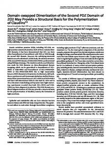
Figure 1 | Three-dimensional structure of PDZ domains. a | Ribbon diagram of the structure of the third PDZ domain of PSD-95 (α-helices in green, β-strands in blue) complexed with its target C-terminal peptide (purple). For details of the structural basis of specific interactions see REFS 1,130. b | Structural model of the PDZ1–PDZ2 domains of PSD-95. The tandem PDZ1 and PDZ2 domains, which bind to NMDA (N-methyl-D-aspartate) receptor (NMDAR) NR2 subunits and Kv1 channels, are arranged in similar orientations8. The structure provides a mechanistic explanation for the synergistic binding of two cytoplasmic tails extending from oligomeric membrane proteins such as receptors and channels.
PDZ DOMAIN
A peptide-binding domain that is important for the organization of membrane proteins, particularly at cell–cell junctions, including synapses. It can bind to the carboxyl termini of proteins or can form dimers with other PDZ domains. PDZ domains are named after the proteins in which these sequence motifs were originally identified (PSD95, discs large, zona occludens 1). POSTSYNAPTIC DENSITY
An electron-dense specialization of excitatory postsynaptic membranes that contains a high concentration of glutamate receptors and associated signalling and cytoskeletal proteins.
772
(T. Nakagawa, T. Walz and M.S., unpublished observations), implying that PSD-95 is not a flexible set of protein-interaction domains linked like ‘beads on a string’. Reinforcing this idea, NMR studies have provided evidence that the first two PDZ domains of PSD-95 are specifically oriented relative to each other in a way that would allow them to interact with C termini coming from the same direction (REF. 8; FIG. 1). PSD-95 forms multimers, and this process seems to be mediated by amino (N)-terminal ‘head-to-head’ interactions9,10. Self-association is a common feature of many PDZ scaffold proteins, and is sometimes mediated by direct interactions between PDZ domains (as in the case of glutamate-receptor-interacting protein (GRIP), described below11). Multimerization of PDZ scaffolds might enhance the clustering of partner proteins in large multimolecular assemblies at specific sites, such as the PSD.
| OCTOBER 2004 | VOLUME 5
Ion channels, receptors and membrane proteins. Electron microscopy immunolocalization and tomography studies indicate that PSD-95 is located close to the postsynaptic membrane (at a mean distance of 12 nm from the postsynaptic membrane), and that it can be labelled by antibodies from both the extracellular and cytoplasmic faces of purified PSDs12,13. It is therefore in a good position to interact with postsynaptic membrane proteins such as receptors, ion channels and celladhesion molecules, as well as with cytoplasmic proteins (FIG. 3; TABLE 1). Such interactions are proposed to be important for the localization and clustering of these proteins at the postsynaptic membrane. In support of this idea, PSD-95 can cluster NMDARs and Shaker-type K+ channels on the surface of heterologous cells14. The best in vivo evidence that PSD-95 clusters postsynaptic proteins comes from Drosophila melanogaster, in which mutations in discs large (Dlg), the D. melanogaster homologue of PSD-95, abolish synaptic clustering of Shaker K+ channels, which bind to the PDZ domains of Dlg15. When studied using light microscopy, the clustering of NMDARs at synapses seems to be independent of PDZ interactions, as clustering of these receptors is not altered by mutations of the cytoplasmic tails of NMDAR subunits, by genetic disruption of PSD-95 or by interfering peptides that disperse synaptic clusters of PSD-95 (REFS 16–18). On the other hand, functional localization of NMDARs in synapses might depend on PSD-95 (REFS 19–22). Clustering at the cell surface might be related to inhibition of receptor internalization. Removing the C-terminal PDZ-binding motif of NR2B, a NMDAR subunit, enhances its internalization in cultured neurons23. Similarly, PSD-95 suppresses the internalization of the Kv1.4 K+ channel24 and attenuates agonistinduced internalization of β1-adrenergic receptors in non-neuronal cells25. PSD-95 also interacts with neuroligin, a postsynaptic membrane protein that interacts trans-synaptically with β-neurexins, which in turn bind to the PDZ domain of CASK/LIN226. CASK, another scaffold of the membrane-associated guanylate kinase (MAGUK) superfamily of proteins (FIG. 2), is enriched on both sides of the synapse and interacts with synaptic membrane proteins such as β-neurexin, syndecan and SynCAM27–29. As well as mediating cell adhesion, the trans-synaptic neuroligin–β-neurexin interaction seems to induce presynaptic differentiation28,30. So, the PSD-95-based scaffold is probably involved in synaptic adhesion and synapse development, a role that resembles that of fasciclin II (FasII), a D. melanogaster cell adhesion molecule that interacts with Dlg in the development of the fly neuromuscular junction31. Although the emphasis has always been on cellbiological functions of PSD-95 (for example, surface delivery or stabilization, synaptic targeting or clustering of other proteins), it is possible that PSD-95 also functionally modulates the activities of membrane proteins to which it binds. For instance, PSD-95 can suppress the activity of the inward rectifier K+ channel (Kir2.3), mainly by reducing its single-channel conductance32.
www.nature.com/reviews/neuro
REVIEWS
PSD-95 PSD-93 SAP102 SAP97 Dlg GRIP1 PICK1 Shank1 SPAR nNOS CASK/LIN2 VELI1/LIN7 MINT1/LIN10 S-SCAM Neurabin-I Spinophilin L-Afadin
Densin-180 Erbin Syntenin-1 ZO-1 Tamalin PDZ domain
SH3
GK
NO synthase PTB
SRC HOMOLOGY 3 DOMAIN
(SH3 domain). A protein–protein interaction domain that binds to PXXP or related peptide sequences.
WW
L27
Ank
Flavodoxin RA
FHA
FABD DIL
SAM NADB Leucine-rich repeat
RapGAP CaM kinase ZU5
Figure 2 | Schematic diagram of PDZ proteins. PDZ domains are often found in scaffold proteins as multiple tandem arrays and/or linked to other kinds of modular protein-interaction domain. PDZ domains are shown as purple ellipses. Other domains are indicated: Ank, ankyrin repeats; CaM kinase, calmodulin-dependent kinase (CaMK)-like domain; DIL, dilute domain; FABD, FADbinding domain; FHA, forkhead-associated domain; GK, guanylate kinase-like domain; L27, domain initially found in LIN2 and LIN7; NADB, NAD-binding domain; NO, nitric oxide; PTB, phosphotyrosine-binding domain; RA, RAS association domain; RapGAP, Rap GTPase-activating protein; SAM, sterile α motif; SH3, Src homology 3 domain; WW, domain with two conserved Trp (W) residues; ZU5, domain present in ZO-1 and UNC5-like netrin receptors. Proteins: Dlg; discs large; GRIP1, glutamate-receptor-interacting protein 1; LIN7, lin7 homologue; LIN10, lin10 homologue; nNOS, neuronal nitric oxide synthase; PICK1, protein interacting with C-kinase 1; PSD-93, postsynaptic density protein 93; PSD-95, postsynaptic density protein 95; SAP97, synapse-associated protein 97; SAP102, synapse-associated protein 102; Shank, SH3 and ankyrin repeat-containing protein; SPAR, spine-associated RapGAP; S-SCAM, synaptic scaffolding molecule; ZO-1, zona occludens protein 1.
GUANYLATE KINASE-LIKE DOMAIN
(GK domain). A protein–protein interaction domain found in the membrane-associated guanylate kinase (MAGUK) superfamily of proteins, which includes PSD-95 and related proteins.
So, PSD-95 can regulate the activity of interacting membrane proteins by influencing their surface delivery, endocytosis, subcellular location, subunit composition and even intrinsic functional properties, such as channel conductance.
RAS, RAP AND RAC
Organization of postsynaptic signalling by PSD-95. Perhaps the most important biochemical function of PSD-95 is to organize signalling complexes at the postsynaptic membrane. In addition to membrane proteins, PSD-95 interacts with a wide variety of cytoplasmic signalling molecules (FIG. 3 and TABLE 1). By physically bringing together cytoplasmic signal-transducing enzymes and surface receptors (such as NMDARs), PSD-95 is thought to facilitate signal coupling in the PSD. An example is the association of PSD-95 with neuronal nitric oxide synthase (nNOS)3,33, a Ca2+/ calmodulin-activated enzyme that produces nitric oxide,
Small monomeric G-proteins that, in their activated GTPbound state, interact with and stimulate their downstream effectors. Hydrolysis of bound GTP by the intrinsic GTPase activity of these proteins terminates their activity. Guanine nucleotide exchange factors (GEFs) stimulate GTP loading and activate these small G-proteins; GTPase-activating proteins (GAPs) inhibit their activity.
NATURE REVIEWS | NEUROSCIENCE
a diffusible ‘transmitter’ that has been implicated in the regulation of neurotransmission and excitotoxicity. As NMDARs are permeable to Ca2+, the ternary NMDAR–PSD-95–nNOS complex might functionally couple NMDAR gating to nNOS activation. This is supported by evidence that disrupting the NMDAR– PSD-95 interaction by introducing a synthetic peptide that mimics the last nine residues of NR2B reduces NMDAR-induced excitotoxicity, without affecting NMDAR function34. An abundant PSD protein that binds to PSD-95 is synaptic RAS GTPase-activating protein (SynGAP), a GTPase-activating protein (GAP) for the Ras small GTPase35,36 (TABLE 1). SynGAP is activated by Ca2+/ calmodulin-dependent protein kinase II (CaMKII)37 and suppresses the Ras–extracellular signal-regulated kinase (ERK) pathway, which regulates synaptic plasticity38. Mice that are heterozygous for a mutant version of
VOLUME 5 | OCTOBER 2004 | 7 7 3
REVIEWS
Table 1 | Proteins that interact with PSD-95 family scaffolds Interacting protein
Comments on the interacting proteins
References
PDZ domains NR2A–D
Subunits of NMDA receptors
131
GluR6
Subunit of kainate receptors
132
δ2 GluR
Subunit of δ-ionotropic glutamate receptors
133
β1-adrenergic receptor
G-protein-coupled receptor
nAChRc
Subunit of neuronal nicotinic acetylcholine receptor
5-HT2A and 5-HT2C Rc
Subunits of 5-HT (serotonin) receptors
ErbB4
A receptor tyrosine kinase for neuregulin
Kv1
Voltage-gated potassium channel
Kir2, Kir3, Kir4 and Kir5
Inward-rectifying potassium channels
Neuroligin
A postsynaptic membrane protein that binds to β-neurexins and regulates synaptic adhesion and development
26,28,30
Stargazin family proteins
Tetra-spanning transmembrane proteins required for surface and synaptic expression of AMPA receptors
64
nNOS
Neuronal nitric oxide synthase
SynGAP
An abundant RasGAP of the PSD that regulates synaptic plasticity
Kalirin-7
A guanine nucleotide exchange factor for Rac1 that regulates spine morphogenesis
Fyn, Lyn, Src and Yes
Src family non-receptor protein tyrosine kinases; might also interact with the SH3 domain of PSD-95
Cypin
A cytosolic protein that regulates dendrite patterning by promoting microtubule assembly
CRIPT
A microtubule-binding protein
Sec8
A subunit of the exocyst complex involved in protein and vesicle trafficking
115
KIF1Bα
A motor of the kinesin superfamily
111
25 76,77 134 135,136 14 32,124
3,33 35,36,39,40 53 48,49
137
18
SH3 domain Pyk2
A non-receptor tyrosine kinase regulated by calcium and PKC and required for LTP induction
50,138
GK domain GKAP/SAPAP
An abundant multi-domain scaffold of PSD that links PSD-95 with Shank
SPAR
A postsynaptic RapGAP that regulates spine morphogenesis
139 57
SH3 and GK domains KA2 GluR
Subunit of kainate receptors
AKAP79/150
An anchoring protein that binds to protein kinase A and protein phosphatase 1
132 41
L27 domain CASK
Mammalian homologue of LIN2
Myosin VI
A minus-end-directed actin-based motor
140,141 113
Only proteins that interact directly with PSD-95 family scaffolds are listed. These interactions might not apply to all members of the PSD-95 family. Owing to space limitations, this list is not comprehensive and not all relevant references are cited. AMPA, α-amino-3-hydroxy-5-methyl-4isoxazole propionic acid; GK, guanylate kinase-like domain; LTP, long-term potentiation; NMDA, N-methyl-D-aspartate; PKC, protein kinase C; PSD-95, postsynaptic density protein 95; Rac, Rap and Ras, small monomeric G-proteins; RapGAP, Rap GTPase-activating protein; RasGAP, Ras GTPase-activating protein; SH3 domain, Src homology 3 domain; Shank, SH3 and ankyrin repeat-containing protein.
774
| OCTOBER 2004 | VOLUME 5
SynGAP show elevated basal activity of ERK in the hippocampus, increased synaptic AMPAR clustering in cortical cultures, reduced LONG-TERM POTENTIATION (LTP) in the CA1 region of the hippocampus and impaired spatial learning39,40. PSD-95 and SAP97, another member of the PSD-95 family of proteins, interact with A-kinase-anchoring protein 79/150 (AKAP79/150) (REF. 41), a scaffold for protein kinase A (PKA), PKC and the Ca2+/calmodulindependent protein phosphatase calcineurin (also known as PP2B). The interaction between PSD-95 and AKAP might bring these kinases and phosphatases close to their specific substrates in the synapse. For instance, the SAP97–AKAP complex facilitates the phosphorylation by PKA of the glutamate receptor GluR1 (REF. 41), a subunit of AMPARs that binds to SAP97 (see below), and is required for the downregulation of AMPAR currents by Ca2+ and PP2B42. As PKA-dependent phosphorylation of GluR1 at Ser845 is involved in the regulation of AMPAR recycling43 and synaptic plasticity44,45, the SAP97–AKAP79 complex might be important for the recruitment of kinases and phosphatases to synaptic AMPARs. Tyrosine phosphorylation regulates NMDAR activity and NMDAR-dependent synaptic plasticity46, including the trafficking of AMPARs47. PSD-95 associates with nonreceptor tyrosine kinases of the Src family48,49 and their upstream activator, proline-rich tyrosine kinase 2 (Pyk2)50, both of which are thought to be important for synaptic plasticity. So PSD-95 might localize the Pyk2–Src signalling cascade close to NMDARs; however, the importance of PSD-95 scaffolds in synaptic regulation by tyrosine phosphorylation has not been directly investigated. Another group of signalling molecules that is attracting growing interest is the group that regulates the assembly and dynamics of F-actin, which is the predominant cytoskeletal element in DENDRITIC SPINES and is important for synaptic morphogenesis and plasticity51,52. PSD-95 binds directly to kalirin-7, a guanine nucleotide exchange factor (GEF) for RAC1 that promotes spine formation53. However, the molecular mechanisms that link activated Rac1 to the postsynaptic actin cytoskeleton are not clear. Kalirin functions downstream of EphB receptors, which have been implicated in the regulation of NMDARs and spine development54,55. In addition to kalirin-7 (which activates Rac), the PSD also contains many regulators of other small GTPases56. Spine-associated RapGAP (SPAR), an inhibitory GAP for RAP, binds to PSD-95 and promotes the growth of dendritic spines. This function depends on SPAR’s GAP domain. SPAR itself contains a PDZ domain (TABLE 1). Degradation of SPAR by the ubiquitin–proteasome pathway leads to loss of PSD-95 and depletion of synapses57. Overexpression of PSD-95 promotes spine growth58, although whether this depends on its interactions with kalirin and SPAR remains to be determined. As well as interacting directly with various signalling enzymes, PSD-95 is also linked by protein interactions to other scaffolds in the PSD, including guanylate kinase-associated protein (GKAP/SAPAP), SH3 and ankyrin repeat-containing protein (Shank/ProSAP) and Homer5,59 (FIG. 2 and TABLE 1). These proteins, which are
www.nature.com/reviews/neuro
REVIEWS
Table 2 | Other synaptic PDZ proteins PDZ protein
Interacting protein(s)
References
Neurabin, spinophilin/neurabin-II Localized in spines; modulates synaptic transmission and spine morphology
Protein phosphatase 1 F-actin
142
Nectin (cell adhesion molecule) F-actin Eph receptors (receptor tyrosine kinases)
143
CaM kinase IIα α-Actinin (F-actin-binding protein) δ-Catenin (N-cadherin-interacting protein)
144
ErbB2 (receptor tyrosine kinase for neuregulin) PSD-95 δ-Catenin
145
Afadin Involved in synapse adhesion and development Densin-180 Abundant PSD protein; member of LAP (leucine-rich repeat and PDZ) family of proteins Erbin LAP protein; suppresses the Ras–MAPK signalling pathway S-SCAM Synaptic multi-PDZ scaffold; might regulate assembly and trafficking of synaptic proteins
NMDAR Neuroligin KIF1Bα β-Catenin (cadherin-associated protein) nRapGEF (guanine nucleotide exchange factor for Rap1)
2
Shank Important scaffold protein of the PSD; promotes morphological and functional maturation of synapse and dendritic spine
GKAP Homer Cortactin (actin regulatory protein) CIRL (calcium-independent receptor for α-latrotoxin) IRSp53 (actin regulatory protein that binds Rac1 and Cdc42) ABP1 (F-actin-binding protein) βPIX (guanine nucleotide exchange factor for Rac1 and Cdc42) Sharpin (multimeric PSD protein)
59,146
Syntenin Small scaffold protein that binds to phosphatidylinositol 4,5-bisphosphate
AMPA, kainate and metabotropic glutamate receptor subunits Syndecan (transmembrane proteoglycan) Neurexin (neuronal surface proteins) SynCAM (synaptic cell adhesion molecule) Ephrin B Neurofascin (neural cell adhesion molecule) Merlin (product of the causal gene for neurofibromatosis type II)
94
Tamalin Possibly involved in trafficking of mGLURs
Group I metabotropic glutamate receptor subunits Cytohesin (guanine nucleotide exchange factor for ARF small GTPases) GKAP S-SCAM
147
Only proteins that directly interact with the indicated PDZ proteins are described. Owing to space limitations, this list is not comprehensive and not all relevant references are cited. AMPA, α-amino-3hydroxy-5-methyl-4-isoxazole propionic acid; Cdc42, Rac, Rap and Ras, small monomeric G-proteins; GKAP, guanylate kinase-associated protein; KIF1Bα, kinesin family member 1Bα; NMDAR, N-methylD-aspartate receptor; PSD-95, postsynaptic density protein 95; Ras–MAPK, Ras mitogen activated protein kinase; S-SCAM, synaptic scaffolding molecule.
found in the deeper (cytoplasmic) part of the PSD12,13, bind to additional signalling and cytoskeletal proteins (TABLE 2). So, PSD-95 is integrated in a large network of signalling and adaptor proteins, many of which also contain PDZ domains. Although PSD-95 interacts
NATURE REVIEWS | NEUROSCIENCE
directly with only a subset of PSD proteins, the central importance of PSD-95 scaffolding in the PSD is reflected by its stoichiometric abundance60. Mass spectrometry analysis indicates that in the PSD, PSD-95 is an order of magnitude more abundant in molar terms than NMDARs, and several times more abundant than GKAP and Shank56. PSD-95 regulates synaptic transmission. As befits an important scaffold of the PSD, PSD-95 has a strong influence on synaptic transmission and plasticity. Overexpression of PSD-95 potentiates AMPAR-mediated excitatory postsynaptic currents (EPSCs), an effect that depends on two palmitoylated N-terminal cysteines in PSD-95 (REFS 58,61–63). Conversely, if PSD-95 is knocked down by RNA INTERFERENCE (RNAi), AMPAR-mediated EPSCs are suppressed (K. Futai, T. Nakagawa,Y. Hayashi & M. S., unpublished observations). NMDAR-mediated EPSCs are unaffected by either gain- or loss-of-function of PSD-95. How does PSD-95 affect AMPAR-mediated EPSCs, given that it does not interact directly with AMPARs? In one current model, PSD-95 recruits the tetraspanning membrane protein stargazin to synapses, where it binds directly to AMPAR subunits. Stargazin and its relatives are essential for the surface expression and synaptic accumulation of AMPARs, and the latter activity depends on an interaction of the stargazin C terminus with the PDZ domains of PSD-95 (REFS 64,65). Synaptic potentiation induced by the overexpression of PSD-95 seems to mimic LTP, in that it converts silent synapses into functional synapses, drives GluR1 into synapses, occludes LTP and enhances LONG-TERM DEPRESSION (LTD)61–63. Moreover, dominant-negative forms of PSD95 can block LTP and experience-driven synaptic potentiation in the barrel cortex63. These overexpression studies indicate that PSD-95 has a central role in the expression of LTP. However, this conclusion needs to be reconciled with the phenotype of PSD-95-deficient mice, which show enhanced LTP and reduced LTD16,66. Dynamic regulation of synaptic PSD-95. If PSD-95 acts as a physiologically important regulator of synaptic strength and structure, then it might be expected that its activity or abundance would be controlled by neural activity. Synaptic accumulation of PSD-95 requires the palmitoylation of two N-terminal cysteines (Cys3 and Cys5)67. Neuronal activity promotes the dispersal of PSD-95 from synapses, in part by depalmitoylating these two residues68. Synaptic stimulation also causes loss of synaptic PSD-95 through the ubiquitin–proteasome pathway57,69. The latter mechanism could involve direct ubiquitylation of PSD-95 (REF. 69) or could be indirect, through the ubiquitylation and degradation of other postsynaptic regulatory proteins such as SPAR57. Activity-dependent dispersal or degradation of PSD-95 is known to correlate with a loss of AMPARs and weakening of synapses, but beyond this its physiological importance is not well understood. The function of PSD-95 is regulated more acutely by phosphorylation. Cyclin-dependent kinase 5 (CDK5), a serine–threonine kinase that is essential for brain
VOLUME 5 | OCTOBER 2004 | 7 7 5
REVIEWS
Presynaptic
LIN10/MINT1
LIN2/CASK
PDZ domain LIN7 Ephrin/ EphR
β-neurexin Neuroligin
NMDAR
AMPAR Stargazin
ErbB2
NMDAR
K ch
GRIP
PSD-95 Src
AMPAR
Erbin
SPAR
AKAP79 PICK1
GKAP nNOS SynGAP GKAP
Kalirin-7 Densin-180 βPIX
Shank IRSp53 Cortactin
CamKIIα
Homer
GKAP
Shank
Shank Homer IP3R
F-actin
LONG-TERM POTENTIATION
(LTP). A long-lasting enhancement of synaptic strength that is elicited by specific patterns of synaptic stimulation (for example, high frequency tetanus). Typically dependent on NMDA-receptor activation, and widely believed to be a means of information storage in the brain.
Neurabin
mGluR
SER
Tamalin
mGluR
Figure 3 | A schematic diagram of the organization of PDZ proteins at a mammalian excitatory synapse. The main PDZ-containing proteins of a glutamatergic synapse are shown, focusing on the postsynaptic density. PDZ domains are indicated by purple circles. The C-terminal cytoplasmic tails of membrane proteins are indicated by black lines. Specific protein–protein interactions are indicated by the overlap of proteins. Only a subset of known protein interactions is illustrated. Although not shown, LIN2, LIN7 and LIN10 are also present postsynaptically, and many of the proteins of the postsynaptic domain are also present in the presynaptic terminal. Green and blue ellipses in PSD-95 represent SH3 and GK domains, respectively. Crooked lines indicate palmitoylation of PSD-95 and GRIP. Grey arrows indicate binding and/or regulatory actions of proteins on the actin cytoskeleton. AKAP79, A-kinase anchor protein 79; AMPAR, AMPA (α-amino-3-hydroxy-5methyl-4-isoxazole propionic acid) receptor; βPIX, PAAK-interactive exchange factor; CaMKIIα, α-subunit of Ca2+/calmodulin-dependent protein kinase II; GK, guanylate kinase-like domain; EphR, ephrin receptor; ErbB2, EGF-related peptide receptor; GKAP, guanylate kinase-associated protein; GRIP, glutamate-receptor-interacting protein; IP3R, IP3 receptor; IRSp53, insulin-receptor substrate p53; K ch, potassium channel; LIN7, lin7 homologue; LIN10, lin10 homologue; mGluR, metabotropic glutamate receptor; NMDAR, NMDA (N-methyl-D-aspartate) receptor; nNOS, neuronal nitric oxide synthase; PICK1, protein interacting with C kinase 1; PSD-95, postsynaptic density protein 95; SER, smooth endoplasmic reticulum; SH3, Src homology 3 domain; Shank, SH3 and ankyrin repeat-containing potein; SPAR, spine-associated RapGAP; SynGAP, synaptic Ras GTPase-activating protein.
DENDRITIC SPINES
Tiny actin-rich protrusions from the dendrite that form the postsynaptic compartment for most excitatory synapses in the brain. RNA INTERFERENCE
(RNAi). A method for suppressing the expression of a specific protein based on targeted hybridization of small interfering RNAs to the mRNA encoding that protein. LONG-TERM DEPRESSION
(LTD). A long-lasting suppression of synaptic strength that is elicited by specific patterns of synaptic stimulation (for example, low frequency stimulation). Typically dependent on NMDA-receptor activation, and widely believed to be a means of information storage in the brain.
776
development, phosphorylates the N-terminal region of PSD-95, inhibiting its multimerization, channel clustering activity and possibly its synaptic localization70. By contrast, phosphorylation by CaMKII of SAP97 in the Nterminal L27 domain promotes synaptic targeting of SAP97, and of its binding partner GluR1 (REF. 71). In D. melanogaster, CaMKII-dependent phosphorylation of the first PDZ domain of Dlg decreases the synaptic localization of Dlg72. Time-lapse imaging has confirmed that PSD-95 tagged with green fluorescent protein undergoes dynamic turnover, although at a slower rate than several other synaptic proteins73. PDZ scaffolds are probably all regulated in a dynamic fashion by subcellular redistribution and protein phosphorylation and degradation. In vivo functions of PSD-95 proteins. Although the PSD-95 family of proteins has been extensively studied in cultured neurons, their functions in vivo are not well established. PSD-95 mutant mice have impaired spatial
| OCTOBER 2004 | VOLUME 5
learning despite enhanced LTP (these animals also have defective LTD)16. PSD-95 deficiency in knock-out mice prevents the maturation of orientation preference in the visual cortex74 and eliminates behavioural sensitization induced by chronic cocaine administration66. These in vivo results indicate that PSD-95 is involved in learning and memory, maturation of cortical circuits and behavioural responses to drugs of abuse, all of which presumably reflect the importance of PSD-95 in synaptic plasticity. Genetic disruption of PSD-93 reduces NMDARmediated postsynaptic responses and blunts NMDARdependent persistent pain 75. In addition, PSD-93 associates with neuronal nicotinic acetylcholine receptors and is required for the normal function and stability of neuronal cholinergic synapses76,77. So, PSD-93 might function as a key scaffold in cholinergic as well as glutamatergic synapses. The in vivo importance of SAP97 and SAP102 for brain function is unclear.
www.nature.com/reviews/neuro
REVIEWS
Box 1 | PDZ scaffolds in trafficking of protein complexes F-actin AMPAR Myosin VI SAP97 Myosin V DLC GKAP PSD-95 NMDAR
NMDAR
Stargazin
AMPAR
LIN2/7/10
GRIP
Liprin-α
PSD-95/SAP97
KIF17
KIF5
KIF1A
KIF1Bα
Microtubule
PDZ-based membrane protein complexes can be moved around the cell as pre-assembled packages. Transport along microtubule tracks is mediated by motor proteins of the KINESIN superfamily (KIFs), whereas transport along actin tracks is carried out by motors of the MYOSIN family. PDZ scaffolds on the surface of cargo vesicles can act as ‘receptors’ for molecular motors by binding to specific kinesins and myosins. For instance, the PDZ domains of PSD-95 (postsynaptic density protein 95), SAP97 (synapse-associated protein 97) and S-SCAM (synaptic scaffolding molecule) interact directly with the C terminus of KIF1Bα (kinesin family member 1Bα), a kinesin motor111. SAP97 can also bind, through its GK (guanylate kinase-like) domain, to KIF13B/GAKIN (kinesin family member 13B)112, and through its N-terminal L27 domain to myosin-VI113. PSD-95 family proteins can also associate indirectly with myosin-V through the PSD-95-binding protein GKAP (guanylate kinase-associated protein)114. Although it is not a motor protein, the Sec8 subunit of the ‘exocyst’ complex (which targets secretory vesicles to the cell surface) also interacts with PSD-95 family members, particularly SAP102 (REF. 115). Dominantnegative Sec8 inhibits NMDA (N-methyl-D-aspartate) receptor (NMDAR) currents in neurons, supporting the involvement of the exocyst complex in synaptic trafficking of an NMDAR–SAP102 complex115. In Caenorhabditis elegans, a complex of PDZ proteins (CASK/LIN2–LIN7–LIN10) (FIGS 2, 3) is important for basolateral targeting of a receptor tyrosine kinase in epithelia116. LIN10 is also required for the synaptic localization of glutamate receptors117. Homologous proteins exist in mammalian neurons on both pre- and postsynaptic sides of the synapse and are probably involved in subcellular targeting of the proteins with which they interact2. The kinesin family motor KIF17 binds directly to the PDZ1 domain of LIN10 and might transport a CASK–LIN7–LIN10–NMDAR complex to synapses118,119. Remarkably, mammalian CASK can redistribute from the plasma membrane to the nucleus to regulate transcription120,121. The GluR2/3-binding protein GRIP interacts directly with conventional kinesin (KIF5) and this association is important for the targeting of AMPA (α-amino-3-hydroxy-5methyl-4-isoxazole propionic acid) receptors (AMPARs) to dendrites122. The GRIPinteracting protein liprin-α also binds to KIF1A, another kinesin-like motor123. As for NMDARs, it is possible that multiple motor proteins contribute to the transport of AMPARs, each interacting in different ways with the AMPAR protein complex. So, beyond their well-known function as organizers of protein complexes at the plasma membrane, there is mounting evidence that PDZ scaffolds have an important role in intracellular protein trafficking in neurons. Indeed, PDZ proteins can act as the ‘motor receptor’, enabling specific motor proteins to bind to and transport the complex. DLC, dynein light chain.
NATURE REVIEWS | NEUROSCIENCE
Differences between PSD-95 family proteins. PSD-95 family proteins are distributed differently from each other both in the brain and in neurons. PSD-95 and PSD-93 are most alike in being highly enriched in the PSD, and this enrichment might be related to the selective palmitoylation of these proteins78. On the other hand, SAP102 and SAP97 are found in dendrites and axons and are abundant in the cytoplasm as well as at synapses79–81. SAP102 is highly expressed early in postnatal development, whereas PSD-95 and PSD-93 predominate at later stages82. Although they show similar specificities of protein interaction in vitro, PSD-95 family members interact with different (but overlapping) sets of proteins in vivo. For instance, PSD-95 is preferentially associated with NR2A in vivo, whereas SAP102 is more associated with NR2B82. The NR2B–SAP102 complex in immature synapses tends to be replaced by the NR2A–PSD-95/ PSD-93 complex in mature synapses82,83. Overexpression of PSD-95 promotes synaptic insertion of NR2A rather than NR2B, thereby modifying the subunit composition and functional properties of synaptic NMDARs84. Stargazin family proteins are selectively associated with PSD-95 and PSD-93 in the brain85. Perhaps most strikingly, SAP97 interacts directly with the AMPAR subunit GluR1 (REF. 86), whereas the other members of the family bind directly to NMDAR NR2 subunits. The SAP97– GluR1 association can be detected early in the secretory pathway, indicating that SAP97 might be involved in the trafficking of GluR1 (REF. 80). Overall, it seems that PSD95 and PSD-93 are more specifically associated with synaptic functions, whereas SAP97 and SAP102 might be more important in trafficking (BOX 1). PDZ scaffolds associated with AMPARs
The PSD-95 family is mainly associated with NMDARs and the PSD, with the exception of SAP97 (see above). AMPAR subunits interact directly with different PDZ proteins, which might account for the more dynamic cell-biological behaviour of AMPARs. The C termini of the AMPAR subunits GluR2 and GluR3 bind to glutamate-receptor-interacting protein/AMPAR-binding protein (GRIP/ABP; encoded by two distinct genes, GRIP1 and ABP/GRIP2) and to protein interacting with C kinase 1 (PICK1). These PDZ-based interactions are important for the synaptic targeting and regulated trafficking of AMPARs (for recent reviews on this subject, see REFS 87,88). Here, we focus on recent progress in this area. GRIP. The GluR2/3 subunit binds specifically to the PDZ5 domain of GRIP, but the PDZ4 domain is also required for a strong interaction. The structure of the tandem PDZ4 and 5 domains reveals that PDZ4 is unlikely to bind to C-terminal peptides but instead stabilizes PDZ5 through interdomain interactions89. GRIP has up to seven PDZ domains (FIG. 2), through which it can interact with many proteins, including Eph receptors and their ephrin ligands90; a RAS guanine nucleotide exchange factor (RasGEF)91; liprin-α92; the transmembrane protein Fraser syndrome 1 (FRAS1)93; and, perhaps, also metabotropic and kainite-type
VOLUME 5 | OCTOBER 2004 | 7 7 7
REVIEWS
Box 2 | Regulation of PDZ interactions by protein phosphorylation The protein interactions of PDZ-based scaffolds should be regulated to allow controlled assembly and disassembly of protein complexes at the synapse. There is widespread evidence that PDZ-ligand interactions are disrupted by phosphorylation, typically on the C-terminal peptide of the ligand. For instance, phosphorylation of the C termini of potassium channels124, β1-adrenergic receptors125 and stargazin126,127 prevents them from binding to the PDZ domains of PSD-95 (postsynaptic density protein 95). Accordingly, phosphorylated stargazin is poorly enriched in PSD fractions126, and phosphorylation-mimicking stargazin mutants fail to cluster at synaptic sites and attenuate synaptic AMPA (α-amino-3-hydroxy-5-methyl-4-isoxazole propionic acid) receptor (AMPAR) currents127. For most of these regulated PDZ interactions, the specific protein kinase that phosphorylates the PDZ-binding C terminus in vivo is unknown. A specific protein kinase has been implicated in regulating interactions between AMPARs and PDZ domains. Phosphorylation of Ser880 in the C terminus of GluR2 (AMPA glutamate receptor 2) by PKC (protein kinase C) prevents it from interacting with GRIP/ABP (glutamate-receptor-interacting protein/AMPAR-binding protein) but not with PICK1 (protein interacting with C-kinase 1), indicating that the phosphorylation might displace AMPARs from GRIP in favour of PICK1. Phosphorylation of GluR2 at Ser880 is correlated with the internalization of AMPARs from synapses and is important for long-term depression (LTD), this mechanism being particularly well-established for cerebellar LTD107,110,128. PDZ–peptide interactions can also be regulated by phosphorylation of the PDZ domain, although this is less common than phosphorylation of the ligand’s C-terminal motif. For example, Ca2+/calmodulin-dependentprotein kinase II (CaMKII)-dependent phosphorylation of the PDZ1 domain of SAP97 (synapse-associated protein 97) disrupts its interaction with the NR2A subunit of NMDA (N-methyl-D-aspartate) receptors (NMDARs), but not with the GLUR1 (glutamate receptor 1) subunit of AMPARs129.
KINESINS
A large family of structurally related motor proteins that use ATP to transport specific cargoes along microtubules. MYOSINS
A large family of structurally related motor proteins that use ATP to transport specific cargoes along actin filaments.
778
glutamate receptors94. GRIP can also dimerize through an interaction between the PDZ6 domains of two monomers11. GRIP is widely expressed in body tissues and in neurons — it is present in both axons and dendrites92. Therefore, the function of GRIP must extend beyond the regulation of AMPARs. This is supported by evidence from knockout mice in which the Grip1 gene is disrupted, which show haemorrhagic blisters and embryonic lethality93,95. Certain splice variants of GRIP can be palmitoylated, like PSD-95 and PSD-93, and palmitoylation of the protein results in it being associated with the plasma membrane and localized at synapses96. Nonpalmitoylated GRIP mostly associates with intracellular membranes97. These differentially modified subpopulations of GRIP might stabilize synaptic and intracellular pools of AMPARs, respectively. GRIP is believed to be involved in synaptic trafficking and/or synaptic stabilization of AMPARs and other interacting proteins. The widespread distribution of GRIP in cells, and its interactions with motor proteins (BOX 1), support the hypothesis that it is involved in trafficking98. GRIP can participate in synaptic function not only by interacting with AMPARs, but also by associating with Eph receptors and their ephrin ligands, which have been implicated in dendritic spine morphogenesis and hippocampal synaptic plasticity99,100. PICK1. PICK1 is present at synaptic and non-synaptic sites in neurons, and its PDZ domain shows relatively promiscuous binding. In addition to PKCα and GluR2/3, it has many other binding partners (both
| OCTOBER 2004 | VOLUME 5
presynaptic and postsynaptic), including the netrin receptor UNC5H101, various metabotropic glutamatereceptor subtypes102,103, the dopamine plasma-membrane transporter104 and the erythroblastic leukaemia viral oncogene homologue 2 (ErbB2) receptor tyrosine kinase105. In many of these cases, the interaction with PICK1 seems to regulate the subcellular localization and/or surface expression of its protein partners. Phosphorylation of the C terminus of GluR2 alters its binding specificity for GRIP and PICK1, and contributes to synaptic plasticity by altering the trafficking of AMPARs (BOX 2). There is some controversy regarding the respective roles of GRIP and PICK1 in the stabilization of synaptic versus intracellular AMPARs106–110. This could be related to the existence of different subpopulations of GRIP and/or PICK1 at synaptic and intracellular locations96, as well as to difficulties in interpreting the results of experimental perturbations of AMPAR trafficking. For instance, increased intracellular accumulation of AMPARs could arise from increased endocytosis of surface receptors or from reduced recycling of intracellular receptors. Moreover, because of overlapping specificities of PDZ–C-terminal interactions, the peptides that are typically used to interfere with the PDZ interactions of GRIP and PICK1 are probably not highly specific for these proteins or for GluR2/3 interactions. Genetic loss-of-function experiments would be helpful to dissect out the functions of GRIP and PICK1. Unfortunately, a generalized GRIP1 knockout is lethal in mice, and ABP/GRIP2 mutants lack an obvious phenotype93,95. PICK1-knockout mice are viable and show normal synaptic transmission in several brain areas. However, cerebellar LTD is abolished, and can be rescued by transient transfection of PICK1deficient Purkinje cells with wild type PICK1, but not mutants of PICK1 with mutations in the PDZ domains (J. Steinberg, J. Xia, K. Takamiya, D. Linden and R. Huganir, unpublished observations). In addition to AMPARs, GRIP and PICK1 have been reported to bind kainate receptors (KARs). Disrupting these PDZ-based interactions with fusion proteins and peptides decreases KAR-mediated synaptic transmission, indicating that GRIP and PICK1 interactions might be required to maintain synaptic KAR function94. Conclusions
PDZ domains were characterized as protein-interaction modules only a decade ago, but they have now come of age. Nowhere is the diversity and function of PDZ proteins better illustrated than at excitatory glutamatergic synapses. Our view of PDZ proteins has evolved from one of static adaptors for clustering interacting proteins to a more dynamic picture in which PDZ scaffolds organize heterogeneous ensembles of proteins, the composition of which changes at different locations in the cell, both during development and in response to neuronal activity. Moreover, PDZ proteins themselves can be mobile within neurons, and their activity and expression levels are regulated by phosphorylation, lipid modification and ubiquityation–degradation. As befits their central role in the organization of glutamate-receptor
www.nature.com/reviews/neuro
REVIEWS complexes, PDZ domain scaffolds have been shown by genetic, electrophysiological and morphological studies to be essential for controlling the structure, strength and plasticity of synapses. The next stage of investigations promises to reveal more insights into the in vivo significance of synaptic PDZ proteins,
1.
2.
3.
4.
5. 6.
7.
8.
9.
10.
11.
12.
13.
14.
15.
16.
17.
18.
19.
20.
21.
22.
Hung, A. Y. & Sheng, M. PDZ domains: structural modules for protein complex assembly. J. Biol. Chem. 277, 5699–5702 (2002). Montgomery, J. M., Zamorano, P. L. & Garner, C. C. MAGUKs in synapse assembly and function: an emerging view. Cell. Mol. Life Sci. 61, 911–929 (2004). McGee, A. W. & Bredt, D. S. Assembly and plasticity of the glutamatergic postsynaptic specialization. Curr. Opin. Neurobiol. 13, 111–118 (2003). Sheng, M. & Sala, C. PDZ domains and the organization of supramolecular complexes. Annu. Rev. Neurosci. 24, 1–29 (2001). Sheng, M. & Kim, M. J. Postsynaptic signaling and plasticity mechanisms. Science 298, 776–780 (2002). McGee, A. W. et al. Structure of the SH3-guanylate kinase module from PSD-95 suggests a mechanism for regulated assembly of MAGUK scaffolding proteins. Mol. Cell 8, 1291–1301 (2001). Tavares, G. A., Panepucci, E. H. & Brunger, A. T. Structural characterization of the intramolecular interaction between the SH3 and guanylate kinase domains of PSD-95. Mol. Cell 8, 1313–1325 (2001). Long, J. F. et al. Supramodular structure and synergistic target binding of the N-terminal tandem PDZ domains of PSD-95. J. Mol. Biol. 327, 203–214 (2003). The structure of the first two PDZ domains of PSD-95 was determined by NMR, suggesting a model in which the tandem PDZ domains are oriented in a similar direction to promote interaction with pairs of C termini extending from oligomeric membrane receptors or ion channels. Hsueh, Y. P. & Sheng, M. Requirement of N-terminal cysteines of PSD-95 for PSD-95 multimerization and ternary complex formation, but not for binding to potassium channel Kv1.4. J. Biol. Chem. 274, 532–536 (1999). Christopherson, K. S. et al. Lipid- and protein-mediated multimerization of PSD-95: implications for receptor clustering and assembly of synaptic protein networks. J. Cell Sci. 116, 3213–3219 (2003). Im, Y. J. et al. Crystal structure of GRIP1 PDZ6-peptide complex reveals the structural basis for class II PDZ target recognition and PDZ domain-mediated multimerization. J. Biol. Chem. 278, 8501–8507 (2002). Valtschanoff, J. G. & Weinberg, R. J. Laminar organization of the NMDA receptor complex within the postsynaptic density. J. Neurosci. 21, 1211–1217 (2001). Petersen, J. D. et al. Distribution of postsynaptic density (PSD)-95 and Ca2+/calmodulin-dependent protein kinase II at the PSD. J. Neurosci. 23, 11270–11278 (2003). Kim, E., Niethammer, M., Rothschild, A., Jan, Y. N. & Sheng, M. Clustering of Shaker-type K+ channels by interaction with a family of membrane-associated guanylate kinases. Nature 378, 85–88 (1995). Gramates, L. S. & Budnik, V. Assembly and maturation of the Drosophila larval neuromuscular junction. Int. Rev. Neurobiol. 43, 93–117 (1999). Migaud, M. et al. Enhanced long-term potentiation and impaired learning in mice with mutant postsynaptic density-95 protein. Nature 396, 433–439 (1998). Sprengel, R. et al. Importance of the intracellular domain of NR2 subunits for NMDA receptor function in vivo. Cell 92, 279–289 (1998). Passafaro, M., Sala, C., Niethammer, M. & Sheng, M. Microtubule binding by CRIPT and its potential role in the synaptic clustering of PSD-95. Nature Neurosci. 2, 1063–1069 (1999). Steigerwald, F. et al. C-terminal truncation of NR2A subunits impairs synaptic but not extrasynaptic localization of NMDA receptors. J. Neurosci. 20, 4573–4581 (2000). Prybylowski, K. et al. Relationship between availability of NMDA receptor subunits and their expression at the synapse. J. Neurosci. 22, 8902–8910 (2002). Barria, A. & Malinow, R. Subunit-specific NMDA receptor trafficking to synapses. Neuron 35, 345–353 (2002). Mori, H. et al. Role of the carboxy-terminal region of the GluR ε2 subunit in synaptic localization of the NMDA receptor channel. Neuron 21, 571–580 (1998).
NATURE REVIEWS | NEUROSCIENCE
beyond their protein interactions and cell-biological functions. With increasing knowledge of their structure and function, PDZ interactions could become plausible targets for pharmaceutical intervention, thereby opening up a wealth of possibilities for the treatment of brain diseases.
23. Roche, K. W. et al. Molecular determinants of NMDA receptor internalization. Nature Neurosci. 4, 794–802 (2001). 24. Jugloff, D. G., Khanna, R., Schlichter, L. C. & Jones, O. T. Internalization of the Kv1.4 potassium channel is suppressed by clustering interactions with PSD-95. J. Biol. Chem. 275, 1357–1364. (2000). 25. Hu, L. A. et al. β1-adrenergic receptor association with PSD-95. Inhibition of receptor internalization and facilitation of β1-adrenergic receptor interaction with N-methyl-Daspartate receptors. J. Biol. Chem. 275, 38659–38666 (2000). 26. Irie, M. et al. Binding of neuroligins to PSD-95. Science 277, 1511–1515 (1997). 27. Biederer, T. et al. SynCAM, a synaptic adhesion molecule that drives synapse assembly. Science 297, 1525–1531 (2002). This paper identifies SynCAM, a homophilic celladhesion molecule of the immunoglobulin superfamily located on both sides of the synapse, which promotes synapse formation by interacting with cytoplasmic PDZ proteins. 28. Dean, C. et al. Neurexin mediates the assembly of presynaptic terminals. Nature Neurosci. 6, 708–716 (2003). β-neurexin This paper reports that the neuroligin–β complex induces presynaptic assembly by two steps: clustering of presynaptic β-neurexin by interaction with oligomeric neuroligin, and recruitment of presynaptic molecules through the PDZ-binding C terminus of β-neurexin. 29. Hsueh, Y. P. et al. Direct interaction of CASK/LIN-2 and syndecan heparan sulfate proteoglycan and their overlapping distribution in neuronal synapses. J. Cell. Biol. 142, 139–151 (1998). 30. Li, Z. & Sheng, M. Some assembly required: the development of neuronal synapses. Nature Rev. Mol. Cell Biol. 4, 833–841 (2003). 31. Packard, M., Mathew, D. & Budnik, V. FASt remodeling of synapses in Drosophila. Curr. Opin. Neurobiol. 13, 527–534 (2003). 32. Nehring, R. B. et al. Neuronal inwardly rectifying K+ channels differentially couple to PDZ proteins of the PSD-95/SAP90 family. J. Neurosci. 20, 156–162 (2000). 33. Brenman, J. E. et al. Interaction of nitric oxide synthase with the postsynaptic density protein PSD-95 and α1-syntrophin mediated by PDZ domains. Cell 84, 757–767 (1996). 34. Aarts, M. et al. Treatment of ischemic brain damage by perturbing NMDA receptor–PSD-95 protein interactions. Science 298, 846–850 (2002). A synthetic peptide that mimics the PDZ-binding C terminus of an NMDAR subunit (NR2B) blocks NMDA-induced excitotoxicity without affecting NMDAR function, suggesting the therapeutic potential of this approach in stroke treatment. 35. Chen, H. J., Rojas-Soto, M., Oguni, A. & Kennedy, M. B. A synaptic Ras-GTPase activating protein (p135 SynGAP) inhibited by CaM kinase II. Neuron 20, 895–904 (1998). 36. Kim, J. H., Liao, D., Lau, L. F. & Huganir, R. L. SynGAP: a synaptic RasGAP that associates with the PSD-95/SAP90 protein family. Neuron 20, 683–691 (1998). 37. Oh, J. S., Manzerra, P. & Kennedy, M. B. Regulation of the neuron-specific Ras GTPase-activating protein, synGAP, by Ca2+/calmodulin-dependent protein kinase II. J. Biol. Chem. 279, 17980–17988 (2004). 38. Zhu, J. J., Qin, Y., Zhao, M., Van Aelst, L. & Malinow, R. Ras and Rap control AMPA receptor trafficking during synaptic plasticity. Cell 110, 443–455 (2002). 39. Komiyama, N. H. et al. SynGAP regulates ERK/MAPK signaling, synaptic plasticity, and learning in the complex with postsynaptic density 95 and NMDA receptor. J. Neurosci. 22, 9721–9732 (2002). 40. Kim, J. H., Lee, H. K., Takamiya, K. & Huganir, R. L. The role of synaptic GTPase-activating protein in neuronal development and synaptic plasticity. J. Neurosci. 23, 1119–1124 (2003). References 39 and 40, studies on SynGAP-deficient mice, report that SynGAP, a PSD-95-binding GAP for Ras, regulates the MAP kinase pathway, synaptic AMPAR clustering, LTP and spatial learning.
41. Colledge, M. et al. Targeting of PKA to glutamate receptors through a MAGUK–AKAP complex. Neuron 27, 107 (2000). 42. Tavalin, S. J. et al. Regulation of GluR1 by the A-kinase anchoring protein 79 (AKAP79) signaling complex shares properties with long-term depression. J. Neurosci. 22, 3044–3051 (2002). 43. Ehlers, M. D. Reinsertion or degradation of AMPA receptors determined by activity-dependent endocytic sorting. Neuron 28, 511–525 (2000). 44. Lee, H. K., Barbarosie, M., Kameyama, K., Bear, M. F. & Huganir, R. L. Regulation of distinct AMPA receptor phosphorylation sites during bidirectional synaptic plasticity. Nature 405, 955–959 (2000). 45. Kameyama, K., Lee, H. K., Bear, M. F. & Huganir, R. L. Involvement of a postsynaptic protein kinase A substrate in the expression of homosynaptic long-term depression. Neuron 21, 1163–1175 (1998). 46. Salter, M. W. & Kalia, L. V. Src kinases: a hub for NMDA receptor regulation. Nature Rev. Neurosci. 5, 317–328 (2004). 47. Ahmadian, G. et al. Tyrosine phosphorylation of GluR2 is required for insulin-stimulated AMPA receptor endocytosis and LTD. EMBO J. 23, 1040–1050 (2004). 48. Kalia, L. V. & Salter, M. W. Interactions between Src family protein tyrosine kinases and PSD-95. Neuropharmacology 45, 720–728 (2003). 49. Tezuka, T., Umemori, H., Akiyama, T., Nakanishi, S. & Yamamoto, T. PSD-95 promotes Fyn-mediated tyrosine phosphorylation of the N-methyl-D-aspartate receptor subunit NR2A. Proc. Natl Acad. Sci. USA 96, 435–440 (1999). 50. Huang, Y. et al. CAKβ/Pyk2 kinase is a signaling link for induction of long-term potentiation in CA1 hippocampus. Neuron 29, 485–496 (2001). 51. Hering, H. & Sheng, M. Dendritic spines: structure, dynamics and regulation. Nature Rev. Neurosci. 2, 880–888 (2001). 52. Bonhoeffer, T. & Yuste, R. Spine motility. Phenomenology, mechanisms, and function. Neuron 35, 1019–1027 (2002). 53. Penzes, P. et al. The neuronal Rho-GEF Kalirin-7 interacts with PDZ domain-containing proteins and regulates dendritic morphogenesis. Neuron 29, 229–242 (2001). 54. Penzes, P. et al. Rapid induction of dendritic spine morphogenesis by trans-synaptic ephrinB-EphB receptor activation of the Rho-GEF kalirin. Neuron 37, 263–274 (2003). Activation of EphB receptors by their ephrinB ligand stimulates Kalirin-7, a PSD-95-binding guanine nucleotide exchange factor (GEF) for Rac1, and promotes spine formation through a signalling pathway that involves Rac1 and PAK. 55. Takasu, M. A., Dalva, M. B., Zigmond, R. E. & Greenberg, M. E. Modulation of NMDA receptor-dependent calcium influx and gene expression through EphB receptors. Science 295, 491–495 (2002). 56. Peng, J. et al. Semiquantitative proteomic analysis of rat forebrain postsynaptic density fractions by mass spectrometry. J. Biol. Chem. 217, 21003–21011 (2004). As well as identifying approximately 400 proteins in the PSD fraction, this paper uses quantitative mass spectrometry to measure the relative abundance in molar terms of key proteins of the PSD, including PSD-95, GKAP, Shank1, NMDAR (NR1) and AMPAR (GluR1 and GluR2). 57. Pak, D. T. & Sheng, M. Targeted protein degradation and synapse remodeling by an inducible protein kinase. Science 302, 1368–1373 (2003). SNK, a protein kinase induced by synaptic activity, binds to and phosphorylates SPAR, a postsynaptic GAP for the small GTPase Rap, leading to degradation of SPAR by the ubiquitin–proteasome pathway, and resulting in activity-dependent loss of synapses and spines. 58. El-Husseini, A. E., Schnell, E., Chetkovich, D. M., Nicoll, R. A. & Bredt, D. S. PSD-95 involvement in maturation of excitatory synapses. Science 290, 1364–1368 (2000). 59. Sheng, M. & Kim, E. The Shank family of scaffold proteins. J. Cell Sci. 113, 1851–1856 (2000).
VOLUME 5 | OCTOBER 2004 | 7 7 9
REVIEWS 60. Cho, K. O., Hunt, C. A. & Kennedy, M. B. The rat brain postsynaptic density fraction contains a homolog of the Drosophila discs-large tumor suppressor protein. Neuron 9, 929–942 (1992). 61. Stein, V., House, D. R., Bredt, D. S. & Nicoll, R. A. Postsynaptic density-95 mimics and occludes hippocampal long-term potentiation and enhances long-term depression. J. Neurosci. 23, 5503–5506 (2003). 62. Beique, J. C. & Andrade, R. PSD-95 regulates synaptic transmission and plasticity in rat cerebral cortex. J. Physiol. 546, 859–867 (2003). 63. Ehrlich, I. & Malinow, R. Postsynaptic density 95 controls AMPA receptor incorporation during long-term potentiation and experience-driven synaptic plasticity. J. Neurosci. 24, 916–927 (2004). References 61–63 demonstrate a central role for PSD-95 in the control of synaptic abundance of AMPARs and synaptic plasticity based on electrophysiological experiments in brain slices transfected with PSD-95 constructs. 64. Chen, L. et al. Stargazin regulates synaptic targeting of AMPA receptors by two distinct mechanisms. Nature 408, 936–943 (2000). Stargazin, an AMPAR- and PSD-95-associated membrane protein, is shown to promote synaptic AMPAR targeting by two distinct mechanisms: surface expression and synaptic targeting. 65. Schnell, E. et al. Direct interactions between PSD-95 and stargazin control synaptic AMPA receptor number. Proc. Natl Acad. Sci. USA 99, 13902–13907 (2002). 66. Yao, W. D. et al. Identification of PSD-95 as a regulator of dopamine-mediated synaptic and behavioral plasticity. Neuron 41, 625–638 (2004). Using gene-expression profiling and PSD-95-deficient mice, this paper implicates changes in the level of PSD-95 in psychostimulant action and addiction to drugs of abuse. 67. Craven, S. E., El-Husseini, A. E. & Bredt, D. S. Synaptic targeting of the postsynaptic density protein PSD-95 mediated by lipid and protein motifs. Neuron 22, 497–509 (1999). 68. El-Husseini Ael, D. et al. Synaptic strength regulated by palmitate cycling on PSD-95. Cell 108, 849–863 (2002). This paper reports that palmitoylation and depalmitoylation of PSD-95 regulate the synaptic localization of PSD-95, and consequently of AMPARs, in an activity-dependent manner. 69. Colledge, M. et al. Ubiquitination regulates PSD-95 degradation and AMPA receptor surface expression. Neuron 40, 595–607 (2003). The authors provide evidence that synaptic activity rapidly breaks down PSD-95 via the ubiquitin–proteasome pathway and that this degradation is required for activity-induced internalization of synaptic AMPARs. 70. Morabito, M. A., Sheng, M. & Tsai, L. H. Cyclin-dependent kinase 5 phosphorylates the N-terminal domain of the postsynaptic density protein PSD-95 in neurons. J. Neurosci. 24, 865–876 (2004). 71. Mauceri, D., Cattabeni, F., Di Luca, M. & Gardoni, F. CaMKIIdependent phosphorylation drives synapse-associated protein 97 into spines. J. Biol. Chem. 279, 23813–23821 (2004). 72. Koh, Y. H., Popova, E., Thomas, U., Griffith, L. C. & Budnik, V. Regulation of DLG localization at synapses by CaMKIIdependent phosphorylation. Cell 98, 353–363 (1999). 73. Inoue, A. & Okabe, S. The dynamic organization of postsynaptic proteins: translocating molecules regulate synaptic function. Curr. Opin. Neurobiol. 13, 332–340 (2003). 74. Fagiolini, M. et al. Separable features of visual cortical plasticity revealed by N-methyl-D-aspartate receptor 2A signaling. Proc. Natl Acad. Sci. USA 100, 2854–2859 (2003). 75. Tao, Y. X. et al. Impaired NMDA receptor-mediated postsynaptic function and blunted NMDA receptordependent persistent pain in mice lacking postsynaptic density-93 protein. J. Neurosci. 23, 6703–6712 (2003). 76. Conroy, W. G., Liu, Z., Nai, Q., Coggan, J. S. & Berg, D. K. PDZ-containing proteins provide a functional postsynaptic scaffold for nicotinic receptors in neurons. Neuron 38, 759–771 (2003). 77. Parker, M. J., Zhao, S., Bredt, D. S., Sanes, J. R. & Feng, G. PSD93 regulates synaptic stability at neuronal cholinergic synapses. J. Neurosci. 24, 378–388 (2004). References 76 and 77 identify PSD-93 as being important for the structure and functions of neuronal cholinergic synapses, in addition to glutamatergic synapses.
780
| OCTOBER 2004 | VOLUME 5
78. El-Husseini, A. E. et al. Ion channel clustering by membraneassociated guanylate kinases. Differential regulation by N-terminal lipid and metal binding motifs. J. Biol. Chem. 275, 23904–23910 (2000). 79. Valtschanoff, J. G. et al. SAP97 concentrates at the postsynaptic density in cerebral cortex. Eur. J. Neurosci. 12, 3605–3614 (2000). 80. Sans, N. et al. Synapse-associated protein 97 selectively associates with a subset of AMPA receptors early in their biosynthetic pathway. J. Neurosci. 21, 7506–7516 (2001). 81. Rumbaugh, G., Sia, G. M., Garner, C. C. & Huganir, R. L. Synapse-associated protein-97 isoform-specific regulation of surface AMPA receptors and synaptic function in cultured neurons. J. Neurosci. 23, 4567–4576 (2003). 82. Sans, N. et al. A developmental change in NMDA receptorassociated proteins at hippocampal synapses. J. Neurosci. 20, 1260–1271 (2000). 83. Townsend, M., Yoshii, A., Mishina, M. & Constantine-Paton, M. Developmental loss of miniature N-methyl-D-aspartate receptor currents in NR2A knockout mice. Proc. Natl Acad. Sci. USA 100, 1340–1345 (2003). 84. Losi, G. et al. PSD-95 regulates NMDA receptors in developing cerebellar granule neurons of the rat. J. Physiol. 548, 21–29 (2003). 85. Dakoji, S., Tomita, S., Karimzadegan, S., Nicoll, R. A. & Bredt, D. S. Interaction of transmembrane AMPA receptor regulatory proteins with multiple membrane associated guanylate kinases. Neuropharmacology 45, 849–856 (2003). 86. Leonard, A. S., Davare, M. A., Horne, M. C., Garner, C. C. & Hell, J. W. SAP97 is associated with the α-amino-3hydroxy-5-methylisoxazole-4-propionic acid receptor GluR1 subunit. J. Biol. Chem. 273, 19518–19524 (1998). 87. Bredt, D. S. & Nicoll, R. A. AMPA receptor trafficking at excitatory synapses. Neuron 40, 361–379 (2003). 88. Malinow, R. & Malenka, R. C. AMPA receptor trafficking and synaptic plasticity. Annu. Rev. Neurosci. 25, 103–126 (2002). 89. Feng, W., Shi, Y., Li, M. & Zhang, M. Tandem PDZ repeats in glutamate receptor-interacting proteins have a novel mode of PDZ domain-mediated target binding. Nature Struct. Biol. 10, 972–978 (2003). 90. Bruckner, K. et al. EphrinB ligands recruit GRIP family PDZ adaptor proteins into raft membrane microdomains. Neuron 22, 511–524 (1999). 91. Ye, B. et al. GRASP-1: a neuronal RasGEF associated with the AMPA receptor/GRIP complex. Neuron 26, 603–617 (2000). 92. Wyszynski, M. et al. Interaction between GRIP and liprina/SYD2 required for AMPA receptor targeting. Neuron 34, 39–52 (2002). 93. Takamiya, K. et al. A direct functional link between the multiPDZ domain protein GRIP1 and the Fraser syndrome protein Fras1. Nature Genet. 36, 172–177 (2004). 94. Hirbec, H. et al. Rapid and differential regulation of AMPA and kainate receptors at hippocampal mossy fibre synapses by PICK1 and GRIP. Neuron 37, 625–638 (2003). 95. Bladt, F., Tafuri, A., Gelkop, S., Langille, L. & Pawson, T. Epidermolysis bullosa and embryonic lethality in mice lacking the multi-PDZ domain protein GRIP1. Proc. Natl Acad. Sci. USA 99, 6816–6821 (2002). 96. DeSouza, S., Fu, J., States, B. A. & Ziff, E. B. Differential palmitoylation directs the AMPA receptor-binding protein ABP to spines or to intracellular clusters. J. Neurosci. 22, 3493–3503 (2002). 97. Fu, J., deSouza, S. & Ziff, E. B. Intracellular membrane targeting and suppression of Ser880 phosphorylation of glutamate receptor 2 by the linker I-set II domain of AMPA receptor-binding protein. J. Neurosci. 23, 7592–7601 (2003). 98. Wyszynski, M. et al. Association of AMPA receptors with a subset of glutamate receptor-interacting protein in vivo. J. Neurosci. 19, 6528–6537 (1999). 99. Contractor, A. et al. Trans-synaptic Eph receptor-ephrin signaling in hippocampal mossy fiber LTP. Science 296, 1864–1869 (2002). This paper shows that the PDZ interaction of EphB receptors with GRIP and trans-synaptic interactions between B-ephrins and EphB receptors are important for the induction of mossy fibre LTP in the CA3 area of the hippocampus. 100. Grunwald, I. C. et al. Hippocampal plasticity requires postsynaptic ephrinBs. Nature Neurosci. 7, 33–40 (2004). 101. Williams, M. E., Wu, S. C., McKenna, W. L. & Hinck, L. Surface expression of the netrin receptor UNC5H1 is regulated through a protein kinase C-interacting protein/protein kinase-dependent mechanism. J. Neurosci. 23, 11279–11288 (2003). 102. Perroy, J. et al. PICK1 is required for the control of synaptic transmission by the metabotropic glutamate receptor 7. EMBO J. 21, 2990–2999 (2002).
103. Hirbec, H. et al. The PDZ proteins PICK1, GRIP, and syntenin bind multiple glutamate receptor subtypes. Analysis of PDZ binding motifs. J. Biol. Chem. 277, 15221–15224 (2002). 104. Torres, G. E. et al. Functional interaction between monoamine plasma membrane transporters and the synaptic PDZ domain-containing protein PICK1. Neuron 30, 121–134 (2001). 105. Jaulin-Bastard, F. et al. The ERBB2/HER2 receptor differentially interacts with ERBIN and PICK1 PSD95/DLG/ZO-1 domain proteins. J. Biol. Chem. 276, 15256–15263 (2001). 106. Braithwaite, S. P., Xia, H. & Malenka, R. C. Differential roles for NSF and GRIP/ABP in AMPA receptor cycling. Proc. Natl Acad. Sci. USA 99, 7096–7101 (2002). 107. Chung, H. J., Steinberg, J. P., Huganir, R. L. & Linden, D. J. Requirement of AMPA receptor GluR2 phosphorylation for cerebellar long-term depression. Science 300, 1751–1755 (2003). By rescue experiments in GluR2-deficient Purkinje cell cultures, the authors show that phosphorylation of Ser880 in GLUR2 is required for the induction of cerebellar LTD. 108. Daw, M. I. et al. PDZ proteins interacting with C-terminal GluR2/3 are involved in a PKC-dependent regulation of AMPA receptors at hippocampal synapses. Neuron 28, 873–886 (2000). 109. Kim, C. H., Chung, H. J., Lee, H. K. & Huganir, R. L. Interaction of the AMPA receptor subunit GluR2/3 with PDZ domains regulates hippocampal long-term depression. Proc. Natl Acad. Sci. USA 98, 11725–11730 (2001). 110. Seidenman, K. J., Steinberg, J. P., Huganir, R. & Malinow, R. Glutamate receptor subunit 2 Serine 880 phosphorylation modulates synaptic transmission and mediates plasticity in CA1 pyramidal cells. J. Neurosci. 23, 9220–9228 (2003). 111. Mok, H. et al. Association of the kinesin superfamily motor protein KIF1Bα with postsynaptic density-95 (PSD-95), synapse-associated protein-97, and synaptic scaffolding molecule PSD-95/discs large/zona occludens-1 proteins. J. Neurosci. 22, 5253–5258 (2002). 112. Asaba, N., Hanada, T., Takeuchi, A. & Chishti, A. H. Direct interaction with a kinesin-related motor mediates transport of mammalian discs large tumor suppressor homologue in epithelial cells. J. Biol. Chem. 278, 8395–8400 (2002). 113. Wu, H., Nash, J. E., Zamorano, P. & Garner, C. C. Interaction of SAP97 with minus-end-directed actin motor myosin VI. Implications for AMPA receptor trafficking. J. Biol. Chem. 277, 30928–30934 (2002). 114. Naisbitt, S. et al. Interaction of the postsynaptic density-95/ guanylate kinase domain-associated protein complex with a light chain of myosin-V and dynein. J. Neurosci. 20, 4524–4534 (2000). 115. Sans, N. et al. NMDA receptor trafficking through an interaction between PDZ proteins and the exocyst complex. Nature Cell Biol. 5, 520–530 (2003). This paper reports that Sec8, a component of the exocyst complex, associates with SAP102, a member of the PSD-95 family, and regulates the surface expression and synaptic localization of the NMDARs. 116. Kaech, S. M., Whitfield, C. W. & Kim, S. K. The LIN-2/LIN-7/ LIN-10 complex mediates basolateral membrane localization of the C. elegans EGF receptor LET-23 in vulval epithelial cells. Cell 94, 761–771 (1998). 117. Rongo, C., Whitfield, C. W., Rodal, A., Kim, S. K. & Kaplan, J. M. LIN-10 is a shared component of the polarized protein localization pathways in neurons and epithelia. Cell 94, 751–759 (1998). 118. Setou, M., Nakagawa, T., Seog, D. H. & Hirokawa, N. Kinesin superfamily motor protein KIF17 and mLin-10 in NMDA receptor-containing vesicle transport. Science 288, 1796–1802 (2000). The KIF17 kinesin motor is shown to interact with the PDZ1 domain of LIN10 and to transport the LIN2/LIN7/LIN10/NMDAR complex to synapses, suggesting that PDZ proteins can function as receptors that link motors to their specific cargoes. 119. Ho, A., Morishita, W., Hammer, R. E., Malenka, R. C. & Sudhof, T. C. A role for Mints in transmitter release: Mint 1 knockout mice exhibit impaired GABAergic synaptic transmission. Proc. Natl Acad. Sci. USA 100, 1409–1414 (2003). 120. Hsueh, Y. P., Wang, T. F., Yang, F. C. & Sheng, M. Nuclear translocation and transcription regulation by the membraneassociated guanylate kinase CASK/LIN-2. Nature 404, 298–302 (2000). 121. Wang, G. S. et al. Transcriptional modification by a CASKinteracting nucleosome assembly protein. Neuron 42, 113–128 (2004).
www.nature.com/reviews/neuro
REVIEWS 122. Setou, M. et al. Glutamate-receptor-interacting protein GRIP1 directly steers kinesin to dendrites. Nature 417, 83–87 (2002). 123. Shin, H. et al. Association of the kinesin motor KIF1A with the multimodular protein liprin-α. J. Biol. Chem. 278, 11393–11401 (2003). 124. Tanemoto, M., Fujita, A., Higashi, K. & Kurachi, Y. PSD-95 mediates formation of a functional homomeric Kir5.1 channel in the brain. Neuron 34, 387–397 (2002). 125. Hu, L. A., Chen, W., Premont, R. T., Cong, M. & Lefkowitz, R. J. G protein-coupled receptor kinase 5 regulates β1-adrenergic receptor association with PSD-95. J. Biol. Chem. 277, 1607–1613 (2002). 126. Choi, J. et al. Phosphorylation of stargazin by protein kinase A regulates its interaction with PSD-95. J. Biol. Chem. 277, 12359–12363 (2002). 127. Chetkovich, D. M., Chen, L., Stocker, T. J., Nicoll, R. A. & Bredt, D. S. Phosphorylation of the postsynaptic density-95 (PSD-95)/discs large/zona occludens-1 binding site of stargazin regulates binding to PSD-95 and synaptic targeting of AMPA receptors. J. Neurosci. 22, 5791–5796 (2002). 128. Perez, J. L. et al. PICK1 targets activated protein kinase Cα to AMPA receptor clusters in spines of hippocampal neurons and reduces surface levels of the AMPA-type glutamate receptor subunit 2. J. Neurosci. 21, 5417–5428 (2001). 129. Gardoni, F. et al. The NMDA receptor complex is altered in an animal model of human cerebral heterotopia. J. Neuropathol. Exp. Neurol. 62, 662–675 (2003). 130. Doyle, D. A. et al. Crystal structures of a complexed and peptide-free membrane protein-binding domain: molecular basis of peptide recognition by PDZ. Cell 85, 1067–1076 (1996). 131. Kornau, H. C., Schenker, L. T., Kennedy, M. B. & Seeburg, P. H. Domain interaction between NMDA receptor subunits and the postsynaptic density protein PSD-95. Science 269, 1737–1740 (1995).
NATURE REVIEWS | NEUROSCIENCE
132. Garcia, E. P. et al. SAP90 binds and clusters kainate receptors causing incomplete desensitization. Neuron 21, 727–739 (1998). 133. Roche, K. W. et al. Postsynaptic density-93 interacts with the δ2 glutamate receptor subunit at parallel fiber synapses. J. Neurosci. 19, 3926–3934 (1999). 134. Becamel, C. et al. The serotonin 5-HT2A and 5-HT2C receptors interact with specific sets of PDZ proteins. J. Biol. Chem. 279, 20257–20266 (2004). 135. Garcia, R. A., Vasudevan, K. & Buonanno, A. The neuregulin receptor ErbB-4 interacts with PDZ-containing proteins at neuronal synapses. Proc. Natl Acad. Sci. USA 97, 3596–3601 (2000). 136. Huang, Y. Z. et al. Regulation of neuregulin signaling by PSD-95 interacting with ErbB4 at CNS synapses. Neuron 26, 443–455 (2000). 137. Akum, B. F. et al. Cypin regulates dendrite patterning in hippocampal neurons by promoting microtubule assembly. Nature Neurosci. 7, 145–152 (2004). 138. Seabold, G. K., Burette, A., Lim, I. A., Weinberg, R. J. & Hell, J. W. Interaction of the tyrosine kinase Pyk2 with the N-methyl-D-aspartate receptor complex via the Src homology 3 domains of PSD-95 and SAP102. J. Biol. Chem. 278, 15040–15048 (2003). 139. Kim, E. et al. GKAP, a novel synaptic protein that interacts with the guanylate kinase-like domain of the PSD-95/SAP90 family of channel clustering molecules. J. Cell. Biol. 136, 669–678 (1997). 140. Chetkovich, D. M. et al. Postsynaptic targeting of alternative postsynaptic density-95 isoforms by distinct mechanisms. J. Neurosci. 22, 6415–6425 (2002). 141. Lee, S., Fan, S., Makarova, O., Straight, S. & Margolis, B. A novel and conserved protein–protein interaction domain of mammalian Lin-2/CASK binds and recruits SAP97 to the lateral surface of epithelia. Mol. Cell Biol. 22, 1778–1791 (2002). 142. Feng, J. et al. Spinophilin regulates the formation and function of dendritic spines. Proc. Natl Acad. Sci. USA 97, 9287–9292 (2000).
143. Takai, Y. & Nakanishi, H. Nectin and afadin: novel organizers of intercellular junctions. J. Cell Sci. 116, 17–27 (2003). 144. Walikonis, R. S. et al. Densin-180 forms a ternary complex with the α-subunit of Ca2+/calmodulin-dependent protein kinase II and α-actinin. J. Neurosci. 21, 423–433 (2001). 145. Kolch, W. Erbin: sorting out ErbB2 receptors or giving Ras a break? Sci. STKE, pe37 (2003). 146. Sala, C. et al. Regulation of dendritic spine morphology and synaptic function by Shank and Homer. Neuron 31, 115–130 (2001). 147. Kitano, J. et al. Tamalin is a scaffold protein that interacts with multiple neuronal proteins in distinct modes of protein–protein association. J. Biol. Chem. 278, 14762–14768 (2003).
Acknowledgements We thank Feng Wei and Mingjie Zhang (Hong Kong University of Science and Technology) for providing the PDZ domain structures for figure 1.
Competing interests statement The authors declare no competing financial interests.
Online links DATABASES The following terms in this article are linked online to: Entrez Gene: http://www.ncbi.nlm.nih.gov/entrez/query.fcgi?db=gene CASK | CDK5 | Dlg | ErbB2 | GKAP | GluR1 | Homer | PICK1 | PSD-95 | SAP97 | Shank | SPAR | Src | syndecan FURTHER INFORMATION Encyclopedia of Life Sciences: http://www.els.net/ AMPA receptors | Dendritic spines | Long-term depression and depotentiation | Long-term potentiation | NMDA receptors Access to this interactive links box is free online.
VOLUME 5 | OCTOBER 2004 | 7 8 1