Diabetologia (2012) 55:1699–1708 DOI 10.1007/s00125-011-2399-7
ARTICLE
Involvement of the RNA-binding protein ARE/poly(U)-binding factor 1 (AUF1) in the cytotoxic effects of proinflammatory cytokines on pancreatic beta cells E. Roggli & S. Gattesco & A. Pautz & R. Regazzi
Received: 17 May 2011 / Accepted: 7 November 2011 / Published online: 13 December 2011 # Springer-Verlag 2011
Abstract Aims/hypothesis Chronic exposure of pancreatic beta cells to proinflammatory cytokines leads to impaired insulin secretion and apoptosis. ARE/poly(U)-binding factor 1 (AUF1) belongs to a protein family that controls mRNA stability and translation by associating with adenosine- and uridine-rich regions of target messengers. We investigated the involvement of AUF1 in cytokine-induced beta cell dysfunction. Methods Production and subcellular distribution of AUF1 isoforms were analysed by western blotting. To test for their role in the control of beta cell functions, each isoform was overproduced individually in insulin-secreting cells. The contribution to cytokine-mediated beta cell dysfunction was evaluated by preventing the production of AUF1 isoforms by RNA interference. The effect of AUF1 on the production of potential targets was assessed by western blotting. Results MIN6 cells and human pancreatic islets were found to produce four AUF1 isoforms (p42>p45>p37>p40). AUF1 isoforms were mainly localised in the nucleus but were partially translocated to the cytoplasm upon exposure of beta cells to cytokines and activation of the ERK pathway. Overproduction of AUF1 did not affect glucose-induced Electronic supplementary material The online version of this article (doi:10.1007/s00125-011-2399-7) contains peer-reviewed but unedited supplementary material, which is available to authorised users. E. Roggli : S. Gattesco : R. Regazzi (*) Department of Cell Biology and Morphology, Faculty of Biology and Medicine, University of Lausanne, Rue du Bugnon 9, CH-1005 Lausanne, Switzerland e-mail:
[email protected] A. Pautz Department of Pharmacology, University Medical Center of the Johannes Gutenberg University Mainz, Mainz, Germany
insulin secretion but promoted apoptosis. This effect was associated with a decrease in the production of the antiapoptotic proteins, B cell leukaemia/lymphoma 2 (BCL2) and myeloid cell leukaemia sequence 1 (MCL1). Silencing of AUF1 isoforms restored the levels of the anti-apoptotic proteins, attenuated the activation of the nuclear factor-κB (NFκB) pathway, and protected the beta cells from cytokineinduced apoptosis. Conclusions/interpretation Our findings point to a contribution of AUF1 to the deleterious effects of cytokines on beta cell functions and suggest a role for this RNA-binding protein in the early phases of type 1 diabetes. Keywords AUF1 . Apoptosis . Cytokine . Diabetes . Heterogeneous nuclear ribonucleoprotein D . Insulin . Islet . RNA . Secretion Abbreviations 3′-UTR 3′-Untranslated region AUBP AU-rich binding protein AUF1 ARE/poly(U)-binding factor 1 BCL2 B cell leukaemia/lymphoma 2 CCL2 Chemokine (C-C motif) ligand 2 CXCL2 Chemokine (C-X-C motif) ligand 2 EGFP Enhanced green fluorescent protein ERK Extracellular regulated MAPK GFP Green fluorescent protein hGH Human growth hormone IκBα Inhibitor of κB iNOS Inducible nitric oxide synthase JNK c-Jun N-terminal kinase MAPK Mitogen-activated protein kinase MCL1 Myeloid cell leukaemia sequence 1 MCP1 Monocyte chemotactic protein 1 MIP2 Macrophage inhibitor protein 2
1700
NFκB qRT-PCR siRNA
Diabetologia (2012) 55:1699–1708
Nuclear factor-κB Quantitative RT-PCR Small interfering RNA
Introduction Type 1 diabetes is an autoimmune disease characterised by a progressive loss of pancreatic beta cells. Insulin, the hormone produced by these cells, plays an essential role in the maintenance of blood glucose homeostasis. During the autoimmune attack, macrophages and lymphocytes infiltrate pancreatic islets and release proinflammatory cytokines with a major impact on the expression of key beta cell genes leading to defective insulin secretion and sensitisation to apoptosis [1, 2]. Most studies investigating the causes of beta cell dysfunction during the early phases of type 1 diabetes have focused on the role of signalling cascades culminating in the activation of transcription factors [1], but, so far, the potential impact of cytokines on mRNA stability has been poorly investigated. Several RNA-binding proteins are known to bind to specific sequences located on the 3′-untranslated region (3′-UTR) of target mRNAs. A family of RNAbinding proteins called AU-rich (ARE)-binding proteins (AUBPs) interacts specifically with adenosine- and uridine-rich regions located in the 3′-UTR of target mRNAs [3]. Under resting conditions, AUBPs are principally located in the nucleus [4] but, upon stimulation, translocate to the cytoplasm and bind to their targets, leading to mRNA protection, stabilisation, degradation or inhibition of messenger translation [5]. AUF1 (ARE/poly(U)-binding factor 1) [6], also known as hnRNP-D (heterogeneous nuclear ribonucleoprotein D), was originally identified as a protein that binds and induces the destabilisation of the mRNAs encoding c-myc and granulocyte–macrophage colony-stimulating factor [7]. AUF1 was later reported to affect the stability of a large variety of mRNAs involved in inflammation, cell cycle control or apoptosis [8, 9]. Although the destabilising function of AUF1 is well documented, some studies suggest that the protein can also exert a positive effect on mRNA stability [10]. Four different isoforms of AUF1 resulting from differential splicing of exons 2 and 7 have been described: p37, p40, p42 and p45 [11]. The production of these isoforms varies between cell types and developmental stages and can be modified in response to different stimuli [12]. Moreover, AUF1 isoforms are subjected to post-translational modifications that affect the activation state of the RNA-binding protein in a cell-type and treatment-dependent manner [13, 14]. Since many proteins involved in stress and immune responses or in cellular growth are encoded by mRNAs
containing ARE sequences, AUF1 is a good candidate for mediating the alterations in gene expression underlying the impairment in beta cell activities observed in the presence of proinflammatory cytokines. The aim of this study was to investigate the possible involvement of AUF1 in cytokine-mediated beta cell dysfunction and in the development of type 1 diabetes. We found that AUF1 is indeed activated upon exposure to cytokines and contributes to beta cell apoptosis elicited by these proinflammatory mediators.
Methods Chemicals The extracellular regulated MAP kinase (ERK) inhibitor, PD98059, was obtained from Calbiochem– Novabiochem (San Diego, CA, USA). The c-Jun N-terminal kinase (JNK) inhibitor, SP600125, was from Enzo Life Sciences (Plymouth Meeting, PA, USA). IL-1β and the p38 mitogen-activated protein kinase (MAPK) inhibitor, SB 239063, were purchased from Sigma (Buchs, Switzerland). Recombinant mouse IFNγ was obtained from R&D Systems (Minneapolis, MN, USA), and TNFα from Alexis Corporation (Lausen, Switzerland). Hoechst 33342 was purchased from Invitrogen (Carlsbad, CA, USA). Culture and transfection of insulin-secreting cell lines and human pancreatic islets The insulin-secreting cell line, MIN6 clone B1 [15], was cultured at a density of 1.5×10⁵ cells/cm2 in DMEM/Glutamax medium (Invitrogen) supplemented with 15% FCS, 50 IU/ml penicillin, 50 μg/ml streptomycin and 70 μmol/l β-mercaptoethanol. The insulinproducing cell line, INS-1E [16], was cultured at the same density as MIN6 cells in RPMI 1640 (Invitrogen) supplemented with 10% FCS, 50 IU/ml penicillin, 50 μg/ml streptomycin, 1 mmol/l sodium pyruvate and 50 μmol/l β-mercaptoethanol. Transient transfections of MIN6 and INS-1E cells were performed with Lipofectamine 2000 (Invitrogen) following the instructions provided by the manufacturer. For a 24-well plate, 60 pmol small interfering RNA (siRNA) duplexes and 0.8 μg plasmids were used. Human pancreatic islets were provided by the Cell Isolation and Transplantation Center at the University of Geneva, School of Medicine, thanks to the Islets for Research distribution programme of the European Consortium for Islet Transplantation (ECIT) sponsored by the Juvenile Diabetes Research Foundation. The purity of islet preparations used for this study ranged from 80% to 95%, and the samples contained 51±7% (mean±SD) insulin-positive cells, as revealed by immunofluorescence analysis using an antibody against insulin (Linco Research, St Charles, MO, USA). The islets were cultured in CMRL medium (Invitrogen)
Diabetologia (2012) 55:1699–1708
supplemented with 10% FCS, 100 IU/ml penicillin, 100 μg/ml streptomycin, 2 mmol/l L-glutamine and 250 μmol/l HEPES. Islet cell monolayers were prepared by treating the islets for 7–9 min with trypsin (0.25 mg/ml)/ EDTA at 37°C. The trypsinisation was terminated by adding serum-containing culture medium. The cells were seeded at a density of 5.5×10⁴ cells/cm2. Human islet cells were transfected using the same conditions as used for MIN6 cells. AUF1 overproduction and downregulation To experimentally increase AUF1 levels, MIN6 cells were transiently transfected with plasmids expressing enhanced green fluorescent protein (EGFP)-labelled constructs of each individual AUF1 isoform [17]. Reduction of the level of the selected isoforms was achieved using siRNA duplexes directed against: exon 2, targeting p45 and p40 (siAUF1p45p40); exon 7, targeting p45 and p42 (siAUF1p45p42); and a sequence astride exon 3 and 4, targeting all isoforms (siAUF1all). The sequences were the following: siAUF1p45p40, 5′-ACU CCU CCC CAC GAC ACA CTT-3′; siAUF1p45p42, 5′-UCA AGG CUA UGG CAA CUA UTT-3′; and siAUF1all, 5′-AGA AAG AUC UGA AGG ACU ATT-3′. An siRNA duplex directed against green fluorescent protein (GFP; 5′-GAC GUA AAC GGC CAC AAG UUC-3′), which has no effect on pancreatic beta cell functions, was used as control. Analysis of the expression of protein-coding genes Total RNA extraction was performed with the RNeasy mini kit (Qiagen, Hilden, Germany). Conventional quantitative RTPCR (qRT-PCR) was carried out as described previously [18]. Real-time PCRs were performed on a Bio-Rad MyiQ Single-Color Real-Time PCR Detection System (Bio-Rad Laboratories, Hercules CA, USA). The list of primers can be found in electronic supplementary material (ESM) Table 1. Samples were tested in triplicate, and the results were normalised using cDNA amplified with 18S primers in the same samples. Secretion assay For assessment of the secretory capacity, MIN6 cells (2×10⁵) plated in 24-well dishes were transiently co-transfected with AUF1-overexpressing plasmids and with a construct encoding the human growth hormone (hGH) (pXGH5; Nichols Institute Diagnostics, San Juan Capistrano, CA, USA). After 72 h, the cells were washed and preincubated for 30 min in Krebs buffer (127 mmol/l NaCl, 4.7 mmol/l KCl, 1 mmol/l CaCl2, 1.2 mmol/l KH2PO4, 1.2 mmol/l MgSO4, 5 mmol/l NaHCO3, 0.1% BSA and 25 mmol/l HEPES, pH 7.4) containing 2 mmol/l glucose. The medium was then discarded, and the cells were incubated for 45 min in either the same buffer (basal condition) or Krebs buffer containing 20 mmol/l glucose (stimulatory condition). After collection of the supernatant fractions, the cells were lysed in PBS containing 0.5% Triton X-100 to
1701
evaluate total cellular hGH content. The amount of hGH in the samples was assessed using an hGH ELISA kit (Roche Diagnostics, Rotkreuz, Switzerland). The same was performed for measurement of insulin secretion except that the cells were not co-transfected with hGH and the cells were lysed in ethanol/acid (75% ethanol, 1.5% HCl and 23.5% water) to evaluate total insulin content. The amount of insulin in the samples was assessed using Insulin ELISA (EIA) (SPI-bio, Montigny-le-Bretonneux, France). Subcellular fractionation and protein extraction Subcellular fractionation was performed as described by Li et al. [19]. Briefly, cells were lysed for 15 min on ice using a Triton X100 lysis buffer (50 mmol/l Tris/HCl, pH 7.5, 0.5% Triton X-100, 137.5 mmol/l NaCl, 10% glycerol, 1 mmol/l sodium vanadate, 50 mmol/l NaF, 10 mmol/l sodium pyrophosphate, 5 mmol/l EDTA and the Protease Inhibitors Cocktail [Sigma, St Louis, MO, USA]). After a 15 min centrifugation at 12,000 g, the supernatant fraction was collected and stored as ‘cytoplasmic fraction’. The pellet was washed and resuspended in Triton X-100 lysis buffer containing 0.5% SDS. After sonication, the tube was centrifuged at 12,000 g for 15 min. The supernatant fraction yielded the ‘nuclear fraction’. To obtain whole cell extracts, the cells were directly scraped into SDS-containing lysis buffer. Western blots Protein extracts (25–50 μg) were separated on polyacrylamide gels and transferred to poly(vinylidine fluoride) membranes. The membranes were incubated overnight at 4°C with primary antibodies. Immunoreactive bands were visualised by chemiluminescence (Amersham Biosciences, Piscataway, NJ, USA) after incubation with horseradish peroxidase-coupled secondary antibodies for 1 h at room temperature. The antibody against AUF1 (07-260) was purchased from Upstate (Temecula, CA, USA). The antibodies against myeloid cell leukaemia sequence 1 (MCL1 [sc-819]) and lamin B (sc-6216) were purchased from Santa-Cruz Biotechnology (Santa-Cruz, CA, USA). The antibodies against B cell leukaemia/lymphoma 2 (BCL2 [2870]), inhibitor of kappa B (IκBα; 4814) and phospho-IκBα (2859) were purchased from Cell Signaling Technologies (Danvers, MA, USA). The antibody against actin (MAB1501) was from Chemicon International (Temecula, CA, USA). Finally, the antibody against α-tubulin (T5168) was obtained from Sigma (Buchs, Switzerland). Apoptosis assay For the assessment of the apoptotic activity, MIN6 cells (1×10⁵) and human dissociated islet cells plated in 24-well dishes were transiently transfected with plasmids overexpressing AUF1 isoforms or with siRNAs reducing their endogenous level. Apoptosis was assessed 2 days later by staining the cells with Hoechst 33342 (Invitrogen) and
1702
Diabetologia (2012) 55:1699–1708
scoring the cells displaying pyknotic nuclei. The experiments were carried out blindly, and at least 800 cells per condition were analysed. Immunohistochemistry Transfected MIN6 cells were incubated overnight at 4°C with primary antibody directed against cleaved caspase 3 (9661; Cell Signaling). Immunolabelled proteins were visualised by incubating the cells for 1 h at room temperature with fluorescent secondary antibodies (Invitrogen). Images were obtained by fluorescence microscopy. Statistical analysis Statistical differences were tested by ANOVA. The experiments including more than two groups were first analysed by ANOVA, and multiple comparisons of the means were then carried out using the post hoc Dunnett’s test, with a discriminating p value of 0.05.
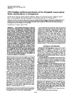
Results We first determined the production pattern of AUF1 isoforms in pancreatic beta cells. We found that the mouse insulin-secreting cell line, MIN6, expresses all four AUF1 isoforms. The two larger isoforms, p45 and p42, are the most abundant, while p40 and p37 are less abundantly produced (Fig. 1a). A similar pattern was observed in human pancreatic islets (Fig. 1b) and in the rat insulin-secreting cell line, INS-1E (not shown). Exposure to IL-1β (10 ng/ml; 5000 U/ml) for 24 h, alone or with a mix of cytokines including IL-1β (0.1 ng/ml; 50 U/ml), TNFα (10 ng/ml; 500 U/ml) and IFNγ (30 ng/ml; 50 U/ml), two conditions that trigger beta cell apoptosis, affected neither the total amount of AUF1 present in MIN6 cells nor the ratio between isoforms (ESM Fig. 1a, b). Similar data were obtained with human islets incubated in the presence of 10 ng/ml IL-1β (ESM Fig. 1c). We then analysed the subcellular localisation of AUF1 isoforms. As demonstrated by the distribution of specific markers (tubulin and lamin B), our fractionation protocol allows a clear separation between cytosolic and nuclear compartments (Fig. 2a). We found that, under resting conditions, the bulk of p45 and p37 is localised in the nucleus, while p40 is detected mainly in the cytosolic fraction. p42 is readily detectable in both fractions and represents the most abundant isoform in the cytosol (ESM Fig. 2a, b). Although, as indicated above, cytokines do not affect the total cellular AUF1 content, exposure to these inflammatory mediators led to redistribution of a fraction of the RNA-binding protein between cellular compartments. Indeed, 24 h of treatment with the cytokine mix increases AUF1 levels in the cytosolic fraction and decreases those detectable in the nuclear fraction, suggesting activation and translocation of the protein from the
Fig. 1 Abundance of AUF1 isoforms in insulin-secreting cells. Total extracts of MIN6 cells (a) or isolated human pancreatic islets (b) were analysed by western blotting using an antibody recognising all AUF1 isoforms. The bands corresponding to each of the AUF1 isoforms were quantified by densitometric scanning of the films. The relative abundance of each isoform was calculated by dividing the intensity of each band by the signal obtained from all AUF1 isoforms; the results shown are the mean±SD of five (MIN6) or three (human islets) independent experiments
nucleus to the cytoplasm (Fig. 2a, b, c). The redistribution of AUF1 isoforms was prevented by a pharmacological inhibitor of the mitogen-activated protein kinase (MAPK), ERK (Fig. 3a, b, c), but not by agents blocking the JNK or p38 kinase pathways (not shown). To assess whether AUF1 activation contributes to cytokine-mediated beta cell dysfunction, AUF1 isoforms were either overproduced individually in MIN6 cells using specific constructs or silenced by RNA interference. Each of the EGFP-tagged AUF1 constructs was overproduced to comparable levels, and taking into account a transfection efficiency with plasmids of ∼30% (determined by scoring EGFP-positive cells) led to an approximately twofold to threefold increase in the cellular level of each individual isoform (ESM Fig. 3). Exogenously produced AUF1 isoforms displayed a subcellular localisation comparable to the endogenous proteins and were mainly localised in the nucleus (ESM Fig. 3). Silencing of AUF1 isoforms was achieved with siRNAs directed against different regions of the transcripts. We used siRNAs directed against exon 2 (shared by isoforms p45 and p40), exon 7 (shared by p45 and p42) or a sequence astride exon 3 and 4 (present in all isoforms) (Fig. 4a). The efficiency of each of the three siRNAs was assessed by western blotting (Fig. 4b, c, d). We first examined whether changes in the level of AUF1 alter the capacity of pancreatic beta cells to synthesise and
Diabetologia (2012) 55:1699–1708
Fig. 2 Impact of proinflammatory cytokines on the subcellular distribution of AUF1 isoforms. a MIN6 cells were incubated for 24 h in the presence or absence of a mixture of cytokines (Cyt Mix) including IL1β (0.1 ng/ml), TNFα (10 ng/ml) and IFNγ (30 ng/ml). Cytosolic and nuclear fractions were prepared as described in the methods section and analysed by western blotting using antibodies against AUF1 and against cytosolic (tubulin) and nuclear (lamin B) markers. The figure shows a representative experiment. Quantification of the subcellular distribution of AUF1 isoforms in MIN6 cells incubated in the presence (black bars) or absence (white bars) of proinflammatory cytokines in the cytosolic (b) and in the nuclear compartment (c). The bands corresponding to each AUF1 isoform were quantified by densitometric scanning of the films from three independent experiments. Significantly different from control, *p