ISSN 0026-8933, Molecular Biology, 2009, Vol. 43, No. 1, pp. 16–23. © Pleiades Publishing, Inc., 2009. Original Russian Text © E.V. Korchagina (Morozova), V.A. Vasyliev, V.I. Korchagin, S.O. Movsessian, S.K. Semyenova, 2009, published in Molekulyarnaya Biologiya, 2009, Vol. 43, No. 1, pp. 19–27.
GENOMICS. TRANSCRIPTOMICS. PROTEOMICS UDC 577.21+576.8
Polymorphism and Structural Features of Two Noncoding Regions of the Liver Fluke Fasciola hepatica (Plathelminthes: Trematoda) Mitochondrial Genome E. V. Korchagina (Morozova)a, b, V. A. Vasylieva, V. I. Korchagina, S. O. Movsessianb, and S. K. Semyenovaa a
Institute of Gene Biology, Russian Academy of Sciences, Moscow, 119334 Russia; e-mail:
[email protected] b Institute of Parasitology, Russian Academy of Sciences, Moscow, 119071 Russia Received May 12, 2008 Accepted for publication May 30, 2008
Abstract—Structural characteristics and polymorphism of the long (LNR) and short (SNR) mitochondrial noncoding regions were studied in the liver fluke Fasciola hepatica. Flukes were sampled from several populations of Russia and Belarus. LNR amplification yielded a set of nine fragments, neighboring ones differing in length by one tandem repeat (85 bp), published for Australian flukes. The LNR amplification products of different lengths were cloned and sequenced. A comparison of the LNR sequences of Australian and Belarussian flukes revealed three nucleotide substitutions and one point heteroplasmy in the first positions of the imperfect repeat and four adjacent perfect repeats. The positions of the three mutations coincided in the perfect and imperfect repeats. The frequency of mutations was 4.0–4.7 %, while the frequency of heteroplasmic sites varied from 0.1 to 1.2%. It was shown that the mutations and the heteroplasmy of one site could change the structure and stability of the putative secondary structures of the perfect and imperfect repeats. SNR amplification in F. hepatica from several populations yielded fragments that differed from the published SNR sequence of Australian F. hepatica by one transversion (T G in position 21). Both noncoding regions had several conserved and potential regulatory sequences. The possible causes of heteroplasmy and a concerted origin of substitutions in different repeats are discussed. DOI: 10.1134/S0026893309010038 Key words: mtDNA, control region, polymorphism, tandem repeats, heteroplasmy, liver fluke Fasciola hepatica, Trematoda
INTRODUCTION
usually contains a short noncoding region (SNR) and a long, or variable, noncoding region (LNR, or VNR), which are separated by one or two tRNA genes [6]. LNRs of most trematodes greatly vary owing to various repeats and are useful as population markers. For instance, LNR variation due to a complex microsatellite repeat has been observed in African and Latin American populations of the blood fluke Schistosoma mansoni [7, 8].
Polymorphism of the mitochondrial genome is widely used in modern molecular genetic reconstructions of the phylogeny and population structure of various eukaryotes. Compared with the nuclear genome, mtDNA is relatively short and is maternally inherited. In addition, the high mutation rate of mtDNA is attributed to the lack of repair and recombination [1]. The mitochondrial genome of most multicellular organisms harbors 36 or 37 genes, which code for 12 or 13 proteins involved in the respiratory chain, two rRNAs, and 22 tRNAs, and has several intergenic spacers of various lengths [2]. One of the longest noncoding regions of vertebrate mtDNA is known as the control region (CR) and contains sequences involved in the regulation of replication and transcription of the mitochondrial genome [3, 4].
The complete mtDNA sequence of the liver fluke Fasciola hepatica from Australia is 14 462 bp and harbors several short (10–15 bp) intergenic spacers and two longer noncoding regions, SNR and LNR, which are separated by the glycine tRNA gene. The SNR is 187 bp. The LNR is 817 bp and contains eight 85-bp GC-rich perfect tandem repeats and one imperfect repeat of 99 bp. A conserved sequence block (CSB) of 42 bp occurs in each of the repeats [9].
The structure of the noncoding sequences is known for only a few parasitic nematodes and flatworms [5]. In flatworms of the class Trematoda, circular mtDNA
The objective of this work was to study the structure of the two noncoding mtDNA regions in liver 16
TWO NONCODING mtDNA REGIONS OF Fasciola hepatica
flukes from several populations of Russia and Belarus. Previously, we studied the polymorphism of two mitochondrial genes, nad1 and cox1, and were the first to find two highly divergent lineages (haplogroups) in Eurasian F. hepatica populations. The lineages differ in distribution through the species range and possibly in origin. The database mtDNA sequence of Australian F. hepatica belongs to lineage I, while both of the lineages are found in many populations of Eurasia [10, 11]. Hence, it is of interest to study the population variation of extended noncoding regions of the mitochondrial genome. Some of these regions may be involved in regulation of complex mtDNA interactions in a parasite–host system. In addition, we checked the hypothesis that heteroplasmy (the presence of different mtDNA molecules in one organism) in F. hepatica results from a change in the number of tandem repeats occurring in a noncoding mtDNA region. Variation of a repeat number is the main cause of common population and individual polymorphism of mtDNA length in certain nematodes, insects, fishes, amphibians, birds, and mammals [12], while heteroplasmy caused by point substitutions is far rarer [13]. EXPERIMENTAL Material. Fasciola hepatica adults (maritas) were isolated from bovine liver in several populations of Russia (Moscow, Kaluga, Bryansk, and Kursk oblasts; n = 19) and Belarus (Brest oblast, n = 8) from 2001 to 2005. Total DNA was isolated from fresh flukes or flukes fixed with 70% ethanol, which were ground in a ceramic mortar with 40 mM Tris-HCl (pH 8.0), 0.5 mM EDTA, 1× SSC, using 500 µl of the buffer per 100 µg of tissue. The mixture was supplemented with 0.5% SDS, incubated in ice for 1 h, supplemented with 1 mg/ml proteinase K, and incubated at 37°C overnight. Deproteinization was performed with equal volumes of phenol, phenol–chloroform, and chloroform. DNA was precipitated with three volumes of 96% ethanol, washed with one volume of 70% ethanol, dried, and dissolved in water. Primers for the polymerase chain reaction (PCR) were directed to the two noncoding regions and were based on the only F. hepatica complete mtDNA sequence available from GenBank (accession no. NC002546), which was obtained for Australian flukes. The SNR was amplified with primers R1 (5'-TGGTTTGGTTTTATTAGGGAGATT-3') and R2 (5'-TAACTCGAAACCACTA-3'), which were respectively complementary to nad5 and the glutamine tRNA gene (regions 13 126–13 149 and 13 438–13 449 in sequence NC002546, respectively) (Fig. 1a). The LNR was amplified using various combinations of seven forward and seven reverse primers. Two primers MOLECULAR BIOLOGY
Vol. 43
No. 1
2009
17
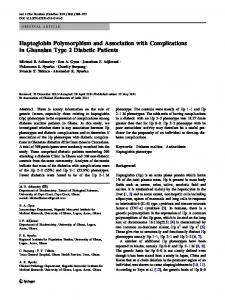
of the set (FD1 and FD10) were used to amplify and clone the LNR fragments. Primer FD1 (5'-TTGTGTTTAGTGGTTTCGGTAGTAT-3') was complementary to the 5' end of the SNR and the 3' end of the glycine tRNA gene, and primer FD10 (5'-TGGAAACTACCCCTAAGAAA-3') was complementary to a cox3 region (regions 13 571–13 595 and 87–106 of sequence NC002546, respectively) (Fig. 1a). PCR. The reaction mixture (25 µl) contained a buffer (62 mM Tris-HCl, pH 8.0, 15.4 mM (NH4)2SO4, 0.01% Tween-20), 2.4 mM MgCl2, 200 µM each dNTP, 0.8 pmol of each primer, 0.6 units of Taq polymerase (Dialat, Moscow) or Pfu polymerase (Fermentas, Moscow), and 10–100 ng of total DNA. Amplification included initial denaturation at 94°C for 2 min; 30 cycles of 94°C for 1.5 min, 50– 53°C for 1 min, and 70°C for 2 min; and last synthesis at 70°C for 10 min. The PCR product was electrophoretically resolved in 1–1.5% agarose gel and stained with ethidium bromide. Cloning was performed with the total amplificate obtained with primers FD1 and FD10 on DNA of a Belarussian fluke. Preliminary analysis of nad1 and cox1 polymorphism showed that the genome belonged to lineage II. Recombinant DNAs were constructed using pMOSBlue and the E. Òoli strain from a pMOSBlue blunt-ended cloning kit as recommended by Amersham Biosciences (United States). Recombinant clones were identified on a selective medium with Xgal. Insert-containing plasmids were examined by PCR with the same primers (FD1 and FD10) and with universal primers T7 and U-19, which were homologous to the ends of the plasmid polylinker. The nucleotide sequence was established for five clones carrying inserts of different lengths (Table 1). Recombinant plasmids were sequenced in both directions with primers T7 and U-19 and an ABI PRISM® BigDyeTM Terminator v. 3.1. kit on an ABI Prism 3100-Avant Genetic Analyzer (Genom Interinstitutional Center, Engelhardt Institute of Molecular Biology, http://www.genome-centre.narod.ru/). Sequence alignments were manually constructed using the Australian F. hepatica mtDNA sequence (NC002546). DNA secondary structures and minimal free energy DG (kcal/mol) were computed using the Mfold program (v. 3.2) [14]. RESULTS LNR Polymorphism LNR amplification for flukes of various populations yielded a set of multiple fragments that formed a ladder with neighboring fragments differing by about 85 bp, the unit size of the tandem repeat known for the LNR of Australian F. hepatica. The pattern with mul-
18
KORCHAGINA et al. (a) R1
R2 LNR
FD1 nad 5
E
SNR
RP RP RP RP RP RP RP RP RPD 1 2 3 4 5 6 7 8
G
(b)
FD10
g a g a 1* 7
t term
TAS
a g 82
t at c 76 83
term
(TA)5 t
a
CSB
g a g a 1* 7
(c)
H
61
RP1–RP8
RPD
cox 3
CSB
g 22
Prom
SNR
–47 bp Fig. 1. (a) Fasciola hepatica mtDNA region including the SNR and LNR. (b, c) Detailed structure of individual elements of the (b) LNR and (c) SNR. Short intergenic spacers are shown in black. RP1–RP8 are the perfect repeats of the LNR. E, G, and H are the genes for glutamine, glycine, and histidine tRNAs, respectively. The nad5 and cox3 genes are indicated. CSB, conserved sequence block; (TA)5, microsatellite repeat. Primer annealing regions are shown with triangular arrows. The positions of the mutations found are shown with vertical arrows. Heteroplasmic sites are asterisked. Prom, putative promoter region; term and TAS, termination-associated signal sequences.
Table 1. Structure and haplotype diversity of the LNR fragments cloned from F. hepatica from Belarus and Australia (NC002546) RP1 (85) Ordinal number of nucleotide in repeat
RP2 (85)
RP3(85)
RP4 (85)
RP5 (85)
RP6 (85)
RP7 (85)
RP8 (85)
RPD (99)
1 7 61 82 1 7 61 82 1 7 61 82 1 7 61 82 1* 7 61 82 1* 7 61 82 1* 7 61 82 1* 7 61 82 1* 7 76 83
NC002546 haplo- g g t a g g t a g g t a g g t a g g t a g g t a g g t a g g t a g g t t type Clone 34
• a a g • a a ? ? ? ? ? ? a a g a a a g a a a g a a a g a a a g a a a c
Clone 28
–
• a a g • a a g • a a g • a a g • a a g • a a g • a a g a a a c
Clone 22
–
• a a g • a a g • a ? ? ? ? ? ? ? ? a g a a a g a a a g • a a c
Clone 5
–
–
Clone 8
–
–
• a a g • a a g • a a g • a a g • a a g • a a g • a a c –
–
–
–
–
• a a g • a a c
Note: The lengths (bp) of the perfect (RP1–RP8) and degenerate (RPD) repeats is shown in parentheses. (*), lack of nucleotide substitution; (?), data unavailable; (–), the repeat is absent from the cloned fragment. Mutant sits are shaded light gray; unknown nucleotides are shaded dark gray; sites displaying heteroplasmy are framed or asterisked. MOLECULAR BIOLOGY
Vol. 43
No. 1
2009
TWO NONCODING mtDNA REGIONS OF Fasciola hepatica
19
Table 2. Structural elements of LNR and SNR of F. hepatica mtDNA Characteristic
LNR
Length, bp AT/GC, % Homology, % Homopolymeric tracts Microsatellite repeats Secondary structure: total hairpins and minimal free energy ∆G, kcal/mol
SNR
RP = 85; RPD = 99; CSB = 42; LNR = 779 187 RP: 48/52; RPD: 56/44; CSB: 35.7/64.3 68/32 RP: RPD = 59.6 RP:RPD (without CSB)=31.7 RP: T3-10, T4-3, T6-1, T8-1, G2-3, G3-2, G4-1. T2-31, G3-2, G5-1, G2-G4-6, C6-1, RPD: T3-2, T4-2, G2-3, G3-2, G4-1, G6-1, C4-1. RP: AT-3; TG-20 RPD: (AT)5; AT-3; TG-10 AT-16; GC-4; AC-3; TG-23. RP haplotypes: G-G-T-A: 3 (∆G = –3.88); 5 (∆G = –6.83) G*- A-Ä-G: 3 (∆G = –5.09); A*- A-A-G: 3 (∆G = –5.09). RPD haplotypes: G-G-T-T: 3 (∆G = –4.88); A*-A-A-C: 4 (∆G = –5.09); G*- A-A-C: 3 (∆G = –4.82).
Note: RP1–RP8, RPD, tandem repeats; CSB, conserved sequence block. Heteroplasmic sites are asteristed. The Australian F. hepatica haplotypes are underlined. Hairpin number is in bold italics.
tiple fragments was stably reproduced with any combination of seven forward and seven reverse primers directed to various LNR-flanking regions (Fig. 1a). Nine major fragments were detected in most cases; 14 and 15 distinct fragments were observed in two flukes from the Kursk and Belarussian populations, respectively (data not shown). The number and lengths of the amplification products remained the same when the hot-start procedure was used, the cycle number was reduced, the annealing temperature was increased, and different DNA polymerases (Taq or Pfu) and/or DMSO were used (data not shown). We cloned the total amplificate obtained with DNA of one Belarussian fluke, whose genome belonged to lineage II. Five clones were selected and sequenced; their inserts were about 200, 700, and 800 bp. Table 1 shows the observed single nucleotide substitutions and their distribution through the five cloned sequences, which were aligned against the known LNR sequence of Australian F. hepatica. Absolutely all of the clones had the terminal degenerate repeat (RPD) of 99 bp, while the number of 80-bp tandem units (RP) was 1, 6, 7, 7, and 8 in clones 8, 5, 22, 28, and 34, respectively. Thus, the LNR pattern of Belarussian F. hepatica included fragments differing in the number of perfect repeats. The largest insert (clone 34) contained nine repeats, as in the LNR sequence of Australian F. hepatica. At the same time, the F. hepatica genomes from the two populations differed by three or four nucleotide substitutions in LNR (Fig. 1b, Table 1). RP1–RP8 of Belarussian F. hepatica had three transitions and one transversion in positions 1 (G A), 8 (G A), 61 (T A), and 82 (A G). RPD had three similar G A mutations in positions 1, 7, and 76 and transition T C in position 83. Point heteroplasmy was observed in perfect and imperfect repeats; i.e., one MOLECULAR BIOLOGY
Vol. 43
No. 1
2009
fluke had mtDNA molecules differing by nucleotide substitution in position 1 (G, A). Heteroplasmy occurred only in some repeats, including RPD (clones 28 and 34) or RP5–RP8 (clones 22 and 34) (Table 1). The positions of three mutations were the same in perfect and imperfect repeats. Two G A transitions arose in the same sites at the 5' ends of RP1 and RPD, and one T A transversion was detected in the same site at the 3' end of CSB. Owing to the concerted origin of homologous substitutions in different repeats of the LNR, the mtDNA populations of the two flukes displayed combinations of three haplotypes of the perfect and imperfect repeats. The Australian F. hepatica genome had one haplotype of RP1–RP8 (G-G-T-A) and one of RPD (G-G-T-T). Point heteroplasmy, resulting from a mutation in the first position of the perfect and imperfect repeats, generated two RP haplotypes (G-A-A-G and A-A-A-G) and two RPD haplotypes (A-A-A-C and G-A-A-C) in Belarussian F. hepatica. The four mutations, their positions, and heteroplasmy at one site differently affected the structure and stability of the putative secondary structures of the perfect and imperfect repeats of the LNR. For instance, the two new RP haplotypes G-A-A-G and A-A-A-G, which differ by one substitution in the first position, formed secondary structures with the same free energy (∆G = –5.09 kcal/mol) and had three small hairpins each. Their structures were less stable as compared to the structure of the known G-G-T-A haplotype of Australian F. hepatica (∆G = –3.88 kcal/mol), which similarly formed three small hairpins and differed by three or four substitutions (Table 2). The four mutations found in RPD did not change the stability of the secondary structures or decreased it, allowing the formation of an additional hairpin. For instance, two similar structures with three hairpins and similar free
20
KORCHAGINA et al.
Table 3. Structure of the putative promoters and known termination-associated sequences (TASs, or terms) of the mtDNA control region in liver fluke F. hepatica, spur-toed frog Xenopus laevis [28], chicken Gallus gallus [29], some mammals [15], and domestic cow Bos taurus [16] Regulatory element Promoters (3'–5')
Species F. hepatica G. gallus B. taurus X. laevis
Termination signals (5'–3')
mammals B. taurus F. hepatica
Sequence SNR: GTGTATATA TT A TTT ATACA ATATA- TT A TT HSP TT TATATA LSP TATAAA TT LSP2 TATATAG-CATA TT GCAC TAS: TACATTAA TAS-A: TACATTAT term: GCCC TAS: RP: (TAS) –TACATATT term: LNR (CSB): GCCCC
Note: HSP and LSP are the known promoters for transcription of the heavy and light mtDNA strands. TACAT and TATA sequences are in italics; hairpin stem regions are underlined; homologous T-containing regions are shaded gray.
energies were formed by the sequences with the Australian haplotype and one of the Belarussian haplotypes that differed by three mutations (∆G = –4.82 for G-A-A-C and ∆G = –4.88 for G-G-T-T). Compared with these sequences, the structure of the sequence with the other Belarussian haplotype (A-A-A-C) was less stable (∆G = –5.09) because of the formation of a fourth hairpin (Table 2). Mutations in the first positions of the repeats did not change the secondary structure at the boundary between two perfect repeats (RP1–RP8). However, such mutations change the RP8–RPD boundary, allowing the formation of a small hairpin with a 2-bp stem. In addition, three substitutions in two small LNR-flanking regions were found in all of the clones examined. One transition (A G) was found upstream of the 5' end of the LNR (position –6), and two similar transitions (T C) were detected in positions +19 and +24 downstream of the 3' end of the LNR (Fig. 1a, vertical arrows). Only one of the T C transitions (position +19) allows a large additional hairpin with a 6-bp stem and an extended 49-nt loop (data not shown). A 42-nt conserved sequence was detectable in each of the repeats (Fig. 1c). The sequence differs by one substitution from the known CSB of the Australian F. hepatica LNR [9]. In addition, RPDs of the Australian and Belarussian F. hepatica sequences have the same (TA)5CATA element, which is involved in one of the stable hairpins and forms a part of its stem and the loop (data not shown). The TACAT pentanucleotide (TAS, Table 3), which is contained in the element, is identical to the vertebrate mtDNA TAS, which is sus-
tained by the secondary structure and is associated with termination of D-loop replication [15] (Table 3). Another element potentially associated with this process is the GCCCC sequence, which occurs in the CSB of each repeat and is homologous to the GCCC terminator tetranucleotide found in many vertebrate genomes [16] (term, Table 3). It should be noted that the perfect and imperfect repeats were similar in nucleotide composition (Table 2). The AT content was 48% in RP1–RP8 and 56.0% in RPD, while GC were prevalent (67.4%) in the CSB. However, the repeat sequences were highly divergent. Homology between RP and RPD was only 59.6%, and the exclusion of the CSB reduced the homology to 31.7%. The perfect and imperfect repeats differed in the content and positions of short homopolymeric tracts and various dinucleotides. For instance, AT dinucleotides clustered to form an extended (AT)5 tract upstream of the CSB in RPD and were found both upstream and downstream of the CSB in RP1–RP8. SNR Polymorphism SNR amplification with DNAs of 20 flukes from several populations of Russia (Moscow, Kaluga, Bryans, and Kursk oblasts) and Belarus (Brest oblast) yielded single fragments, which were similar in size (187 bp) to the known SNR sequence of Alustralian F. hepatica (NC002546). Transversion T G in position 21 was found in ten flukes and had no effect on the SNR secondary structure. MOLECULAR BIOLOGY
Vol. 43
No. 1
2009
TWO NONCODING mtDNA REGIONS OF Fasciola hepatica
While the LNR had comparable AT and GC contents (52 and 48%, respectively), the SNR was enriched in AT (68%) (Table 2). Homopolymeric guanine and thymidine tracts were found in each SNR but differed in length and location. As in the LNR, most of the repeats in the SNR were di-, tri-, and tetranucleotides, AT dinucleotides being the most abundant. The AT density in the SNR was substantially higher than in the LNR. The SNR was found to contain six palindromes, which are potentially capable of forming six small hairpins with the stem length varying from 2 to 9 bp and the loop length varying from 4 to 16 nt. An ATrich segment was found in the 3'-terminal region of the SNR (positions from –23 to –50) and included two TATA motifs (italicized in Table 3), which were involved in a cruciform structure formed by two hairpins on the two strands (the hairpin stems are underlined in Table 3). DISCUSSION Tandem repeats are a common feature of the mtDNA CRs of most animals. Such repeats are found in almost all taxa examined, vary in length from microsatellites (3 bp) to extended repeats (777 bp), and usually occur in the most variable regions that flank the CR [13]. Heteroplasmy for the repeat number has been observed upon amplification of the mtDNA CR in vertebrates and some invertebrates [17, 18]. We cloned the total product amplified from DNA of one F. hepatica fluke and were the first to observe the simultaneous presence of several DNA molecules differing in the number of perfect repeats in the LNR, suggesting heteroplasmy. This is indirectly supported by the finding of two PCR patterns with 14 and 15 amplified fragments and the fact that the mtDNA length substantially varies among individual F. hepatica flukes, the difference reaching 19 kb [19]. The same LNR pattern with multiple fragments was obtained in several PCR variants used to improve the specificity of amplification. However, nonspecific amplification cannot be excluded. The exact mechanism that causes and sustains heteroplasmy for mtDNA length is unknown. Changed copies are thought to result from polymerase slippage during DNA replication [20], mispairing [21], and, in some cases, DNA recombination [18]. These factors may cause heteroplasmy for individual sites and/or mtDNA length both in vivo and in vitro. Tandem repeats are capable of forming secondary structures, which hinder the function of DNA polymerases in amplification and sequencing of individual genomic regions [22–24]. Hence, special experiments should be performed to rule out artifactual LNR amplification resulting from premature termination of Taq polymerase [25]. Such termination may occur in each MOLECULAR BIOLOGY
Vol. 43
No. 1
2009
21
repeat at the CSB sites corresponding to vertebrate termination-associated signal sequences (terms; Fig. 1, Table 3). The results are quite reliable in the case of the four point substitutions and heteroplasmy for one of the mutations found in the LNR repeats. These findings cannot be explained by mistakes introduced by Taq polymerase, since the average frequency of such mistakes ranges from 2 × 10⁻⁵ to 2 × 10⁻⁴ [26, 27]. In our experiments, the frequency of heteroplasmic sites was 1.0–1.2% in individual repeats (one mutation in 99 bp and one in 85 bp) and 0.1–0.6% in the clones (two mutations in 85 + 99 bp and five in 779 bp). The frequency of mutations was 4.0–4.7% in LNR repeats (four mutations in 99 or 85 bp) and 4.3–4.6% in the clones (eight mutations in 85 + 99 bp to 36 mutations in 779 bp). These estimates are comparable with the mutation frequency (4.1%) of a coding mtDNA region (cox1) examined previously in two F. hepatica haplogroups [11]. It is unclear whether the mutation-induced changes in the thermodynamic stability of potential DNA secondary structures are functionally significant (Table 2). The four mutations found in RP1–RP8 increase the minimal free energy from ∆G = –3.88 to ∆G = –5.09, although no apparent change in the secondary structures is detectable. The mutation of the first nucleotide in RPD changes the thermodynamic stability to a greater extent, from ∆G = –4.82 and –4.88 to ∆G = −5.09. It is also unclear whether the mutations affect the formation of secondary structures in native mtDNA, increasing the likelihood of erroneous slippage and DNA strand pairing. It is possible that these mutations in the CR confer some selective advantages and are associated with divergence of the two F. hepatica mtDNA haplogroups in early speciation. Such an association is supported by the fact that three of the mutations occur exclusively in the genome of one of the haplogroups (lineages), which have earlier been isolated on the basis of two coding regions of F. hepatica mtDNA [11]. Several interesting features of the organization of the two noncoding regions were observed by comparing the mitochondrial genome for F. hepatica and several vertebrates. For instance, analysis of spatial and structural homologies indicates that the two close TATA boxes may correspond to the two neighboring unidirectional promoters that are responsible for transcription of the two strands in one direction. Unidirectional transcription is presumably characteristics of the CR in many nematodes, annelids, mollusks, brachiopods, and cnidarians, including Fasciola [9]. It is possible, however, that the putative promoter region is more intricately organized. This conclusion is based on comparisons with the known motifs characteristic of differently directed (HSR and LSR) pro-
22
KORCHAGINA et al.
moters of mammals (cow Bos taurus) and bidirectional promoters of the mtDNA CR of vertebrates, including amphibians (spur-toed frog Xenopus laeivis) and birds (domestic chicken Gallus gallus) (Table 3). Based on these comparisons, the two TATA boxes found in F. hepatica can be regarded as two unidirectional promoters, one of which has the canonical 3'-TATATATT sequence, and the other, neighbor promoter has the 3'-TATTTATA sequence. On the other hand, this fragment of the F. hepatica CR can be regarded as one bidirectional promoter similar to the chicken ATATA–TTATT or bass TATTTACA–TTATTAAAAT bidirectional promoter. The F. hepatica sequence has certain similarity to the B. taurus HSP heavy-chain promoter (3'TTTATATA) and one of the X. laevis mtDNA light-chain promoters. We did not observe any apparent homology in the arrangement of conserved and variable domains or individual signal sequences of the two noncoding regions between F. hepatica and vertebrates. The vertebrate mtDNA CR usually has a conserved central domain and peripheral 5'- and 3'-terminal domains. The 5'-terminal domain includes one or several transcription termination-associated sequences (TASs). Tandem repeats usually occur in the CR region that coincides with the 5' end of the D-loop, where replication is initiated, and with its 3' end, where its termination takes place [30, 31]. When the two extended noncoding regions of F. hepatica are considered as one sequence, its most variable part, that containing the tandem repeats, is close to the 3' end. It is this part (RPD) that harbors one of the termination signals. Conserved domains cannot be identified until data on the sequences of close fluke species become available, but the comparison of the two F. hepatica forms demonstrates that their boundaries are changed in the most conserved block, the CSB. In the future, large-scale complete genome sequencing will provide insight into mtDNA evolution in invertebrates, including digenetic flukes. ACKNOWLEDGMENTS We are grateful to V.V. Gorokhov, I.A. Arkhipov, and G.G. Khrisanfova for F. hepatica specimens and to A.P. Ryskov for valuable comments and advice. Experiments were carried out in the Laboratory of Genome Organization (Institute of Gene Biology) and were supported by the Russian Foundation for Basic Research (project nos. 06-04-49073 and 08-0412204), the program Leading Scientific Schools, and the program Molecular and Cell Biology and subprogram Gene Pool Dynamics of the Presidium of the Russian Academy of Sciences.
REFERENCES 1. Brown W., George M., Wilson A. 1979. Rapid evolution of animal mitochondrial DNA. Proc. Natl. Acad. Sci. USA. 76, 1967–1971. 2. Boor J. 1999. Survey and summary of animal mitoghondrial genomes. Nulceic Acids Res. 27, 1767–1780. 3. Brown G.G., Gadaleta G., Pepe G., Saccone C., Sbisa E. 1986. Structural conservation and variation in the Dloop-containing region of vertebrate mitochondrial DNA. J. Mol. Biol. 192, 503–511. 4. Clayton D.A. 1991. Replication and transcription of vertebrate mitochondrial DNA. Annu. Rev. Cell. Dev. Biol. 7, 453–476. 5. Le T., Blair D., Agatsuma T., et al. 2000a. Phylogenies inferred from mitochondrial gene orders: A cautionary tale from the parasitic flatworms. Mol. Biol. Evol. 17, 1123–1125. 6. Le T., Blair D., McManus D. 2002. Mitochondrial genomes of parasitic flatworms. Trends Parasitol. 18, 206–213. 7. Despres L., Imbert-Establet D., Monnerot M. 1993. Molecular characterization of mitochondrial DNA provides evidence for the recent introduction of Schistosoma mansoni into America. Mol. Biochem. Parasitol. 60, 221–230. 8. Pena H.B., de Souza C.P., Simpson A.J., Pena S.D. 1995. Intracellular promiscuity in Schistosoma mansoni: Nuclear transcribed DNA sequences are part of a mitochondrial minisatellite region. Proc. Natl. Acad. Sci. USA. 92, 915–919. 9. Le T.H., Blair D., McManus D.P. 2001. Complete DNA sequence and gene organization of the mitochondrial genome of the liver fluke, Fasciola hepatica L. (Platyhelminthes; Trematoda). Parasitology. 123, 609–621. 10. Morozova E.V., Chrisanfova G.G., Archipov I.A., Semyenova S.K. 2004. Polymorphism of the ND1 and COI mitochondrial genes in populations of liver fluke Fasciola hepatica. Russ. J. Genet. 40, 817–820. 11. Semyenova S.K., Morozova E.V., Chrisanfova G.G., Gorokhov V.V., Arkhipov I.A., Moskvin A.S., Movsessyan S.O., Ryskov A.P. 2006. Genetic differentiation in Eastern European and Western Asian populations of the liver fluke, Fasciola hepatica, as revealed by mitochondrial nad1 and cox1 genes. J. Parasitol. 92, 523–530. 12. Lunt D.H., Whipple L.E., Hyman B.C. 1998. Mitochondrial DNA variable number tandem repeats (VNTRs): Utility and problems in molecular ecology. Mol. Ecol. 7, 1441–1455. 13. Moritz C., Dowling T., Brown W. 1987. Evolution of animal mitochondrial DNA: Relevance for population biology and systematics. Ann. Rev. Ecol. Syst. 18, 269– 292. 14. Zuker M. 2003. Mfold web server for nucleic acid folding and hybridization prediction. Nucleic Acids Res. 31, 3406–3415 (http://www.bioinfo.rpi.edu/applications/mfold/). 15. Zardoya R., Meyer A. 1996. The complete nucleotide sequence of the mitochondrial genome of the lungfish (Protopterus dolloi) supports its phylogenetic position as a close relative of land vertebrates. Genetics. 142, 1249–1263. MOLECULAR BIOLOGY
Vol. 43
No. 1
2009
TWO NONCODING mtDNA REGIONS OF Fasciola hepatica 16. Hiendleder S., Zachartchenko V., Wenigerkind H., Reichenbach H.-D., Bruggerhoff K., Prelle K., Stojkovic M., Wolf E. 2003. Heteroplasmy in bovinae fetuses prodused by intra- and inter-subspecies somatic cell nuclear transfer: Neutral segregation of nuclear donor mitochondrial DNA in various tissues and evidence for recipient cow mitochondria in fetal blood. Biol. Reproduction. 68, 159–166. 17. Zhang D.-X., Hewitt G.M. 1997. Insect mitochondrial control region: A review of its structure evolution and usefulness in evolutionary studies. Biochem. Syst. Ecol. 2, 99–120. 18. Lunt D.H., Hyman B.C. 1997. Animal mitochondrial DNA recombination. Nature. 387, 247. 19. Agatsuma T., Arakawa Y., Iwagami M., Honzako Y., Cahyaringsih U., Kang S.Y., Hong S.J. 2000. Molecular evidence of natural hybridization between Fasciola hepatica and F. gigantica. Parasitol. Int. 49, 319–327. 20. Levinson G., Gutman G.A. 1987. Slipped-strand mispairing: A major mechanism for DNA sequence evolution. Mol. Biol. Evol. 43, 203–221. 21. Buroker N.E., Brown J.R., Gilbert T.A., O’Hara P.J., Beckenbach A.T., et al. 1990. Length heteroplasmy of sturgeon mitochondrial DNA: An illegitimate elongation model. Genetics. 124, 157–163. 22. Hoelzel A.R., Lopez J.V., Dover G.A., O’Brien S.J. 1995. Rapid evolution of a heteroplasmic repetitive sequence in the mitochondrial DNA control region of carnivores. J. Mol. Evol. 39, 191–199. 23. Landre P.A., Gelfand D.H., Watson R.M. 1995. The use of cosolvents to enhance amplification by the polymerase chain reaction. In: PCR Strategies. Eds Innis
MOLECULAR BIOLOGY
Vol. 43
No. 1
2009
24.
25. 26. 27.
28. 29.
30.
31.
23
M.A., Gelfand D.H., Sninsky J.J., San Diego, SD: Academic Press, 3–16. Kupriyanova N.S., Shibalev D.V., Voronov A.S., Ryskov A.P. 2004. PCR-generated artificial ribosomal DNAs from premature termination at Alu sequences. Biomol. Engin. 21, 21–25. Campball N.J.H., Sturm R.A., Barker S.C. 2000. Large mitochondrial repeats multiplied during the polymerase chain reaction. Mol. Ecol. Notes. 1, 336–340. Keohavong P., Thilly W.G. 1989. Fidelity of DNA-polymerases in DNA amplification. Proc. Natl. Acad. Sci. USA. 86, 9253–9257. Lundberg K.S., Shoemaker D.D., Adams M.W.W., Short J.M., Sorge J.A., Mathur E.J. 1991. High-fidelity amplification using a thermostable DNA-polymerast isolated from Pyrococcus furiosus. Gene. 108, 1–6. Antoshechkin I., Bogenhagen D.F. 1995. Distinct roles for two purified factors in transcription of Xenopus mitochondrial DNA. Mol. Cell. Biol. 15, 7032–7042. L’Abbe D., Duhaime J.-F., Lang B.F., Morais R. 1991. The transcription of DNA in chicken mitochondria initiates one major bidirectional promoter. J. Biol. Chem. 266, 10844–10850. Saccone C., Pesole G., Sbisa E. 1991. The main regulatory region of mammalian mitochondrial DNA: Structure–function model and evolutionary pattern. J. Mol. Evol. 33, 83–91. Randi E., Liccini V. 1998. Organization and evolution of the mitochondrial DNA control region in the avian genus Alectoris. J. Mol. Evol. 47, 149–157.