May 9, 2018 - University of Oklahoma Health Science Center, Oklahoma City, Oklahoma 73104, .... By using ENPs in biological studies, the vast ...... Investigators applied this strategy ... approach; start with the smallest unit and build up until the ...... (120) Sadat Tabatabaei Mirakabad, F., Nejati-Koshki, K., Akbarzadeh,.
Review pubs.acs.org/bc
Cite This: Bioconjugate Chem. XXXX, XXX, XXX−XXX
Probing Cellular Processes Using Engineered Nanoparticles †,§ ́ Md Nazir Hossen,†,§ Brennah Murphy,§ Lorena Garcıa-Hevia, Resham Bhattacharya,‡ ,†,§ and Priyabrata Mukherjee* †
Peggy and Charles Stephenson Cancer Center, ‡Department of Obstetrics and Gynecology, and §Department of Pathology, University of Oklahoma Health Science Center, Oklahoma City, Oklahoma 73104, United States
ABSTRACT: Nanoparticles, the building blocks of nanotechnology, have been widely utilized in various biomedical applications, such as detection, diagnosis, imaging, and therapy. However, another emerging, albeit under-represented, area is the employment of nanoparticles as tools to understand cellular processes (e.g., oxidative stress-induced signaling cascades). Such investigations have enormous potential to characterize a disease from a different perspective and unravel some new features that otherwise would have remained a mystery. In this review, we summarize the intrinsic biological properties of unmodified as well surface modified nanoparticles and discuss how such properties could be utilized to interrogate biological processes and provide a perspective for future evolution of this field.
■
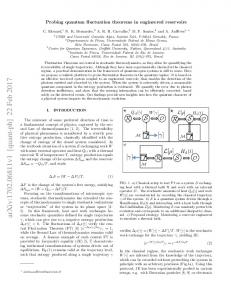
INTRODUCTION Nanotechnology is a means by which engineered nanoparticles (usually 1 to 100 nm in dimension) are utilized to develop materials, structures, devices, and systems for various purposes.1−4 Currently, there are a wide variety of nanomaterials, including metals, metal oxides, and quantum dots, which exhibit unique optical, electrical, and magnetic properties, and have been widely used for a number of biomedical and nanotechnological applications. Other material types, such as polymers, lipids, small molecules, and organic molecules, can be assembled into carriers incorporated with contrast agents and drugs for applications in molecular and cellular labeling, tracking, detection, drug delivery, medical imaging, and therapy.2,4,5 These engineered nanoparticles (ENPs) can enter cells by various intracellular uptake pathways such as phagocytosis, macropinocytosis, clathrin-dependent endocytosis, caveolin-dependent endocytosis, or the crossing of membranes by diffusion (Figure 1).6−16 Thus, these ENPs may serve as unique baits to catch and identify new adapter molecules that characterize uptake pathways and broaden our understanding of how malignant cells tweak their intracellular uptake pathways to survive and thrive under nonconducive environments. Some nanomaterials, particularly nanoparticles of gold, possess intrinsic biological activity. Intrinsic biological activity is dependent on many parameters, such as physical character© XXXX American Chemical Society
istics (size, shape, surface, porosity, agglomeration state, and critical structure) and chemical properties (solubility, chemical compositions, and surface modification).17−19 Accumulated evidence shows that the majority of ENPs induces oxidative stress (OS) upon the activation of an array of cell signaling cascades and produces reactive oxygen species (ROS) and cytotoxicity.19−21 Several ways by which ENPs can mediate ROS production are (i) they can catalyze oxidation−reduction reactions through their surfaces; (ii) they can interact with cellular components and ROS production machineries, such as mitochondria and the NADPH oxidase system; and (iii) they can decrease cellular antioxidant defense mechanisms by deactivating or decreasing the production of antioxidants (Figure 1).17 The level of ROS generated within the cell directly influences the cellular response. At low levels, the cell generates an antioxidant defense response, whereas at higher levels, cells induce an inflammatory response and proliferate. However, at very high ROS levels, cells undergo either programmed cell death (apoptosis, autophagy, pyropoptosis, or programmed necrosis) or nonprogrammed death (accidental necrosis).17,22 Utilizing gold nanoparticles (AuNPs) of various surface charges, our group recently demonstrated that ovarian Received: January 10, 2018 Revised: May 7, 2018 Published: May 9, 2018 A
DOI: 10.1021/acs.bioconjchem.8b00026 Bioconjugate Chem. XXXX, XXX, XXX−XXX
Review
Bioconjugate Chemistry
Figure 1. Engineered nanoparticle entry, intracellular appearance, ROS generation, and ROS-mediated signaling cascades. Engineered nanoparticles (ENPs) can enter animal cells by phagocytosis, pinocytosis, macropinocytosis, clathrin/caveolin-independent endocytosis, or diffusion and appear in the cytosol. Subsequently, ENPs can generate and induce the production of reactive oxygen species (ROS) in the mitochondria via intrinsic (formation of reactive groups) and cell-mediated (triggering cellular mechanisms for the damage or activation of mitochondria) pathways. Activation of nicotinamide adenine dinucleotide phosphate (NADPH) oxidase by ENPs can also regulate the intracellular ROS content. The produced ROS can interact with critical signaling molecules, which modulate cell signaling pathways and subsequently affect a variety of cellular processes (e.g., proliferation, metabolism, differentiation, and survival).
the reported mechanistic cellular responses upon exposure to various ENPs, including metallic, metallic oxide, polymeric, and lipid nanoparticles. Additionally, we will address how interrogations utilizing ENPs can help us to better understand these cellular processes and provide opportunities to identify new molecules responsible for poor pathological outcome. Effects of Engineered Nanoparticles on Cellular Processes. By using ENPs in biological studies, the vast complexity of different cellular processes can be elucidated and consequently modified. A growing number of nanoparticles are being investigated for their use in intracellular applications, such as labeling, tracking, detection, drug delivery, medical imaging, and therapy.1,2,4,5 Based on their compositions, these nanoparticles may be classified into two categories: inorganic and organic. Inorganic nanoparticles are mostly metallic (Au, Ag, Cu, Pt, and Pd), metallic oxides, and sulfides (Ti, Fe, Ce, SiO2, Zn, Al, Cu+, Co, Se, etc.), whereas organic nanoparticles are mainly liposomes, polymeric micelles, dendrimers, polymers, and lipids, as shown in Figure 2. In general, the activities of inorganic ENPs depend on the catalytic property of the core metal/metal oxide, toxicity of the shell components, intrinsic biological properties, and other aspects. On the other hand, organic ENPs elicit their effects on the basis of their compositions and physicochemical properties. Based on the activity, we herein discuss each ENP and provide information on how to probe cellular signaling mechanisms using ENPs. Metallic Nanoparticles. Gold Nanoparticles (AuNPs). AuNPs are biocompatible, easy to synthesize and characterize, and are functionalizable via thiol-metal chemistry.4,41 As shown in Figure 2A, AuNPs can act as a self-therapeutic agent or be
cancer cells overcome the cytotoxic effects of positively charged gold nanoparticles (+AuNPs) by exploiting overexpression of mitochondrial uptake protein 1 (MICU1) as a negative regulator of Ca2+ entry to the mitochondria.23−25 Previously, we also demonstrated how the protein-corona formation around the surface of functionalized and nonfunctionalized nanoparticles could be exploited to identify new therapeutic targets in ovarian cancer and identified HDGF and SMNDC1 as potential therapeutic targets.26,27 Recently, Rotello’s groups reported how the protein-corona around +AuNPs could be tailored for selective recognition by macrophages.28 In addition, AuNPs without surface functionalization possess intrinsic therapeutic properties that had been used for the treatment of cancer,29,30 rheumatoid arthritis,31 ocular disease,32,33 and obesity.34 Recently, AuNPs had also been reported to promote osteogenic differentiation of mesenchymal stem cells (MSCs) through activation of the p38-MAPK pathway.35,36 Mirsadeghi et al. also probed the inhibitory effects of AuNPs on the fibrillation process of amyloid beta (Aβ) proteins and observed how the protein-corona around AuNPs affects the fibrillation process.37 Previously, Rotello’s groups reported that aminoacid-functionalized AuNPs effectively interacting with the enzyme α-chymotrypsin via surface complementary charges can make supramolecular complexes, resulting in a decrease in its enzymatic activity. The structural diversity of amino acids allowed them to probe the role of many functional groups on regulating enzymatic function.38,39 Recently, our group reported how unique chemical affinities of AuNPs could be exploited to reprogram the tumor microenvironment in pancreatic cancer.40 In this review, we will attempt to provide B
DOI: 10.1021/acs.bioconjchem.8b00026 Bioconjugate Chem. XXXX, XXX, XXX−XXX
Review
Bioconjugate Chemistry
localization in the nucleus and cytoplasm was mediated by the neutral AuNP. Moreover, it was notable that induction of apoptosis was observed by the treatment with charged AuNPs while necrosis was induced by neutral AuNPs. Rotello’s group51 also reported the cytotoxicity of 2 nm cationic AuNPs with various hydrophobicities in HeLa cells using mitochondrial, ROS, and comet assays, which measures DNA damage. The results strongly suggested that hydrophobicity was linearly correlated with the observed acute toxicity and increased DNA damage. Clearly, these AuNPs can generate significant amounts of ROS that oxidatively impair DNA at doses where mitochondrial activity was not affected.51 Taken together, AuNPs, irrespective of normal or diseased cells, may have the potential to activate and/or down-regulate the cellular signaling cascades which can then be utilized as a probe for intracellular processes. Silver Nanoparticles (AgNPs). AgNPs exhibited antiangiogenic effects, as reported by Eom and colleagues.52 They showed that 40 nm AgNPs had anti-angiogenic effects in normal bovine retinal epithelial cells (BREC) in vitro as well as an in vivo matrigel plug assay. The report stated that the proliferation and migration of VEGF-induced angiogenesis of BRECs could be prevented by the treatment of AgNPs, thus suggesting that AgNPs are in some way related to the activation of the PI3K/Akt signaling pathway.52 Therefore, they went on to a conclusion that AgNPs may act as a suppressor of the formation of new blood vessels in vivo. This group has also been found the antitumor effects of 50 nm AgNPs in vitro and in vivo. Moreover, incubation of Dalton’s lymphoma ascites (DLA) cell lines with AgNPs executed toxicity in a dosedependent manner which was related to the activation of caspase-3 and inhibition of cellular proliferation. Furthermore, injection of AgNPs into tumor harboring mice displayed a 65% decrease in ascites production and tumor growth compared to mice treated with sham.53 AgNPs treatment has been displayed a considerable amount of apoptosis in squamous cell carcinoma through TLR2. The outcome of their findings showed that nanoparticle-mediated cell death was significantly decreased via the targeted inhibition of TLR-2.54 AgNPs also exerted a strong apoptosis in normal murine dendritic cell lines in a ROSdependent manner.55 AgNPs have been shown the cytotoxicity in lung cancer cells (A549) which was strongly correlated with the ROS levels and early apoptosis-mediated mitochondrial damage. Moreover, the production of ROS also seems to be an arbitrator of genotoxicity. However, such nanocarrier-mediated cytotoxicity can greatly be reduced via the pretreatment of cells with an antioxidant.56 In addition, treatment of MDA-MB-231 breast cancer cells with AgNPs has also been shown apoptosisinduced cytotoxicity in a dose-dependent manner via caspase-3 activation and ROS generation.57 Finally, AgNPs induced ER stress-dependent apoptosis.58 Such apoptosis-operated cell death may be helpful to consider AgNPs as a potential selftherapeutic agent for human breast cancer therapy. Moreover, increased intracellular Ca2+ by AgNPs treatment resulted in the inhibition of the proliferation of IMR-90 and human glioblastoma cells (U251).58 Therefore, AgNPs could have the potential to inhibit the signaling cascades and the initiation of the cellular apoptosis pathways. Platinum Nanoparticles. This type of nanoparticle is implicated in the improvement of bone loss after estrogen deficiency. PtNPs can damage the receptor activator of nuclear factor-κB (NF-κB) ligand signaling and inhibit osteoclast formation.59 Moreover, pectin coated bimetallic nanoparticles
Figure 2. Schematic illustration of common nanomaterials used in various biomedical applications.
used to deliver small molecules such as anticancer drugs, proteins, DNA, or RNA.29,41 AuNPs bind to heparin-binding growth factors (HB-GFs), such as VEGF165 and bFGF, through the HB domain, leading to the unfolding of their protein structure and inhibition of their function in normal cell lines, including HUVEC and NIH3T3.42,43 Since HB−GFs are critically important for angiogenesis and epithelial-mesenchymal transition (EMT), a mechanism that confers metastatic potential to tumor cells, AuNPs may find wide applications as therapeutic agents in angiogenesis-dependent disorders and in preventing tumor growth and metastasis.44 Furthermore, AuNPs can enhance apoptosis via p53, bax/bcl-2, and caspase pathways in MCF-7 human breast cancer cells in a dosedependent manner, indicating potential application of AuNP as a therapeutic molecule in breast cancer.45 Moreover, Tsai et al. reported that AuNPs induced apoptosis in K562 cells through ER-stress.46 There is also exciting evidence indicating that treatment with AuNPs can alter gene expression levels in normal human lung fibroblast cells (IMR-90) via up- and down-regulation of microRNA-155 and PROS-1 genes, respectively, while observation using TEM suggested no change in the DNA methylation of the PROS-1 gene and induction of chromatin condensation in the nucleus.47 A similar study by Li and colleagues48 reported that 20 nm AuNPs can trigger OS and autophagy in human lung fibroblasts.48 AuNPs treatment also not only upregulated many autophagy related genes (e.g., ATG-7) but also improved lipid peroxidation levels, as confirmed by the formation of malondialdehyde (MDA) adducts. Furthermore, authors detected formed autophagosome and a notable increase in the inflammatory enzyme gene production of cyclooxygenase-2 (COX-2) and PNK (polyneucleotide kinase 3′-phosphatase).49 The biological effect of 60 nm AuNPs on normal murine macrophage cells was also studied by the Goering group.49 Although AuNPs appeared in intracellular vesicles, as evidenced by TEM images, no proinflammatory response was induced by the cells. Schaeublin et al.50 showed that treatment of keratinocytes (HaCaT) with various charges (positive, negative, or neutral) of 1.5 nm AuNPs distinctly disrupted cell morphology in a dose dependent manner.50 In addition, it should be noted that mitochondrial distress was only observed in cells which were exposed to charged AuNPs. Both an enhanced caspase-3 expression and nuclear localization of p53 were found by using the charged AuNPs; however, only an increase in p53 C
DOI: 10.1021/acs.bioconjchem.8b00026 Bioconjugate Chem. XXXX, XXX, XXX−XXX
Review
Bioconjugate Chemistry
of pro-apoptotic proteins into the cytosol.76 They have also shown cytotoxicity in vitro in Hep-2 cells and genotoxicity in human lung epithelial cells, and have caused mitochondrial dysfunction, oxidative DNA damage, and cell death in the A549 cell line.77−79 Cerium Oxide Nanoparticles. Through the ROS-mediated stimulation of ERK1/2, JNK, and p38/MAPK, cerium oxide nanoparticles trigger apoptosis in hepatoma SMMC-7721 cells. ROS scavengers dramatically reduced activation of kinases and simultaneously decreased apoptotic rate.80 Cerium oxide nanoparticles induced lung inflammation and alveolar macrophage apoptosis in vivo and apoptosis via caspase-3 activation in vitro.81 Chromatin condensation in BEAS-2B cells was also observed.82,83 In addition, they have been shown to activate HO-1 induction via the p38-Nrf-2 signaling pathway in vitro in BEAS-2B cells and induce lipid peroxidation and membrane damage in vitro in lung cancer cells.84 Aluminum Oxide Nanoparticles. Aluminum oxide (Al2O3) nanoparticles are notable inducers of mitochondria-mediated OS and cytotoxicity in human mesenchymal stem cells. Moreover, they have been shown to decrease the expression of antiapoptotic protein Bcl-2.85,86 Al-NPs exposure also modulate the gene and protein expressions of MAPK and their activities in the brain.87 Cobalt Oxide Nanoparticles. Cobalt oxide nanoparticles produced OS-induced ROS in human hepatocarcinoma (HepG2) cells that resulted in a significant reduction in GSH and a concomitant increase in OS markers, such as superoxide dismutase, lipid hydroperoxide, and catalase activity. Both the initiation and execution of caspase-3 dependent apoptosis was caused by the alteration of these intracellular enzyme levels.88 Furthermore, it was found that chitosan-coated cobalt oxide nanoparticles stimulated TNFα-mediated apoptosis in Jurkat cells.89 Selenium Nanoparticles. There is an exciting report indicating that modification of selenium nanoparticles with folic acid (FA-Se-NPs) helps nanoparticles to the entrance of mitochondrial compartments in MCF-7 human breast cancer cells, resulting in the induction of ROS generation, and finally damaged mitochondria. These cells underwent mitochondriadependent apoptosis, therefore suggesting that selenium nanoparticles could be a potential targeted therapy for cancer.90 Nickel Oxide Nanoparticles. Nickel oxide nanoparticles activate apoptosis-mediated cell signaling cascades in human epithelial airway cells, A549 cells, MCF10A cells, HepG2 cells, and WI38 cells.91,92 Moreover, nickel ferrite nanoparticles were able to induce OS in A549 cells which led to ROS generation and GSH depletion. Additionally, nanoparticle-treated cancer cells displayed an increase in apoptotic mediators Bax, caspase3, and caspase-9 expression, while survivin and Bcl-2 were downregulated.93 Zinc Oxide Nanoparticles. Zinc oxide nanoparticles execute apoptosis via both ROS generation and mitochondria-dependent intrinsic pathway activation. Treatment with these nanoparticles triggered the mitochondria-mediated apoptosis pathway via a reduction in the membrane potential of mitochondria and an enhancement in the ratio of Bax/Bcl-2 in human fetal lung fibroblasts, rat retinal ganglion cells, and HepG2 cells. In cancer cells, including human skin melanoma cell line (A375), human alveolar adenocarcinoma cells, human colon carcinoma, and human amnion epithelial (WISH) cells, apoptosis is mediated via p53, survivin, and bax/bcl-2 pathways.94−102
of Pt and Au (CP-Au/Pt) also show mitochondrial NADH: ubiquinone oxidoreductase60 and free radical scavenging activities,61,62 suggesting its implication in OS-induced diseases such as Parkinson’s disease. Metallic Oxide Nanoparticles. Similar to metallic nanoparticles, metallic oxide nanoparticles (MONPs) can also generate and induce ROS and subsequently activate the signaling cascades, as shown in Figure 1. MONPs can form reactive groups due to the structural defects and transition metals on the particle surface.19 In addition to their inherent ROS producing properties, MONPs are also indirectly able to generate ROS by triggering cellular mechanisms through damage or activation of mitochondria. 19,63 Moreover, MONPs can regulate the intracellular ROS content by activating nicotinamide adenine dinucleotide phosphate (NADPH) oxidase.63 The produced ROS can then interact with critical signaling molecules, which modulate cell signaling pathways and subsequently, affect a variety of cellular processes (e.g., proliferation, metabolism, differentiation, and survival).17,19 Titanium Dioxide Nanoparticles. Treatment with titanium dioxide nanoparticles (TiO2-NPs) was associated with the OSinduced ROS and malondialdehyde (MDA) generation with a decrease in the activity of catalase and glutathione (GSH) in A549 cells, resulting in an increase of the expression of p53, p21, and cleaved caspase-3 with the suppression of Bcl-2 at both mRNA and protein levels.64 In transformed cells (Bax/ Bak deficient cells), TiO2-NPs showed lysosomal damagemediated cell death.65 In addition, apoptotic cell death was also found via the upregulation of Bax and Fas.66 Moreover, TiO2NPs could increase the intracellular concentrations of Ca2+ through the opening of L-type Ca2+ channels on normal mast cells and the nonspecific influx of extracellular Ca2+ by the permeation of the plasma membrane via an OS-dependent mechanism.67 Recently, it has been reported that a sustained elevation of Ca2+ was achieved by inducing the release of Ca2+ from ER stores, therefore, leading to histamine secretion. Mucin secretion in normal human bronchial chaGoK1 epithelial cells is also induced by TiO2-NPs treatment by inducing extracellular Ca2+ influx and calcium release from the endoplasmic reticulum.68 Iron Oxide Nanoparticles. Iron oxide nanoparticles (IONPs) are strong inducers of ROS generation. IONPs have shown peroxidase-like activity in the acidic environment of lysosomes,69 resulting in the production of ROS and activation of cell-signaling cascades.70−72 They have also been shown to induce necrosis and apoptosis in murine macrophages and increase human microvascular cell permeability.21 IONPs can also activate NF-κB and AP-1, inflammation in human epidermal keratinocytes (HEK) and murine epidermal cells (JB6 P (+)).73 Even bare Fe3O4 has shown toxicity in A549 cells in terms of cell death, mitochondrial damage, DNA damage, and acute cytotoxicity.74 Furthermore, Raucht et al. demonstrated that superparamagnetic iron oxide nanoparticles (SPIONs) can stimulate EGFR and downregulate ERK and Akt signaling independent of ROS production in colon cancer cells (HCT116 and HKE3) and breast cancer cells (MCF10CA1), while less activity was observed in nonmalignant cells (MCF10A).75 Copper Oxide Nanoparticles. Cytosolic cuprous oxide nanoparticles can be nonspecifically taken up by the mitochondria, therefore, resulting in the damage of the mitochondrial membrane and leading to the subsequent release D
DOI: 10.1021/acs.bioconjchem.8b00026 Bioconjugate Chem. XXXX, XXX, XXX−XXX
Review
Bioconjugate Chemistry Silica Oxide Nanoparticles. Several studies showed that cytotoxicity, inflammation, and apoptosis were mediated through nanoparticle-dependent activation of p53, Bax, cell cycle arrest, caspase pathway, and cytokine inflammatory responses in various cells, including human embryonic kidney cells, human hepatic cells, Kupffer cells, human epithelial carcinoma, WI-38, myocardial H9c2 (2−1) cells, peripheral blood mononuclear cells (PBMC), and lung cancer cells. These nanoparticles have also been shown to execute inhibitory effects by interfering with the MAPK/ERK1/2 and Nrf2/ARE signaling pathways.103−111 Polymeric Nanoparticles. Polymeric nanoparticles, as shown in Figure 2D, are mainly composed of biodegradable synthetic cationic, anionic, neutral, and/or water insoluble polymers that can either covalently attach or encapsulate therapeutic payloads.112 Examples of these polymers include chitosan, polyethylene amines (PEI),(cationic), poly(2-propylacrylic acid), heparin, poly(dicarboxylatophenoxyphosphazene) (anionic), polyethylene glycol (PEG), pluronic acid (neutral), and poly(D,L-lactide-coglycolide) (PLGA) (water insoluble polymers).113 These polymers have been used extensively as delivery vehicles for various biomedical applications.112 Among these polymers, nanoparticles having a positive charge exhibit major toxic effects. This is mainly due to the activation of several signaling pathways, including apoptosis (type 1 cell death), autophagy (type 2 cell death), necrosis (type 3 cell death), and pyroptosis.114 Interestingly, it was recently reported that incorporation of a low concentration of silver nanoparticles into chitosan nanocarriers induced apoptosis in human colon cancer (HT 29) cells, via an intrinsic apoptotic pathway and increased production of intracellular ROS.115 PEI is a polymer (high in positive charge) which forms an extremely branched network116 and cellular toxicity (e.g., stress-induced toxicities) is mainly dependent on geometry (linear or branched), shape, and size.117,118 In addition, it was found that PEI−PLGA copolymers caused OS on cells by producing ROS that damaged DNA with apparently no effect on cell viability.119,120 Bexiga et al. also reported that a human brain astrocytoma cell line showed a rapid increase in ROS generation when treated with cationic polystyrene nanoparticles.121 Therefore, an emerging area of interest is how scientists can exploit the intrinsic biological effects of these nanomaterials to characterize a disease or to identify molecular machineries responsible for these effects. These identified machines may then be exploited as potential targets to modulate disease outcome. Additionally, identifying proteins or biomolecules around NPs, from disease/ normal plasma, subcellular fractionation (cytosol, mitochondria, nucleus), or whole cell lysates from normal and malignant cells may provide a representative signature of the disease, which may be exploited as a diagnostic biomarker and therapeutic intervention. In conclusion, we can say that polymeric nanoparticles involve in mediating cellular signaling mechanisms, especially ROS generation. Dendrimers. Dendrimers are well-defined, homogeneous, and monodisperse polymeric structures that are composed of a branch of repeating units around a central core and a multivalent functional group containing outer layer, as shown in Figure 2C. Polyamidoamine (PMAM) dendrimers exhibit apoptosis of KB cells via lysosomal damage as well as ROS and autophagy flux in neuronal cells.122−125 However, in human lung cells, these ENPs have been shown to be toxic to the mitochondria. In addition, 60 nm polystyrene nanoparticles can
mediate their toxicity through both charge-dependent and lysosomal damage in RAW264.7 cells.126 Liposomes. Liposome are spherical vesicles having a bilayer membrane structure that is made of one or more concentric amphiphilic phospholipids and an aqueous core,127 as shown in Figure 2F. They are capable of carrying payloads not only in hydrophilic compartments but also in hydrophobic layers, which protects the payload from the external environment.127 There is no direct evidence that bare liposomes can augment any cellular signaling pathways. However, while cationic lipids are used in either liposome or nucleic acid/lipid complexes, toxicity may arise from both high positive charge and large size of complexes crucial for facilitating the cellular uptake.128−132 Moreover, both the administered dose of lipoplexes and nitrogen/phosphate (N/P) charge ratio of cationic lipids and nucleic acids are normally associated with such toxicity.133 Recently, there is an interesting piece of evidence showing that thermosensitive liposomes containing functionalized protease substrates and magnetic nanoparticles can be a promising tool to remotely probe tumor specific activity profiles of matrix metalloproteinases.134 Protein Corona. Protein corona offers a new biological identity to ENPs. Corona is formed via adsorption of plasma, serum, or cellular proteins on nanoparticle surface either in vitro and in vivo or both.135 Mainly two types of protein coronas are formed, soft and hard corona.136 Initially formed soft corona usually turns into hard corona within a few minutes, where proteins remain strongly adsorbed to the surface of ENPs by various affinity forces (e.g., van der Waals interactions, electrostatic and hydrophobic interactions, and hydrogen bonding).135−141 Protein corona around nanoparticles could be modulated by engineering surface properties of the nanoparticles and their sizes, and thus, each type of nanoparticles could have a unique corona signature. This coronic signature might influence nanoparticle properties in vivo including clearance and toxicity. Another unique ex vivo application is to exploit the corona formation to identify disease specific signatures from patient samples. This approach is not only help understanding how nanoparticles interact with proteins and provide guiding principles for protein nanoparticle interaction; it may also be used to identify new therapeutic targets, e.g., in cancer, as recently demonstrated. Formation of protein corona could be modulated by surface modifications of the nanoparticle, as for example, PEGylation reduces adsorption of proteins around nanoparticles and thus decrease uptake by reticulo endothelial systems (RES) and increase circulation time. Controlling of blood resident time is essential to deliver nanotherapeutics through passive targeting exploiting the enhanced permeability and retention effect. Evidence demonstrated that apolipoproteins were the most abundant class of proteins in the corona, and that can be reduced by PEG-coating on the surface of either lipid or polymeric nanoparticles and/or metallic nanoparticles. One group found that among different lengths of PEG chain, PEG2 kDa is the best candidate to avoid such serum protein−ENPs interaction.142 Bertrand et al. showed that the surface PEG density of poly(ethylene glycol)-b-poly(lactic co glycolic acid) (PEG− PLGA) nanoparticles plays a pivotal role for their early clearance in vivo. This finding identified a PEG density threshold value (approximately 20 poly(ethylene glycol) chains (MW 5 kDa) per 100 nm2) below which blood clearance is rapid, while PEGylation beyond this value does not significantly extend the circulation times, irrespective of various sizes of E
DOI: 10.1021/acs.bioconjchem.8b00026 Bioconjugate Chem. XXXX, XXX, XXX−XXX
Review
Bioconjugate Chemistry
coronas around partially covered and completely covered nanoparticles.172,173 Effect of ENPs Size and Composition on Signaling Mechanisms. The effect of physicochemical properties, particularly size and surface charge of ENPs, plays a pivotal role in the activation of signaling cascades. In general, owing to a high surface area, smaller ENPs are more biologically active and induce adverse chemical reactions, such as ROS generation, compared to larger ENPs.153 It was reported that 1.4 nm AuNPs caused rapid cell death by necrosis, while larger, 15 nm AuNPs showed low toxicity irrespective of cell types.154 Sykes et al. reported that tumor accumulation of actively targeted 60 nm AuNPs was 5-fold faster and 2-fold higher than that of passively targeted 60 nm AuNPs, while using other sizes of AuNPs, including 15, 30, and 100 nm, showed no difference in tumor accumulation.155 Arvizo et al. reported that among various sizes of AuNPs including 5, 10, 20, 50, and 100 nm, 20 nm AuNPs showed highest antiproliferative activity against cancer cells whereas normal cells remained unaffected.29,156 Park et al. reported that 4 nm AgNPs induced excessive levels of ROS production and interleukin-8 secretion than 20 and 70 nm AgNPs.157 Others showed size dependent toxicity when treated with 50 and 200 nm SiO2158 and 45 and 90 nm polymer nanoparticles.159 Thus, the size of ENPs plays a critical role in mediating intrinsic biological activities. However, such an effect is dependent not only on size but also on surface properties such as surface charge, hydrophobicity, hydrophilicity, and so forth. In general, cationic ENPs exhibit more toxic effects than neutral or anionic ones, probably because of their high affinity toward the negatively charged plasma membrane and induction of various signaling cascades.153 Arvizo et al. demonstrated that positively charged AuNPs depolarized the plasma membrane potential of both cancer and noncancer cells that induced influx of extracellular Ca2+ leading to Ca2+-overload. Interestingly, normal bronchial epithelial cells (BECs) and airway smooth muscle cells did not survive upon such treatment, while there was no effect on cancer cells. Investigating how cancer cells survived such high Ca2+-overload led to the identification of MICU1 as a glycolytic switch that helped cancer survive not only Ca2+-overload but cytotoxic effects of chemotherapeutics such as cisplatin.23,164 In addition, both cationic and anionic ENPs (e.g., liposomes) have a short blood circulation time that increases complement activation and macrophage uptake. They also exhibit poor tumor penetration properties. In comparison, neutral ENPs are relatively stable, exhibiting longer blood circulation time and high in tumor penetration properties.160−164 Collectively, these results suggest that ENP size and composition play a vital role in generating cellular response. Thus, identification of biomolecules associated with different types of nanoparticles may provide molecular insight in their mechanisms of actions as well as identifying new molecules to interrogate the disease. Exploring Engineered Nanoparticles as a Probe for Cellular Processes. Recently we demonstrated how a basic understanding of cell−nanomaterial interactions led to the identification of MICU1 as a glycolytic switch in ovarian cancer, which is responsible for tumor growth and therapy resistance. + AuNPs depolarize the plasma membrane of normal and cancer cells, thereby allowing the entry of extracellular Ca2+ into the cytosol and inducing ER stress. Ovarian cancer cells exploit the overexpression of MICU1, that functions as a gatekeeper, to prevent Ca2+ entry into the mitochondria and thereby shunts the metabolic requirement toward glycolysis.164 Therefore, it is
nanoparticles, such as nanoparticles with diameters of 55, 90, and 140 nm.143 Hossen et al. also showed that a combination coating of a shorter PEG2 kDa as a biostabilizer and a longer PEG5 kDa as spacer for ligand attachment on the surface of liposomes demonstrated promising biostability, active targeting efficiency, and accumulation.144 Walkey et al. also reported that by applying various PEG densities from low (0−0.32 PEG/ nm2) to high (>0.96 PEG/nm2) on AuNPs, around 147 serum proteins were identified and then categorized into cluster A, B, C, and D. Among these clusters, cluster A and B proteins were involved in mediating macrophage uptake and combined with complement component C3 that increases clearance of pathogens, apoptotic cells, and particles via binding with macrophage complement receptors.145 In addition, in a similar approach, we also identified new targets such as SMNDC1, PPA1, PI15, and HDGF and in cellular lysate proteins using AuNPs.23,26,27 Thus, ENPs can be utilized as a probing tool to identify the fate associated signaling proteins for rational design of delivery systems and discovering new targets for cancer therapeutics. Nanoendocytosis. Nanoendocytosis is a common process by which ENPs can enter cells using various proteins associated pathways, including phagocytosis, macropinocytosis, clathrinmediated endocytosis, caveolae-mediated endocytosis, and clathrin- and caveolae-independent endocytosis,6−16 as shown in Figure 1. Phagocytes (e.g., macrophages, neutrophils, and monocytes) utilize the phagocytosis process to take up relatively large ENPs (typically a few micrometers) via the complexation with opsonic proteins, e.g., immunoglobin (IgM and IgG), complement component (C3, C4, and C5), and blood serum proteins. These opsonized ENPs bind to cell surface, internalize via the formation of phagosomes, which subsequently fuse with lysosomes and are destroyed by acidification or enzymolysis in the lysosomes.146 Macropinocytosis is an actin-driven endocytosis which involves the internalization of a large quantity of extracellular fluids and ENPs via plasma membrane ruffling. The membrane ruffling forms a large vesicle called a macropinosome. Membrane ruffling is associated with the activation of signaling cascade such as the receptor tyrosine kinases which play an imperative role in the uptake process.147,148 In clathrin-dependent endocytosis, ENPs binding with receptors activate the formation of clathrin-coated pits on the cytosolic side of the plasma membrane, which move inside the cell upon the function of GTPase activity of dynamin and actin.149 Caveolindependent endocytosis is also involved in the formation of caveolin coats (enrich in caveolin protein) on the cytosolic side of the plasma membrane upon interaction with receptors. The entry of ENPs through this pathway is proceeded with complex biochemical signaling cascades that still remain poorly understood. However, ENPs following this route could bypass lysosomes, thereby considered as a beneficial pathway for the improvement of therapeutic efficacy.150−152 Bhattacharyya et al. demonstrated that nanoconjugation modulates antibody mediated receptor endocytosis and endocytosis pathway can be switched by nanodesign. They demonstrated that AuNPs partially coated with anti-EGFR antibody cetuximab enters the cells through macropinocytosis pathway whereas nanoconjugates with complete coverage on AuNPs endocyotosed via dynamin 2 dependent caveolar uptake. Such switching of endocytosis pathway by nanodesign is interesting and may be exploited to identify new adapter molecules for macropinocytosis and caveolar uptake pathway by identifying protein F
DOI: 10.1021/acs.bioconjchem.8b00026 Bioconjugate Chem. XXXX, XXX, XXX−XXX
Gold Nanoparticles (AuNPs)
Metallic
Platinum Nanoparticles (PtNPs) Hybrid Nanoparticles (AuNPs +PtNPs)
Silver Nanoparticles (AgNPs)
compositions
nanoparticles
Unknown Positive Negative Negative
Neutral/Positive/Negative Positive
Unknown 1−3, 5−6, and 15−20 20 20
1.5
Unknown Unknown
Negative Unknown
Unknown
>100
2, 4.7
Unknown
2.3
20
Negative
56
Negative
Unknown
50
121
Unknown
50
2
Negative
surface charge (mV)
5, 10, and 20
size (nm)
G
Damages the receptor activator of NF-κB ligand signaling and inhibits osteoclast formation Antioxidant like activities via scavenging free radicals
in vitro activity measurements without cells
Inhibits cell proliferation by increasing intracellular Ca2+
Induces ROS, ER-stress, and consequential apoptosis
Induces ROS-mediated apoptosis
Induces ROS-mediated apoptosis
Activates TLR-2 to induce apoptosis in a dose dependent manner
Halts angiogenesis via inhibiting cell proliferation and migration, perhaps by targeting the PI3K/Akt signaling pathway Activates caspase-3 and inhibits cellular proliferation;
Increases ROS production and damages DNA
Enhanced apoptosis via p53, bax/bcl-2, and caspase pathways Induces apoptosis through ER-stress Upregulates microRNA-155, downregulates PROS-1, and induces chromatin condensation Triggers oxidative stress and autophagy; enhances lipid peroxidation levels and upregulates autophagy-related genes, stimulates autophagosomes formation, and increases inflammatory enzyme gene production Disrupts cell morphology in dose dependent manner; increases caspase-3 and nuclear localization of p53; induces mitochondrial stress and apoptosis
Bind and unfold heparin growth factors, thereby inhibiting their functions
main signaling mechanisms
Osteoclasts
MDA-MB-231 breast cancer IMR-90 and U251 glioblastoma
A549 lung cancer
Bovine Retinal Endothelial cells (PEDF) Dalton’s Lymphoma Ascites Human juvenile costal chondrocytes and PDL cells Murine dendrites
Hela
HaCaT keratinocytes
HUVEC and NIH3T3 normal cell lines MCF-7 breast cancer K562 leukemia IMR-90 lung fibroblasts IMR-90 lung fibroblasts
cell name and type
Table 1. Summary of Signaling Processes Mediated by Metallic Nanoparticles
Unknown
Dose effect (Metallic composition Unknown
Core component Core component Concentration
Dose
Surface charge and Core size Intrinsic biological properties Shell component
Surface Charge
Metallic core size Unknown Unknown unknown Adsorbed protein
reasons for action
refs
60−62
59
58
57
56
55
54
53
52
51
50
45 46 47 48
42, 43
Bioconjugate Chemistry Review
DOI: 10.1021/acs.bioconjchem.8b00026 Bioconjugate Chem. XXXX, XXX, XXX−XXX
H
Folic Acid-Mediated Selenium (FA-SeNP) Nickel Oxide
Cobalt Oxide
Aluminum Oxide (Al2O3)
Cerium Oxide
Copper Oxide
Iron Oxide (IONP)
compositions
Titanium Dioxide (TiO2-NP)
nanoparticles
Metallic Oxides
Negative
Unknown Negative
70
151 and 97 311 1nd 350
Unknown
62
Unknown Unknown
15, 30 and 45 160 Unknown Negative
Unknown Unknown
Unknown 30
100−400 265
Unknown
Unknown Unknown
42 and 3000 30
20−30
Negative
66
Negative
9 and 15 Unknown
Unknown
15, 20, 50
40−110
Negative
Unknown