DOI: 10.1590/0004-282X20130007
ARTICLE
Programmable valve represents an efficient and safe tool in the treatment of idiopathic normal-pressure hydrocephalus patients Válvula de pressão programável representa uma ferramenta eficaz e segura no tratamento de pacientes com hidrocefalia de pressão normal idiopática Matheus Fernandes de Oliveira1, Felippe Saad2, Rodolfo Casimiro Reis1, José Marcus Rotta3, Fernando Campos Gomes Pinto2
ABSTRACT Idiopathic normal pressure hydrocephalus (iNPH) is characterized by gait disturbance, dementia and /or urinary incontinence, dilation of the ventricular system and normal opening cerebrospinal fluid pressure. Shunt surgery is the standard treatment of iNHP. Diversions with programmable valves are recommended, once drainage pressure can be changed. However, well-defined protocols still lack guiding the steps to attain proper pressure for each patient. Methods: In our study, we reported the experience of shunting 24 patients with iNPH using Strata® (Medtronic) valve, following a protocol based on a positive Tap Test. Results: We observed clinical improvement in 20 patients and stability/ worsening in 4 patients. Complications occurred in five patients, including one death. The results display improvement, and complications occurred at a lower rate than reported in other studies. Conclusions: The Strata® valve used in the proposed protocol represents an efficient and safe tool in the treatment of iNPH.
Key words: hydrocephalus, normal pressure, cerebrospinal fluid shunts, treatment. RESUMO A hidrocefalia de pressão normal idiopática (iNPH) é caracterizada por alterações na marcha, demência e/ou incontinência urinária, além de dilatação dos ventrículos com pressão normal de abertura no líquido cefalorraquidiano. A cirurgia de derivação é o principal tratamento da iNHP. São recomendadas válvulas programáveis, pois a pressão de drenagem pode ser alterada. Embora as válvulas programáveis sejam utilizadas, não há protocolos para atingir a pressão adequada de cada paciente. Métodos: Neste estudo, relatamos nossa experiência com 24 pacientes com iNPH que usaram a válvula Strata® (Medtronic), seguindo protocolo baseado em um Tap test positivo. Resultados: Observamos melhora em 20 pacientes e estabilidade ou piora em 4. Ocorreram complicações em cinco pacientes, tendo um deles falecido. Houve importante melhora clínica, e as complicações ocorreram em taxa mais baixa do que as relatadas em outros estudos. Conclusões: A válvula Strata® utilizada no protocolo proposto representa uma ferramenta eficiente e segura no tratamento de iNPH.
Palavras-Chave: hidrocefalia de pressão normal, derivações do líquido cefalorraquidiano, tratamento.
Idiopathic normal pressure hydrocephalus (iNPH) is a condition characterized by gait disturbance, dementia and/or urinary incontinence without causative disorders, as well as dilation of ventricular system due to disturbance of cerebrospinal fluid (CSF) circulation with normal CSF pressure and no secondary cause1-3. It is a differential diagnosis for most of the dementia syndromes and occurs mainly in elderly populations4. The known incidence is approximately 6 per 100,000 and it has a prevalence of 22 per 100,000 for suspected iNPH.
There are associations with hypertension, cerebrovascular and Alzheimer diseases1-3. The clinical evolution of iNPH is not clear5. Without surgery, most iNPH patients present early clinical deterioration. A small number of patients might improve without shunting; however, the extent of improvement is not clear3. Ventricular CSF shunting is the main treatment in the management of iNPH and its results in reducing the caregiver’s burden when caring for iNPH patients are well-established6.
Neurosurgery Residency Program, Department of Neurosurgery, Hospital do Servidor Público Estadual de São Paulo, São Paulo SP, Brazil;
1
Post Graduation Program, Hospital das Clínicas, Faculty of Medicine, University of São Paulo (USP), São Paulo SP, Brazil.
2
Department of Neurosurgery, Hospital do Servidor Público Estadual de São Paulo, São Paulo SP, Brazil;
3
Correspondence: Matheus Fernandes de Oliveira; Avenida Pedro de Toledo 1800; 04039-004 São Paulo SP - Brasil; E-mail:
[email protected] Conflict of interest: There is no conflict of interest to declare. Received 25 July 2012; Received in final form 24 October 2012; Accepted 31 October 2012.
229
Shunting with programmable valves is indicated, as the drainage pressure can be adjusted. Theoretically, after shunting, there is less pressure in ventricle walls, determining better perfusion and potentially re-establishing functions. Hemodynamic evaluations have already shown a higher cerebral blood flow in patients with iNPH7,8. Another theoretical statement is the linkage between perfusion and function, but also depending in increasing complexity of the neural network after shunting9-11. However, the surgical approach to iNPH may result in complications, especially mechanical malfunction, infections and overdrainage. When the valve pressure is too low, there is excessive drainage, creating negative pressure inside the skull, sometimes causing rupture of veins in the subdural space, creating nontraumatic subdural hematomas or hygromas12-15. This study aimed to describe the experience with patients with iNPH at the stages of admission, diagnosis, surgery (programmable valve Strata® – Medtronic) and postoperative follow-up and to propose a routine of follow-up and a standard protocol for valve adjustment.
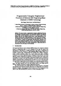
METHODS We prospectively selected patients with a diagnosis of iNPH from January 2010 to January 2012 in a Brazilian tertiary hospital. They were users of this medical facility screened
Patient with Clinical + Radiological findings
MMSE, TUG and JSINPH
Tap Test (TT)
MMSE, TUG and JSINPH (3 hours after TT)
MMSE, TUG and JSINPH (72 hours after TT)
Clinical improvement
No improvement
Surgery+ follow-up
Differencial diagnosis
MMSE: Mini-Mental State Examination; TUG: Time Up and Go; JSINPH: Japanese Scale for Idiopathic Normal Pressure Hydrocephalus; TT: Tap Test.
Fig 1. Protocol design. The scheme displays the protocol consisting in patients selection and undergoing the tests MMSE, TUG and JSINPH before and after TT, in order to determine the suitability of diversion surgery.
230
Arq Neuropsiquiatr 2013;71(4):229-236
in the hospital wards and forwarded by neurologists, geriatricians and other physicians. These patients underwent elective ventriculoperitoneal shunt with Strata® programmable valve system. It is an interventional, non-controlled, study with prospective features. The project was approved by the Research and Ethics Committee. The accepted diagnostic criteria were: clinical syndrome consistent with Adams-Hakim (gait apraxia, urinary incontinence and dementia) associated with ventricular dilatation documented by cranial computed tomography (CT) and magnetic resonance imaging (MRI) scans. Inclusion criteria: diagnosis of iNPH, absence of malignant disease and well-controlled clinical comorbidities (hypertension, diabetes mellitus, hormonal disorders, etc.). Exclusion criteria: diagnosis of secondary NPH, inability to walk, malignancy and uncontrolled clinical comorbidities. To select the patients, we considered typical clinical settings, such as urinary incontinence, gait disturbance and dementia, as well as compatible image studies associated to clinical response to Tap Test (TT). All patients underwent clinical evaluation, which consists of the Mini-Mental State Examination (MMSE)16 and Time Up and Go (TUG)17 tests and the application of Japanese Scale for Idiopathic Normal Pressure Hydrocephalus (JSINPH)2 in three stages: prior to the TT, 3 hours after the TT and 72 hours after the TT (Figs 1 and 2). All patients were submitted to imaging studies (CT scan and MRI of the skull with CSF flow study) in order to screen for secondary causes of hydrocephalus. The standard measure used to determine the concept of hydrocephalus was the Evans Ratio (ER), which consists in dividing the distance between frontal horns of lateral ventricles to maximal biparietal diameter. An ER value >0.3 is compatible with ventriculomegaly. JSINPH GAIT DISTURBANCE 0.......................................absent 1.......................................unstable, but independent gait 2.......................................walking with one cane 3.......................................walking with two canes or a walker frame 4.......................................walking not possible DEMENTIA 0.......................................absent 1.......................................no aparent dementia, but apathetic 2.......................................socially dependent, but independent at home 3.......................................partially dependent at home 4.......................................totally dependente URINARY INCONTINENCE 0.......................................absent 1.......................................absent, but with pollakisuria or urinary urgency 2.......................................sometimes only at night 3.......................................sometimes, even during the day 4.......................................frequent
Fig 2. Japanese Scale for Idiopathic Normal Pressure Hydrocephalus (JSINPH) is a recognized tool to evaluate and grade the three main symptoms in Idiopathic Normal Pressure Hydrocephalus (iNPH).
Once the diagnosis of hydrocephalus is established and secondary causes are excluded, the testing phase was carried out to verify whether the diversion procedure with programmable valves would be adequate. Each patient was submitted to a protocol, which consists of two steps: • Step 1 – (a) performance of the MMSE; (b) Time Up and Go; (c) application of the Japanese Scale for Idiopathic Normal Pressure Hydrocephalus; (d) lumbar puncture with drainage of 40–50 mL of CSF and manometry (TT). • Step 2 – three hours after the puncture, steps (a), (b) and (c) were repeated. According to the results, patients were taken back to the clinic or submitted to peritoneal shunt with Strata® adjustable valve (Medtronic). The MMSE16 is a standard test introduced by Folstein in order to evaluate the general patterns of dementia disorders. The Time Up and Go17 is a test that consists of measuring the time it takes for an individual to rise from a chair, walk three meters and sit back, assessing gait independence. The cutoff is approximately 12 seconds, although some authors diverge. Thus, patients able to perform the route in less than 12 seconds are considered independent with respect to gait, while those who take longer conceptually have a gait dysfunction. It should be noted that patients with orthopedic and neurological sequelae automatically take more time to complete the route. The Japanese grading scale for iNPH2 is a tool to access the patients’ clinical background. It measures the three main symptoms in different degrees of presentation, and can be easily performed in the preoperative period and as an evaluation questionnaire and in the follow-up of the patients. The TT1-3,18 is a recognized test that consists in the removal of varying amounts of CSF (40 to 50 mL) by lumbar puncture, after which the patient with suspected iNPH has hypothetically symptom improvement. It is believed that the removal of the excessive cerebrospinal fluid CSF by puncturing allows a transient improvement in clinical pattern. Usually, the element that responds better to the TT is gait, followed by urinary incontinence. As there is a learning bias in the application of MMSE, we considered an improvement after TT in patients with any better scores in TUG and/or JSINPH. The subjective opinion was also considered, added to TUG and/or JSINPH. Ventricular peritoneal shunt surgery was indicated for patients with improvement in at least one of the classic triad of symptoms, in any degree. As there is a learning bias for the MMSE, we decided to associate their improved score to an improvement in some other parameter (urinary incontinence or gait). The subjective
improvement reported by patients was also considered, but as it was difficult to measure it and due to the low degree of reliability of some reports, even because of dementia, we chose to always associate an objective parameter. In this manner, patients with indication for surgery underwent ventricular peritoneal shunt through strict technique, with the right frontal trepanation point of Kocher. The valve applied in the study was the Strata® (Medtronic), which is an externally adjustable magnetic radiopaque with anti-siphoning valve programmed to one of five performance level settings, from 0.5 to 2.5, with 0.5 increments, with each level setting being correspondent to a drainage pressure19 (Table 1). The Strata® valve (Medtronic) was then adjusted to 2.5 (140 mmHg), and the patient was discharged with monthly follow-up in the first three months and then quartlely follow-up. Our final follow-up time was one year. In this protocol, we proposed the adjustment of the valve according to symptoms, assessed with the JSINPH, Time Up and Go, MMSE and pattern of hydrocephalus. The valve level should start in 2.5 and be adjusted on a quarterly basis, with reductions of 0.5 per occasion towards the best clinical outcome, avoiding complications and symptoms of overdrainage. The objective was to achieve the lowest performance level in order to reduce the hydrocephalus and its symptoms, and the limits were the lowest level (0.5) or a level which caused new symptomatology, suggesting overdrainage (headache, dizziness, slit ventricle syndrome and signs of dural detachment). The team involved in the execution of the preoperative tasks consists of two neurosurgeons guided by a senior neurosurgeon. The evaluations of each patient before and after TT were performed by the same professional. The operative and follow-up tasks were conducted by the same team with the help of two more neurosurgeons. Statistics In this study, numerical data are presented as mean±standard deviation (SD) or median with range when appropriate. Categorical data are presented as percentages. When comparing groups, the level of significance is considered when p0.05). As our hospital is reference for a population of approximately 2 million inhabitants, the incidence found was 1.2 per 100,000. All 24 patients presented with some sort of symptoms from the Hakim’s triad. As expected, the dementia symptoms were presented for more time, for an average of 35 months previously to surgery. Gait apraxia was presented for a mean of 25 months, and urinary incontinence for 22 months. The diagnostic and preoperative mean Evan’s ratio was 0.37 with SD of 0.03, with no statistically difference between females and males. The preoperative MMSE mean was 19 before TT and 21 after TT, with statistical difference (p=0.01). The mean preoperative Japanese Scale was 6 before TT and 5 after TT, with statistical difference (p=0.001). Mean preoperative TUG was 41 seconds before and 36.41 seconds after TT, with statistical difference (p=0.003). The CSF mean pressure was 152 mmHg before and 29 mmHg after the TT. The initial valve status performance was 2.5 until follow-up. The patients were routinely discharged in the second postoperative day and were followed-up monthly in the first three months and, then, quartely. The parameters used for adjustment of the valve were radiological findings and clinical symptoms. Our time of follow-up was one year. Outcome After one year of follow-up, the average number of adjustments in valve pressure was 3.125, varying from a single adjustment in four patients to six adjustments in other four patients (Table 2). The final performance mean setting adjustment was 1.5. All the patients had their settings between 1 and 2. The objective clinical and radiological results were astounding. The mean MMSE changed from 19 to 21 (p=0.03). JSINPH changed from 6 to 4.63 (p=0.016). TUG, which before
232
Arq Neuropsiquiatr 2013;71(4):229-236
surgery was 41 seconds, became 35.77 seconds in the follow-up (p=0.01). The Evans Ratio in CT scans changed from 0.37 to 0.33 (p=0.0001), as illustrated in Fig 3. Four patients (three males e one female) did not show clinical improvement. One presented worsening of gait and dementia instead of radiological improvement (Evans changed from 0.45 to 0.38); other patient presented stroke during the course of the follow-up, resulting in confusional state and hemiparesis; another one developed subdural hematoma followed by subdural empyema, resulting in septic shock and death; the only female referred worsening of gait instead of surgery. The radiological results revealed that in 23 patients the Evans ratio decreased after shunting. Only one male patient (subject 2) presented increase in Evans ratio ( from 0.36 to 0.40), instead of clinical improvement. Complications There were eight complications in five patients (three males and two females). We consider major complications those requiring surgical correction, including subdural hematomas and empyemas, permanent shunt obstruction and shunt infection. We consider minor complications those in which the adjustment of pressure and clinical measures were sufficient and effective for the control, including wound dehiscence, slit ventricle syndrome, transient shunt obstruction and resetting after MRI procedures20,21 (Table 2). In the follow-up, malfunctioning of the valve forced the valve revision in two patients. One patient presented wound dehiscence and valve exposure after six months of the surgery, and it was necessary to remove the valve. There was no significant difference in complications rate between males and females (p