Pt/SiO2 Model Catalyst: Metal Support Interaction Studied by TEM and EELS. D. Wanga, S. Pennerb, D. S. Sua, G. Rupprechterc, K. Hayekb, R. Schlögla.
Pt/SiO2 Model Catalyst: Metal Support Interaction Studied by TEM and EELS D. Wanga, S. Pennerb, D. S. Sua, G. Rupprechterc, K. Hayekb, R. Schlögla a
Department of Inorganic Chemistry, Fritz Haber Institute of the Max Planck Society, Faradayweg 4-6, D-14195 Berlin, Germany b Institut für Physikalische Chemie, Leopold-Franzens-Universität, A-6020 Innsbruck, Austria c Department of Chemical Physics, Fritz Haber Institute of the Max Planck Society, Faradayweg 4-6, D-14195 Berlin, Germany Dispersed metal particles supported on porous substrates are widely applied in heterogeneous catalysis. The catalysts usually show activities and selectivities strongly depending on the pretreatment (cleaning/activation) conditions. The noble metal may interact with both reducible (e.g. TiO2) and non-reducible (e.g. SiO2 and Al2O3) oxide supports [1, 2]. This metal support interaction may have drastic effects on the chemisorptive and catalytic properties of the metal phase. Therefore, the investigation of structural changes under various treatment conditions is of great importance for elucidating the mechanism of the metal support interaction. In the previous investigations, well-oriented and regularly shaped platinum particles were grown on a thin supporting film of amorphous silica as a model catalyst using the method reported in [3]. After an oxidising treatment (O2, 673K, 1h) the samples were exposed to H2 at 873K for 1h. Electron diffraction, bright field images, and high-resolution transmission electron microscope (HRTEM) images were used to reveal the formation of platinum silicide Pt3Si with Cu3Au structure, monoclinic Pt3Si and tetragonal Pt12Si5 after the reductive treatment [4]. In the present work, sophisticated process of particles coalescence is observed by HRTEM images. In addition, electron energy-loss spectroscopy (EELS) is applied to show the Si electronic structural change in bonding. Fig. 1a shows the beginning stages of a coalescence process of three particles with platelet shape. It can be seen that each two particles coagulate at the corners. The contacting area exhibits curved edges and shows either a contrast differing from those of the particles (arrowed A) or lattice fringes showing a grain boundary (arrowed B). This indicates the rearrangement and the diffusion of atoms at the corners leading to the formation of the curved edges. Fig. 1b shows a coalescence process at an advanced stage. The particle obviously consists of two grains that may stem from two particles. Fourier transforms from these two areas are shown in two different insets. Their lattice fringes have slightly different spacing and orientation and an interface is formed between the two crystallites. Near the interface and the surface of the coalesced particle, a small area with different contrast (within the rectangle in Fig. 1b) can be seen, also showing the rearrangement of atoms. This may be due to the metal-support interaction with the presence of H2 at high temperature, which leads to the flattening of the particles and subsequently the increase of contacting area with the substrate. Moreover, the silicide formation also increases the particle size. These effects may cause the asgrown particles with very close spacing to contact with each other and the surface diffusion at high temperature may lead to their coalescence. On both as-grown sample and that after heating in hydrogen at 873 K, the spectra of Si L energyloss near edge structure (ELNES) were taken from the free silica substrate and from areas covered with Pt particles. After background subtraction and multiple scattering correction, they are plotted in
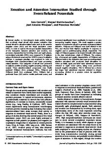
Fig. 2. The spectra from silica for both the as-grown and the reduced samples exhibit the typical Si L ELNES of SiO2, identical with the one measured from the area with Pt particles for the as-grown sample (Fig. 2). After the treatment, some new features appear in the Si L ELNES spectrum obtained from particle (Fig. 2). The normalized ELNES spectrum from the silica substrate is subtracted from it and the difference curve is also shown in Fig. 2. Up to about 140 eV, the ELNES spectrum from particle is similar to that from SiO2, indicating the unchanged SiO4 tetrahedron in the substrate under the supported particles. However, an additional energy loss upon the SiO2 spectrum is observed, starting from about 141 eV, increasing to the highest point at about 150 eV and gradually decreasing again. This is a signature of silicon in a changed chemical environment compared to SiO4, due to the formation of silicide, which causes a considerable change in near edge fine structure of Si [5]. Therefore, the EELS also indicates the reduction of silica and the formation of silicide proceeding through the interface between the Pt particle and the silica substrate [6]. References: [1] G.L. Haller, D.E. Resasco, Adv. Catal. 36 (1989) 173. [2] G.J. den Otter, F.M. Dautzenberg, J. Catal. 53 (1978) 116. [3] G. Rupprechter, K. Hayek, L. Rendón, M. José-Yacamán, Thin Solid Films 260 (1995) 148. [4] D. Wang, S. Penner, D.S. Su, G. Rupprechter, K. Hayek, R. Schlögl, Mater. Chem. Phys. in press. [5] S.J. Naftel, A. Bzowski, T.K. Sham, D.-X. Xu, S.R. Das, J. Phys. IV 7 (1997) C2-1131. [6] This work was in part supported by the Austrian Science Foundation (Project S 8105).
Fig. 1. High-resolution images of particles after the reduction, showing (a) the coalescence of three particles, (b) the coalescence of two crystallites with an interface formed in between.
Fig. 2. The Si L ELNES spectra from the free substrate for both the as-grown and the reduced samples, and the spectrum from areas with particles for the as-grown sample exhibit typical SiO2 features. The spectrum from particle for the reduced sample exhibits new features indicating Si in a changed chemical environment. The difference curve between them is presented.