Jun 5, 1989 - decrease in Tlco, could be explained by a loss of pulmonary ... medical examination required by the Norwegian. Directorate ..... training effect because of raised breathing resistance. .... Effect of gas density on mechanics of.
242
British Journal of Industrial Medicine 1990;47:242-247
Pulmonary mechanical function and diffusion capacity after deep saturation dives E Thorsen, K Segadal, E Myrseth, A Pasche, A Gulsvik
Abstract To assess the effects of deep saturation dives on pulmonary function, static and dynamic lung volumes, transfer factor for carbon monoxide (Tlco), delta-N2, and closing volume (CV) were measured before and after eight saturation dives to pressures of 3-1-46 MPa. The atmospheres were helium-oxygen mixtures with partial pressures of oxygen of 40-60 kPa. The durations of the dives were 14-30 days. Mean rate of decompression was 10-5-13-5 kPalhour. A total of 43 divers were examined, six ofwhom took part in two dives, the others in one only. Dynamic lung volumes did not change significantly but total lung capacity (TLC) increased significantly by 4-3% and residual volume (RV) by 14-8% (p < 005). CV was increased by 16-7% (p < 0 01). The Tlco was reduced from 13-0i 16 to 11'8± 1-7 mmollminl kPa (p < 0 01) when corrected to a haemoglobin concentration of 146 g/l. Effective alveolar volume was unchanged. The increase in TLC and decrease in Tlco were correlated (r = - 0 574, p < 0-02). A control examination of 38 of the divers four to six weeks after the dives showed a partial normalisation of the changes. The increase in TLC, RV, and CV, and the decrease in Tlco, could be explained by a loss of pulmonary elastic tissue caused by inflammatory reactions induced by oxygen toxicity or venous gas emboli.
capacity. During long exposures, toxic effects of oxygen in this concentration range cannot be excluded and during decompression the venous gas microemboli filtered in the pulmonary circulation may also induce inflammatory reactions in the lungs and gas exchange abnormalities.23 An increase in vital capacity has in some cases been reported after saturation dives, attributed to a training effect of respiratory muscles.' Hyacinthe et al7 and Cotes et al6 have also shown reduced transfer factor for carbon monoxide (Tlco) immediately after two deep dives to a pressure equal to 3 1 MPa. This could be an effect of oxygen toxicity but effects of microembolisation cannot be excluded. Probably all decompressions from those depths will produce venous gas emboli filtered in the pulmonary circulation but not necessarily associated with clinical decompression sickness.8 The possibility of long term effects on pulmonary function of professional diving is still controversial. The cross sectional studies of divers' lung function by Watt,9 Davey et al,'0 and Crosbie et al" show larger than predicted vital capacities of divers but lower than predicted maximal flow rates at low lung volumes. This may indicate an airflow limitation but it is not known whether it is an adaptive response to diving or a progressive deterioration of lung function. We have measured pulmonary function before and after eight deep saturation dives in Norway during the period 1983-6 to evaluate the magnitude and functional significance of changes in pulmonary During deep saturation dives, the lungs are exposed function. to an artificial atmosphere with high density and usually a raised partial pressure of oxygen of 40-60 Methods kPa during decompression to facilitate inert gas THE DIVES elimination. The increased work of breathing Eight dives numbered 1 to 8 (table 1) in the depth imposed by the increased density of the breathing gas range equivalent to a pressure of 3-1-46 MPa (300is depth related' and the ventilatory capacity will 450 metres of sea water-msw)* were studied. Two eventually become a limiting factor for physical work were open sea dives (dives 5 and 6) whereas the others were simulated in the NUTEC onshore hyperbaric chamber complex. Welding trials were performed in Norwegian Underwater Technology Centre AIS dives 2 and 8. Equipment and operational procedures (NUTEC), Gravdalsveien 255, 5034 Ytre Laksevag, were tested both in the dry and wet. The durations of and Department ofThoracic Medicine, University of the dives were 14-30 days. The mean rate of decomBergen, Norway pression was 10 5-13 5 kPa/h. The atmosphere was
E Thorsen, K Segadal, E Myrseth, A Pasche, A Gulsvik
*1 MPa= 100 msw= 10 bar, 100 kPa= 10 msw= 1 bar.
Pulmonary function after saturation dives
243
helium-oxygen mixtures with partial pressures of oxygen of 40-60 kPa. Table 1 gives the characteristics of each dive. The number of divers in each saturation dive was four to nine, giving a total of 49 man dives. The diving procedures and the protocol for medical and physiological monitoring of the divers were approved by the Regional Ethical Review Committee (dives 1-5) and the ethical committee of the Norwegian Research Council for Science and the Humanities (dives 6-8). THE DIVERS
Forty three professional divers participated in the dives. Their average age was 30 3 years (range 2339), weight 78-3 kg (range 67-91), and height 180-2 cm (range 168-193). Ten were current smokers, four ex-smokers, and 29 non-smokers. Their experience as saturation divers was on average 5-8 years (range 1-9) and their total number of days in saturation on average 270 days (range 5-600). On the first examination before the dives, they had all passed the annual medical examination required by the Norwegian Directorate of Public Health for offshore diving. The time since their last routine saturation dive (less than 1 9 MPa) was at least four weeks. Six divers participated in two of the deep saturation dives described here and the time between their deep dives was from five months to two years. PROTOCOL
The divers were first examined four to six weeks before the dives (predive) and re-examined one to three days (first postdive) and four to six weeks (second postdive) after the dives. Three divers were examined at other institutes predive and their results are not included in this study. A second postdive examination was not done after dives 1 and 5. From 1983 to 1984 (dives 6-8) the examinations included clinical examination, dynamic lung volumes, and diffusion capacity. Later (dives 1-5), static lung volumes and distribution of ventilation were also included. Chest radiographs were taken in connection with dives 1 and 4 only. Table I Characteristics of the dives Partial pressure
Pressure
Duration*
Dive
No of divers
(MPa)
(days)
1 2 3 4
6 6 6 6
37 37 37 46
2+ 12+13
5
4 9 6
31 31 36 36
6 7 8
6
2+3+13 2+10+13 2+10+18 1+3+10 1+9+9 2+11+11 2+6+11
of oxygent (kPa)
40-50 40-50 40-50 40-50 40-50 40-60 40-60 40-50
+ bottom time + decompression time. tBottom phase and decompression phase.
*Compression time
ASSESSMENT OF PULMONARY FUNCTION
Static lung volumes The multibreath nitrogen washout technique was used to measure functional residual capacity (FRC). Combined with the measurements of expiratory reserve volume (ERV) and inspiratory vital capacity (IVC), total lung capacity (TLC) and residual volume (RV) were calculated.
Dynamic lung volumes A minimum of three satisfactory forced vital capacity manoeuvres were performed.'2 The forced expiratory vital capacity (FEVC), forced expired volume in one second (FEVy), and peak expiratory flow rate (PEF) were taken as the highest readings obtained. The forced mean mid-expiratory flow rate (FEF2,75.,,) and forced expiratory flow rates at 50°o and 750o of FEVC expired (FEF,,,,., FEF75.. ) were taken as the highest readings from flow volume curves not differing by more than 5%h from the highest FEVC. The forced inspiratory vital capacity (FIVC), forced inspired volume in one second (FIVI), forced inspiratory flow rate at 50% of FIVC (FIF50,,,), and peak inspiratory flow rate (PIF) were taken as the highest readings obtained. Maximum voluntary ventilation (MVV) was measured as the highest ventilation sustained for 12 seconds.
Diffusion capacity Tlc, was measured by the single breath holding method.'2 Effective alveolar volume (VA) was then measured simultaneously by helium dilution and transfer per unit effective alveolar volume (Kco) was calculated. Tlco was corrected to a haemoglobin concentration of 146 g/l. Distribution of ventilation During the multibreath nitrogen washout test for measuring FRC, the nitrogen washout time (NWT) (time to bring expired N2 concentration below 2%) and the lung clearance index (LCI) (volume ventilated to bring expired N2 concentration below 2% relative to FRC) were measured. The slope of phase 3 of the single breath 02 test-delta N2-was also measured along with closing volume (CV) at the point of inflection between phases 3 and 4. Closing capacity (CC) was calculated as the sum of RV and CV. The predive and postdive examinations were performed with the same equipment and technicians on each occasion at least two hours after breakfast without tea or coffee and with no smoking in the last two hours before the examination. Volume and test gas calibrations were done before each test and the results were corrected to the BTPS condition. STATISTICS
For comparison of results between predive and
244
Thorsen, Segadal, Myrseth, Pdsche, Gulsvik
postdive examinations paired Students t test was applied. Least squares linear regression analysis was done for correlation analysis. Differences between examinations were calculated as difference from the mean. All data are expressed as mean ± 1SD. A p value less than 0 05 was considered significant.'3
Results On the predive examinations none of the divers reported pulmonary symptoms and the clinical examinations of the heart and lungs were considered normal. Immediately after the dives retrosternal discomfort was reported by 20 divers, including nine with a non-productive cough provoked by deep inspirations. Otherwise non-specific symptoms of weakness, general fatigue, and insomnia were reported. Clinical examinations were still normal and in the two dives where chest radiographs were taken they were also normal. All symptoms had gradually disappeared during the first two weeks after the dives.
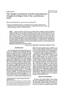
10-
8-
1.-
X
ITLC
|
X; 64.0 -0 E
>
4
CC~~~~~~~R cc
2-
J~~1RV
1.
Predive
1st postdive
2nd postdive
Fig I Static lung volumes on predive and postdive examinations. *Significantly different from predive.
STATIC LUNG VOLUMES In 24 divers diving to
3-7 and 4-6 MPa there was a significant increase in TLC of 4-3%, FRC of 11 -6%, and RV of 14-8% from the predive to first postdive examination. The IVC and ERV were unchanged (table 2). On the second postdive examination the TLC and its subdivisions were partially normalised (fig 1).
were pooled (table 2). In dives 2, 3, and 4 FEVC increased significantly from the predive to first postdive examination by 38%, 5-6%, and 8-1% respectively, whereas FEV, and flow rates at other lung volumes were unchanged (fig 2). As shown in figure 3, the outline of the flow volume curves did not differ, only their positions related to absolute lung DYNAMIC LUNG VOLUMES volume, which means that at the same absolute lung There were no changes from the predive through the volume, flow rate was lower postdive. The change in first and second postdive examinations when all dives FEVC (all dives) did not correlate with diver's age
Table 2 Results of selected pulmonary function tests. n refers to number of examinations
Dynamic lung volumes: FVC (1) FEV, (1) FEF2575"0 (l/s) PEF (1/s) FIV, (1) PIF (1/s) MVV (1/min) Static lung volumes: TLC (1) FRC (1) RV (1) CV (0. VC) CC (1) Diffusion capacity;
Tlco (mmol/min/kPa) K(o (mmol/min/kPa/l) VA
(I) Hb (g/l) Packed cell volume (volume fraction)
*Significantly different from predive p < 0-05. **Significantly different from predive p < 0-01.
Predive
First postdive
Second postdive
(n = 46)
(n = 46)
(n = 38)
6-10±0-68 4-81 ±055
6 17±0 64 4-78±0-56 4-41 ± 115 11-71±1-48 5-25±0-78 8-40± 2-20
6-16±0-60 4-69±0 55 4-54± 120 11-77±1-37 5-31±0-80 8-63± 2-34 171±24
4-57+ 1*13 11-95±1-43 5-52±0-85 8-81 ± 2-27 174±19 n=24
7-70± 0-85
3-77±0-74
1-56±0-36 11-0±1-6 2-35±0-41 n=46 13-00± 162
1-60±0-20 8 13±0 80 147±11 0-44 ± 0-02
182±24 n=24 8.04 ± 0-67* 4-11 ±0.61* 1-81 ±0-37*
n=24 7-86±0 70
13.0± 1-8* 2-74 ± 0-37*
11-2±2-1 2-50+0-40
n=46 11 -82± 1*66** 1-44 ± 0-17*
n=38 12-62± 166
8-20±0-73 138±15 0-42 ± 0-02
8-24±0-84 139±13 0-42 ± 0-03
3-79±0-68 1-69±0-34
1-53±0-21
245
Pulmonary function after saturation dives 6-8
5'61
FEVC
FEV1
52-
6-4
-
®
4-8
-
640
w
w
U-
LL
5*64
4.4-~
Predive
1st postdive
Predive
2nd postdive
1 st postdive
2nd postdive
Fig 2 FEVC and FEV, predive and postdive. Means for each dive, Nos 1-8, are plotted. *Significantly different from predive (p < 005).
(r = 0 139) or depth (r = 0 196) or rate of decompres- ing volume was significantly increased from 11 0 ± sion (r = 0 224) but correlated positively with dura- 1-6 to 13-0 ± 1-8% of VC (p < 0-05). Since both RV tion of the dives (r=0-359, p < 0 02). The dives and CV were increased, closing capacity was also where welding was performed showed no specific increased (table 2). trends compared with the other dives. TRANSFER FACTOR FOR CARBON MONOXIDE
Tlco was significantly reduced by 9-5% immediately There were no changes in the distribution indices after the dives. Kco was reduced by the same delta-N,, LCI, or nitrogen washout time. The clos- magnitude (9 0%). Figure 4 shows the results of Tlco
DISTRIBUTION OF VENTILATION
14-
- - Predive 1st postdive
TLCO
12c
0.1 -0
0
E E -J
0
-
0
LL
C-,
1-. 10-
0
10
Predive
1st postdive
2nd postdive
Volume (btps)
Fig 3 Flow volume loops at predive and first postdive examinations related to absolute lung volume (n = 24). 1 SD is shown in only one direction.
Fig 4 Tlco predive and postdive. Means for each dive, Nos 1-8, are plotted. All means at first postdive examination differ significantly from predive (p < 0-05S).
Thorsen, Segadal, Myrseth, Pdsche, Gulsvik
246 130_
y =
r =
-041 x +141-8 0-574
120aL)
0
1100
100-
0* 0
9070
80
90 TLCO % predive
100
Fig 5 Correlation plot between change in TLC and Tlco from predive to first postdive examination.
in each dive. The reduction in Tlco did not correlate with duration of the dive (r = 0-123), diver's age (r = - 0 060), or depth (r=0 099) but there was a significant negative correlation between change in TLC and change in Tlco (r = - 0-574, p < 0 02) (fig 5). Tlco was still reduced on the second postdive examination four to six weeks after the dives (dives 1 and 5 not examined), the reduction being 5 6% (NS). The reduction in Tlco of divers decompressed at a partial pressure of oxygen of 50 kPa and 60 kPa were 9 7% and 9 1% respectively (NS). -
Discussion Twenty of the 43 divers reported chest symptoms persisting for up to two weeks after the dives. The symptoms were consistent with a tracheobronchitis that may be induced by raised partial pressure of oxygen. A reduction in VC, which is a characteristic finding in oxygen toxicity, was not shown. The results of the assessment of static and dynamic lung volumes indicate an expansion of the lungs. Some reports indicate an increase in VC immediately after deep dives56 but this was seen in only three of our dives. The TLC and VC may be increased by a training effect because of raised breathing resistance. The results of training of respiratory muscles by loaded breathing'4 and swimming'5 have shown an increase in FRC, TLC, and VC but unchanged RV. An increase in residual volume has also been shown after a saturation dive to 1 86 MPa.4 The significant correlation between duration of the dive and increase in FEVC could reflect a training effect. There was no increase in PEF, MVV, or maximal inspiratory flow rates, which are effort dependent, to support this. Maximal respiratory pressures were not measured in this study but Cotes et al did not find changes in maximal respiratory pressures after the dive to 3 1 MPa where there was an increase in FEVC of 6-8%.6 The capillary endothelial cells are probably the most vulnerable structure to hyperoxic injury with
disruption of the endothelial lining and occlusion of capillaries and small arterioles.'6 Venous gas embolisation will also result in a microvascular injury with occlusion of capillaries and increased permeability of the endothelium.'7 8 Reduced static lung volumes, which are characteristic of oxygen toxicity, will not be seen unless the lymphatic drainage capacity of the lungs is overloaded, forming oedema. Existing oxygen tolerance tables indicate an oxygen concentration of 50 kPa as being harmless,'9 but there exists no experience to support this when exposure time is more than two weeks. These tables are based on changes in vital capacity as the measure of oxygen toxicity but other lung function variables such as the Tlco would probably detect changes at an earlier stage. The inflammatory processes associated with oxygen toxicity and gas embolism injury may destroy pulmonary elastic tissue through the mechanisms of oxygen radicals. In a study by Riley et al in mice exposed to hyperoxia a degradation of collagen was shown with a reduced pulmonary recoil pressure and histopathological evidence of an emphysematous lesion eight weeks after exposure.20 Oxygen radicals are also involved in the process of injury after air embolisation.2'22 The pattern of changes in static lung volumes in our study may be explained by that mechanism resulting in a loss of pulmonary elastic tissue. The increase in closing capacity and reduction of Tlco is also consistent with this. The characteristics of divers' lung function described by Crosbie et al indicates a slight bronchial obstruction in divers." It is not known whether it is a real obstruction or a result of the divers' significantly higher than predicted lung volumes. Our study indicates that a slight obstruction might be induced by the dives when looking at both the increase in RV and TLC and maximum flow rates at absolute lung volumes. At a given absolute lung volume, maximum expiratory flow is lower postdive. The functional significance of changes in pulmonary function after deep dives is indicated by the striking reduction of Tlco. The transfer of CO from alveoli to haemoglobin depends on the area available for diffusion, the condition of the membrane over which diffusion takes place, the capillary area with its blood volume, and haemoglobin concentration. The measurement of static lung volumes as well as VA show that the lung volume and thereby alveolar area available for diffusion is almost unchanged. The haemoglobin concentration has been corrected for and does not explain the difference between predive and postdive results. The explanation is then reduced to an increased thickness of the diffusive membrane or a reduced capillary area available for diffusion. Reductions in Tlco have been shown after exposure to both normobaric and hyperbaric oxygen67 2324 and the time for complete recovery may
Pulmonary function after saturation dives
be several weeks. In the study by Puy et al the Tlco was partitioned into the membrane and blood components, and the main reduction in TL6 was in the blood component, indicating changes in the pulmonary capillary bed.24 Hyacinthe et alhave shown a 13% reduction of Tlco after a decompression from 3-1 MPa at a partial pressure of oxygen of up to 80 kPa.' This is definitely a toxic oxygen concentration. The Tlco was still reduced at two weeks after this dive. In the study by Cotes et al after a dive to 3-1 MPa, a reduction in Tlco of 9-6% was shown and the recovery was complete at four weeks postdive.6 Our data indicates a recovery time of more than five weeks depending on the initial reduction. Tlco does not significantly correlate with duration of the dives, but seems in some way to be related to the change in TLC as reflected by the negative correlation between change in Tlen and TLC. Other significant correlations between changes in lung function parameters and characteristics of the hyperbaric exposure or the divers themselves were not found, such as age, depth, rate of decompression, or partial pressure of oxygen. The individual response to the environmental challenges differ considerably and with so many aetiologic factors acting simultaneously makes it difficult to draw conclusions. The range of variation for oxygen concentrations and rates of decompression in these dives are also small. Systematic studies of routine operational saturation diving to less than 2 MPa should be carried out, making the correlations over a wider range for the independent variables. It may be concluded that significant pulmonary changes after deep saturation diving are induced by the dive and that the recovery may take several weeks. The findings of this study support the findings in cross sectional studies which indicate the development of airflow limitation in professional divers. So long as effects on long term health are mostly unknown, efforts should be taken to assure that a complete recovery has taken place before other dives are done, not only deep dives but also routine diving. The Tl60 test and static lung volumes should be considered in the follow up examinations of divers. This work was supported by Norsk Hydro, Statoil, and the Royal Norwegian Council for Scientific and Industrial Research (NTNF). K Segadal was supported by grants from the Norwegian Research Council for Science and the Humanities (NAVF) from 1986 to 1988 and the Hyperbaric Medical Research Programme (grant No 13.91.99-118).
247 1 Maio DA, Fahri LE. Effect of gas density on mechanics of breathing. J Appl Physiol 1967;23:687-93. 2 Hlastala MP, Robertson HT, Ross BK. Gas exchange abnormalities produced by venous gas emboli. Respir Physiol
1979;36:1-17.
3 Neuman TS, Spragg RG, Wagner PD, Moser KM. Cardiopulmonary consequences of decompression stress. Respir
Physiol 1980;41:143-53. Hayashi E, Yelverton C. Hana Kai II: a 17-day dry saturation dive at 18-6 ATA. Cardiopulmonary functions. Undersea Biomed Res 1977;4:267-81. 5 Yamasaki M, Taya Y, Fujiie K, Seki K, Sasaki T, Nakayama H.
4 Smith RM, Hong SK, Dressendorfer RH, Dwyer HJ,
Effect of a simulated saturation dive to 31 ATA on pulmonary function. Ann Physiol Anthropol 1986;5:191-6. 6 Cotes JE, Davey I S, Reed JW, Rooks M. Respiratory effects of a single saturation dive to 300 m. Br J Ind Med 1987;44:76-82. 7 Hyacinthe R, Giry P, Brousolle B. Development of alterations in pulmonary diffusing capacity after a deep saturation dive with high oxygen level during decompression. In: Bachrach AJ, Matzen MM, eds. Underwater physiology VII. Proceedings of seventh symposium on underwater physiology. Bethesda: Undersea Medical Society, 1979:75-83. 8 Spencer MP, Clark HF. Precordial monitoring of pulmonary gas embolism and decompression sickness. Aerospace Med
1972;43:762-7.
9 Watt S. Effect of commercial diving on ventilatory function. Br J Ind Med 1985;42:59-62. 10 Davey IS, Cotes JE, Reed JW. Relationship of ventilatory capacity to hyperbaric exposure in divers. J Appl Physiol 1984;56: 1655-8. 11 Crosbie WA, Reed JW, Clarke MC. Functional characteristics of the large lungs found in commercial divers. J Appl Physiol
1979;46:639-45. Europ Physiopathol Respir 1983;19(suppl 5). Rosner B. Fundamentals of biostatistics. Boston: Duxbury Press, 1982. Leith DE, Bradley M. Ventilatory muscle strength and endurance training. J Appl Physiol 1976;41:508-16. Clanton TL, Dixon GF, Drake J, Gadek JE. Effects of swim training on lung volumes and inspiratory muscle conditioning. J Appl Physiol 1987;62:39-46. Jones R, Zapol WM, Reid L. Pulmonary artery remodelling and pulmonary hypertension after exposure to hyperoxia for 7 days. Am JPathol 1984;117:273-80. Hall JE, Hofman WF, Ehrhart IC. Venous occlusion pressure and vascular permeability in the dog lung after air embolization. J Appl Physiol 1988;65:34-40. Ohkuda K, Nakhara K, Binder A, Staub NC. Venous air emboli in sheep: reversible increase in lung microvascular permeability. J Appl Physiol 1981;49:887-94. Harabin AL, Homer LD, Weathersby PK, Flynn ET. An analysis of decrements in vital capacity as an index of pulmonary oxygen toxicity. J Appl Physiol 1987;63:1130-5. Riley DJ, Cramer MJ, Kerr JS, Chae CU, Yu SY, Berg RA. Damage and repair of lung connective tissue in rats exposed to toxic levels of oxygen. Am Rev Respir Dis 1987;135:441-7. Flick MR, Perel A, Staub NC. Leucocytes are required for increased lung microvascular permeability after microembolization in sheep. Circ Res 1981;48:344-51. Flick MR, Milligan SA, Hoeffel JM, Goldstein IM. Catalase prevents increased lung microvascular permeability during air emboli in unanesthetized sheep. J Appl Physiol 1988;64:
12 Quanjer PhH, ed. Standardized lung function testing. Bull 13 14
15 16
17 18 19 20 21 22
929-35. 23 Caldwell PRB, Lee WL Jr, Schildkraut HS, Archibald ER. Changes in lung volume, diffusing capacity and blood gases in men breathing oxygen. J Appl Physiol 1966;21:1477-83. 24 Puy RJM, Hyde RW, Fisher AB, Clark JM, Dickson J, Lambertsen CJ. Alterations in the pulmonary capillary bed during early O2 toxicity in man. J Appl Physiol 1968;24: 537-43.
Accepted 5 June 1989