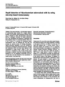
Clinical Chemistry 46:5 631– 635 (2000)
Molecular Diagnostics and Genetics
Rapid Detection of Point Mutations by Fluorescence Resonance Energy Transfer and Probe Melting Curves in Candida Species Juergen Loeffler,1* Lars Hagmeyer,1 Holger Hebart,1 Norbert Henke,1 Ulrike Schumacher,2 and Hermann Einsele1
Background: The LightCyclerTM combines rapid amplification of nucleic acids in glass capillaries with melting curve analysis based on fluorescence resonance energy transfer for the sensitive detection of point mutations in various settings, such as drug resistance and hereditary diseases. Point mutations leading to an altered structure of lanosteroldemethylase, the target enzyme of the fungistatic azoles, are an important mechanism of acquired resistance in Candida albicans. Methods: We screened 13 fluconazole-resistant C. albicans and 21 fluconazole-resistant C. tropicalis strains (minimum inhibitory concentration >128 mg/L), isolated from patients with AIDS, for the presence of defined point mutations by comparing conventional cycle sequencing with a newly designed LightCyclerbased assay. Results: In C. tropicalis, 5 of 21 isolates showed the wild-type sequence, and 8 of 21 showed the homozygous nucleotide exchange thymine to cytosine at position 1554 (T1554C). A heterozygous genotype was detected in 8 of 21 isolates by the LightCycler, but in only 3 of 21 isolates by conventional cycle sequencing. In 2 of 13 C. albicans isolates, a homozygous point mutation leading to an amino acid exchange at position 464 (glycine to serine) was detected in both assays. Conclusion: The LightCycler technique offers standardized, fast, sensitive, and reproducible detection of point mutations in different Candida spp.
Azole antifungal agents have therapeutic activity against different Candida species. Azole antifungal agents are commonly used for prophylaxis and treatment of superficial candidiasis, and more recently, for invasive Candida infections in transplant, leukemic, and AIDS patients (1 ). As the number of patients treated with azoles and the duration of antifungal therapy have increased during the last decade, azole resistance has emerged as a major problem (2 ). Despite many studies, the molecular mechanisms of azole resistance are still not fully understood. Point mutations in the gene ERG11, which encodes the target enzyme of the azoles, the sterol 14␣-demethylase, have been described that may mediate azole resistance through decreased drug access or by reducing binding capacity of the azoles to the active site of the enzyme (3 ). To screen clinical C. albicans and C. tropicalis isolates rapidly and cost-effectively for the presence of point mutations associated with azole resistance, we established an assay based on the LightCyclerTM technique (Roche Molecular Systems). The amplicons are hybridized in real time to oligonucleotide probes binding to the target genome region that contains mutation sites. The assay allows amplification of 32 samples in parallel within 45 min. Previously, we demonstrated that in C. albicans, the amino acid substitution G464S, observed in the hemebinding domain of the enzyme, is associated with perturbation of the heme environment (4, 5 ). This substitution causes resistance by substantially reducing the inhibitory effect of fluconazole. In C. tropicalis, cycle sequencing with an ABI 373A (Perkin-Elmer) showed that the nucleotide exchange T1554C was present in 16 of 21 fluconazole-resistant isolates. Although this point mutation does not influence the amino acid sequence, we used this common point mutation to establish the method on the LightCycler instrument.
© 2000 American Association for Clinical Chemistry
Eberhard-Karls-Universita¨t, 1 Medizinische Klinik, Abteilung II, and 2 Hygieneinsitut, Abteilung Medizinische Mikrobiologie, 72076 Tuebingen, Germany. *Address correspondence to this author at: Medizinische Klinik, Abteilung II, Labor Prof. Dr. Med. H. Einsele, Otfried-Mueller-Strasse 10, 72076 Tuebingen, Germany. Fax 49-7071-293179; e-mail
[email protected] gen.de. Received December 17, 1999; accepted February 29, 2000.
631
632
Loeffler et al.: FRET and Melting Curves to Detect Candida Mutations
Materials and Methods fungal isolates In total, 13 fluconazole-resistant C. albicans isolates (CAR1–CAR13; 5 from the Hygieneinstitut Innsbruck, Austria; 8 from the Rudolf-Virchow-Klinikum, Berlin, Germany) and 21 fluconazole-resistant C. tropicalis isolates (CTR1–CTR21; 11 from the Veterans Affairs Hospital, San Antonio, TX; 8 from the North Manchester General Hospital, Manchester, United Kingdom; 2 from the Hygieneinstitut Innsbruck, Austria) were analyzed.
patients Thirty-four Candida strains analyzed were isolated from patients who did not respond to fluconazole therapy. All patients were HIV positive with intermediate (CD4⫹ lymphocyte counts, 200 –500/L) or advanced (CD4⫹ lymphocyte counts, ⬍200/L) immune depletion and oropharyngeal or esophageal candidiasis. In addition, these patients had also been hospitalized because of the following diseases: Burkitt lymphoma, polytrauma, Lyell syndrome, acute myeloid leukemia, Kaposi sarcoma, mycobacterial infections, cytomegalovirus retinitis, and Pneumocystis carinii pneumonia. All patients had received fluconazole prophylaxis or treatment at a dosage of 100 – 800 mg/day for up to 3 years. Fluconazole-sensitive strains (CAS, CTS) were isolated from patients with hematological malignancies (Medizinische Klinik Tu¨bingen, Germany).
susceptibility testing The minimum inhibitory concentrations of fluconazole for all isolates were determined by the NCCLS M27-A broth microdilution method for yeasts (6 ) as well as by E-test (AB Biodisk) according to the instructions of the manufacturer.
dna extraction Fungi were subcultured on Sabouraud glucose agar for 48 h at 30 °C and suspended in sterile 9 g/L NaCl solution at a concentration of 106 colony-forming units (CFU)/mL (McFarland A530, 0.5 corresponds to 106 cells). DNA was extracted as described previously (7 ) using recombinant lyticase and the QIAamp Tissue Kit procedure (Qiagen). Briefly, fungal suspensions were centrifuged for 10 min at 3000g, and 1 mL of lyticase buffer was added to generate spheroplasts [10 kU/L recombinant lyticase (Sigma), 50 mmol/L Tris, 1 mmol/L EDTA, 2 mL/L -mercaptoethanol]. After incubation for 45 min at 37 °C, samples were centrifuged for 10 min at 3000g, followed by incubation of the spheroplasts in ATL and AL lysis buffers from the QIAamp Tissue Kit according to the protocol. DNA was isolated using QIAamp spin columns and stored immediately after elution at ⫺0 °C. For sensitivity testing, fungal suspensions were serially diluted from 105 cells to 101 cells. Specificity testing was performed with DNA extracted
previously from fungal cultures (C. albicans, C. tropicalis, and C. glabrata).
dna amplification by conventional thermocycling DNA was amplified using standard conditions as described previously (4 ). The following primers that bound to the Candida ERG11 gene were selected: for C. albicans, forward primer CAL-PRF (5⬘-TCT CCA GGT TAT GCT CAT ACT A-3⬘) and reverse primer CAL-PRR (5⬘-AAC AAT CAG AAC ACT GAA TCG AA-3⬘); for C. tropicalis, forward primer CTR-PRF (5⬘-TCA TAC CAG TGA TAG ATG G-3⬘) and reverse primer CTR-PRR (5⬘-TTT TCT AGC TAC TCC ATG G-3⬘), all from Fa. Roth, Karlsruhe, Germany. The primers are shown in Table 1.
cycle sequencing Before cycle sequencing, amplicons were purified by using the QIAquick kit (Qiagen) according to the manufacturer’s protocol. PCR was performed in a Perkin-Elmer GeneAmp 9600 thermocycler using AmpliTaq FS and the BigDye Terminator Mix (Perkin-Elmer). Cycle sequencing was performed in an ABI 373A automatic sequencer (Applied Biosystems) at 29 W for 14 h using a 6% urea-polyacrylamide gel. Cycle sequencing was performed by forward and reverse priming, respectively.
gel electrophoresis For optimization of the amplification process, conventional gel electrophoresis was performed using a 2% agarose gel (Sigma) in 40 mmol/L Tris, 1 mmol/L EDTA (Life Technologies) for 2 h at 90 V, followed by ethidium bromide staining.
dna amplification with the LightCycler The LightCycler PCR and detection system was used for amplification and online mutation detection. The PCR mixture contained Taq Polymerase, LightCycler Hybridization 1⫻ reaction buffer, dNTP mixture (with dUTP instead of dTTP), 3 mmol/L magnesium chloride, and 10 pmol of each primer, respectively.
Table 1. Oligonucleotide sequences. Name
Oligonucleotide sequence
Tm,a ° C
CAL-PRFb CAL-PRR CAL-FL CAL-RED CTR-PRF CTR-PRR CTR-FL CTR-RED
5⬘-TCTCCAGGTTATGCTCATACTA-3⬘ 5⬘-AACAATCAGAACACTGAATCGAA-3⬘ 5⬘-ATCTATGTCTACCACCACCAAATGGTAAA-3⬘c 5⬘-AGGTGAAGAAACCCCTTTAGAAACTTTCC-3⬘ 5⬘-TCATACCAGTGATAGATGG-3⬘ 5⬘-TTTTCTAGCTACTCCATGG-3⬘ 5⬘-GTTGATTACCAATCCATGGTTACCTTAC-3⬘ 5⬘-ATTAGAACCTGCTGAAATTGTTTGG-3⬘
49.8 54.1 60.2 63.2 43.7 46.9 59.2 57.5
a
Tm, melting temperature of the oligonucleotide. CAL, C. albicans; PRF, forward primer; PRR, reverse primer; FL, fluorescein labeled; RED, LC Red 640 labeled; CTR, C. tropicalis. c Point mutations are in bold. b
Clinical Chemistry 46, No. 5, 2000
Samples were amplified by 60 cycles of repeated denaturation (0 s at 95 °C), annealing (C. albicans, 15 s at 54 °C; C. tropicalis, 15 s at 50 °C), and enzymatic chain extension (20 s at 72 °C). The PCR was completed within 45 min, including melting curve analysis.
LightCycler-based melting curve analysis For mutation detection, the LightCycler DNA Master Hybridization Probes Kit was used as described by the manufacturer. Briefly, hybridization probes consisted of two different oligonucleotides that hybridized to an internal sequence of the ERG11 gene of C. albicans or C. tropicalis. Hybridization probe 1 was labeled at the 3⬘ end with fluorescein spanning the point mutation, hybridization probe 2 was labeled at the 5⬘ end with LightCycler Red 640 functioning as an anchor probe. Probes were obtained from Tibmolbiol. Both probes could hybridize in a head-to-tail arrangement, bringing the two fluorescent dyes into close proximity. During fluorescence energy resonance transfer (FRET), fluorescein was excited by the light source of the LightCycler instrument. The excitation energy was transferred to the acceptor fluorophore, LightCycler-Red 640, and the emitted fluorescence was measured by the photohybrids of the instrument. The fluorescence was monitored while the temperature was slowly increased; the fluorescence decreased when one of the probes melted off and the two fluorescent dyes were no longer in close proximity. A hybridization probe spanning one mismatch could still hybridize to the target sequence but melted off at lower temperature than a hybridization probe with a perfect match.
633
melting temperature (Tm) than the sequence that has a mismatch with the mutation probe (Fig. 1). In heterozygous samples containing both sequences, in addition to the maximum there were two peaks or shoulders in the curve at exactly the same temperatures as the respective homozygous samples (Fig. 2). In the study presented, all 13 fluconazole-resistant C. albicans and 21 fluconazole-resistant C. tropicalis isolates were analyzed by using the LightCycler instrument. In 2 of 13 C. albicans isolates, a homozygous point mutation leading to the amino acid exchange at position G464S was detected (CAR5 and CAR9; Fig. 1). Oligonucleotides CAL3-FL and CAL3-RED showed a difference of 6 °C in the melting temperature profile between isolates CAR5 and CAR9 and all other isolates In C. tropicalis, 8 homozygous and 8 heterozygous mutations (T1554C) were detected with a 100% reproducibility by using the probes CTR-FL and CTR-RED (Fig. 2).
detection limit of the assay Analysis of serially diluted cells from C. tropicalis (105–101 CFU/mL) showed that for LightCycler-based analysis, a
Results susceptibility of the yeast pathogens The minimum inhibitory concentrations of fluconazole for C. albicans and C. tropicalis isolated from patients not responding to therapy (CAR, CTR) were between 128 and 256 mg/L. Fluconazole-sensitive strains (CAS, CTS) had minimum inhibitory concentrations ⬎4 mg/L.
cycle sequencing Thirteen fluconazole-resistant C. albicans and 21 fluconazole-resistant C. tropicalis isolates were screened for the presence of point mutations in the ERG11 gene. In C. albicans, 2 of 13 isolates showed point mutations leading to the amino acid substitution G464S (CAR5 and CAR9); 11 of 13 did not show this point mutation. In C. tropicalis, 8 of 21 showed the homozygous nucleotide exchange T1554C, 3 of 21 were heterozygous for T1554C, and in 5 of 21, sequencing data did not permit discrimination between homozygous and heterozygous mutations. In 5 of 21, no point mutation could be detected.
LightCycler-based detection of point mutations In LightCycler-based analysis, the melting peaks indicated that the fully homologous sequence has a higher
Fig. 1. Nucleotide exchange in fluconazole-resistant C. albicans isolates at position 1537, detected by melting curve analysis. Wild-type genotype CAR1 (A) and fluconazole-sensitive isolate CAS (B) were included to demonstrate the high reproducibility of the assay. For specificity control, C. tropicalis and C. glabrata isolates were included. (A), f, C. albicans isolate CAR5, fluconazole resistant, homozygous genotype; ⫹, C. albicans isolate CAR9, fluconazole resistant, homozygous genotype; F, C. albicans isolate CAR1, fluconazole resistant, wild-type genotype; ⫻, C. tropicalis isolate 4; 〫, C. glabrata isolate. (B), f, C. albicans isolate CAR5, fluconazole resistant, homozygous genotype; ⫹, C. albicans isolate CAR9, fluconazole resistant, homozygous genotype; F, C. albicans isolate CAS, sensitive to fluconazole; ⫻, C. tropicalis isolate 4; 〫, C. glabrata isolate.
634
Loeffler et al.: FRET and Melting Curves to Detect Candida Mutations
duplicate and showed identical results, indicating the high reproducibility of the assay (Figs. 1 and 2).
Discussion
Fig. 2. Nucleotide exchange in fluconazole-resistant C. tropicalis isolates at position 1554, detected by melting curve analysis. Melting curves of homozygous and heterozygous isolates, a resistant isolate that does not carry a point mutation at position 1554, and a fluconazole-sensitive isolate. For specificity control, C. tropicalis and C. glabrata isolates were included. I, C. tropicalis isolate 1, fluconazole resistant, heterozygous genotype; F, C. tropicalis isolate 2, fluconazole resistant, homozygous genotype; f, C. tropicalis isolate 4, fluconazole resistant, wild-type strain; ⫹, C. tropicalis isolate CTS, sensitive to fluconazole; ⫻, C. albicans CAR9; 〫, C. glabrata isolate.
detection limit of 101 fungal cells could be achieved (Fig. 3).
specificity of the assay The oligonucleotide detecting the point mutation leading to the amino acid substitution G464S in C. albicans did not cross-react with DNA extracted from C. tropicalis or C. glabrata. The oligonucleotide detecting the nucleotide exchange T1554C in C. tropicalis did not hybridize with DNA extracted from C. albicans or C. glabrata.
reproducibility of the assay All LightCycler-based assays detecting point mutations in C. albicans and C. tropicalis were performed at least in
Fig. 3. Analysis of serially diluted C. tropicalis cells (105–101) demonstrating a detection limit of 10 CFU/mL. E, C. tropicalis isolate 4, fluconazole resistant, 105 cells; F, C. tropicalis isolate 4, fluconazole resistant, 104 cells; 䡺, C. tropicalis isolate 4, fluconazole resistant, 103 cells; f, C. tropicalis isolate 4, fluconazole resistant, 102 cells; ⫻, C. tropicalis isolate 4, fluconazole resistant, 101 cells.
Point mutations are known to be associated with resistance to antiviral (8, 9 ), antibacterial (10 ), and antifungal (3 ) drugs, as well as with hereditary diseases, such as coagulation disorders (11 ), hemochromatosis (12 ), and -thalassemia (13 ). Therefore, a fast, standardized, sensitive, and highly reproducible method for the detection of point mutations might be of general interest. Several methodological approaches for mutation detection have been described, e.g., single-strand conformation polymorphism analysis (14 ), denaturing gradient gel electrophoresis (15 ), and heteroduplex analysis. With these techniques, mutations are detected by time-consuming electrophoretic separation (15 ). Other techniques, such as restriction fragment length polymorphism and enzyme mismatch cleavage, apply enzymatic reactions to detect genome mutations (16 ). Moreover, screening of clinical isolates by conventional cycle sequencing to detect point mutations is time- and labor-intensive and thus expensive. In contrast, real-time fluorescence genotyping by rapid-cycle PCR (17 ) offers fast, easy point mutation analysis. Previously published reports (3, 4 ) described that in C. albicans, resistance against azole antifungal drugs may be caused by point mutations in the gene ERG11, which encodes the target enzyme of the azoles, lanosteroldemethylase. Lamb et al. (3 ) showed that the amino acid exchange T315A in lanosteroldemethylase reduces enzyme activity and, consequently, increases fluconazole resistance through reduced affinity. For G464S, we have shown that this amino acid exchange causes resistance through substantially reducing the inhibitory effect of fluconazole and that the substitution is associated with perturbation of the heme environment (5 ). In clinical specimens such as blood samples, fungal pathogens might be present in low numbers. Conventional diagnostic tests such as blood culture often show low sensitivity and specificity and require an incubation of 24 – 48 h (18 ). In contrast, amplification and melting curve analysis by the LightCycler ensures highly sensitive and specific detection of the fungal pathogen with a detection limit of 10 CFU/mL (Fig. 3). Species-specific amplification and hybridization of C. albicans and C. tropicalis DNA could be achieved using the species-specific primer pairs CAL-PR and CTR-PR and the oligonucleotides CAL 3-FL/CAL 3-RED and CTR-FL/CTR-RED, respectively. The LightCycler allows the analysis of 32 samples per run. DNA is amplified in closed glass capillaries, which ensures rapid equilibration between the air and the reaction components because of the high surface-to-volume ratio of the capillaries. Amplification and melting curve analysis are performed within 45 min. Consequently, the technique described allows rapid screening of specimens
Clinical Chemistry 46, No. 5, 2000
in the routine laboratory (19 ). In addition, the assays is cost-effective because consumable costs are below $5 US per sample. In C. albicans, the results of the LightCycler-based analysis corresponded to the results found by conventional cycle sequencing. In C. tropicalis, five identical isolates showed the wild-type sequence without a point mutation at position 1554 by conventional cycle sequencing and LightCycler-based analysis. In 5 of 21 isolates, cycle sequencing did not allow analysis of the DNA sequence, whereas the LightCycler-based assays detected a heterozygous point mutation. Interpretation of cycle sequencing data in these cases was difficult because of background fluorescence, even when PCR and cycle sequencing conditions were optimized (20 ). In contrast, the LightCycler technique allowed reliable discrimination between wild-type sequences and homozygous and heterozygous mutations. In fungi, point mutations can lead to acquired resistance against fluconazole, itraconazole, and ketoconazole. Furthermore, it has been demonstrated that certain resistance mechanisms lead to cross-resistance of the same isolate to fluconazole and itraconazole (amino acid exchange at position G464S), whereas others only influence resistance to one of the azoles described above [amino acid exchange at position F105L or overexpression of the MDR1 gene, (21 )]. Thus, LightCycler-based detection might also be used to discriminate between single- and multiple-resistance isolates. In conclusion, the LightCycler offers a commercially available, fast, accurate, and reproducible tool for in vitro amplification of DNA combined with sensitive and specific detection of point mutations by melting curve analysis. Thus, LightCycler-based assays can be valuable tools for the screening of clinical isolates in numerous acquired and hereditary diseases in microbiology and clinical chemistry.
We thank M. Klose (Roche Diagnostics, Mannheim, Germany) and O. Landt (Tibmolbiol, Berlin, Germany) for excellent technical support; M. Rinaldi and A. Fothergill (Veterans Affairs Hospital, San Antonio, TX), D. Denning (North Manchester General Hospital, Manchester, United Kingdom), and C. Lass-Floerl (Hygieneinstitut Innsbruck, Innsbruck, Austria) for providing clinical fungal isolates; and J. Kun (Tropeninstitut der Universita¨t Tu¨bingen, Tu¨bingen, Germany) for cycle sequencing. We also thank S. Kelly (University, of Wales, Aberystwyth, United Kingdom) for comments on the English text.
3.
4.
5.
6.
7.
8. 9.
10.
11.
12.
13.
14.
15. 16. 17. 18. 19.
20.
References 1. Como JA, Dismukes WE. Oral azole drugs as systemic antifungal therapy. N Engl J Med 1994;330:263–72. 2. Law D, Moore CB, Joseph LA, Keaney MG, Denning D. High
21.
635
incidence of antifungal drug resistance in Candida tropicalis. Int J Antimicrob Ag 1996;7:241–5. Lamb DC, Kelly D, Schunck WH, Shyadehi AZ, Akhtar M, Lowe DJ, et al. The mutation T315A in Candida albicans sterol 14␣demethylase causes reduced enzyme activity and fluconazole resistance through reduced affinity. J Biol Chem 1997;272: 5682– 8. Lo¨ffler J, Kelly S, Hebart H, Schumacher U, Lass-Floerl C, Einsele H. Molecular analysis of CYP51 from fluconazole-resistant Candida albicans strains. FEMS Microbiol Lett 1997;151:263– 8. Kelly S, Lamb D, Loeffler J, Einsele H, Kelly D. The G464S amino acid substitution in Candida albicans sterol 14␣-demethylase causes fluconazole resistance in the clinic through reduced affinity. Biochem Biophys Res Commun 1999;262:174 –9. National Committee for Clinical Laboratory Standards. Reference method for broth dilution antifungal susceptibility testing of yeasts. NCCLS Document M-27A. Villanova, PA: NCCLS, 1997. Lo¨ffler J, Hebart H, Schumacher U, Reitze H, Einsele H. Comparison of different methods for extraction of DNA of fungal pathogens from cultures and blood. J Clin Microbiol 1997;35:3311–2. Mayers DL. Prevalence and incidence of resistance to zidovudine and other antiretroviral drugs. Am J Med 1997;102:70 –5. Visse B, Dumont B, Huraux JM, Fillet AM. Single amino acid change in DNA polymerase is associated with foscarnet resistance in a Varicella zoster virus strain recovered from a patient with AIDS. J Infect Dis 1998;178(Suppl 1):S55–7. Takenouchi T, Sakagawa N, Sugawara M. Detection of gyrA mutations among 335 Pseudomonas aeruginosa strains isolated in Japan and their susceptibilities to fluoroquinolones. Antimicrob Agents Chemother 1999;43:406 –9. Barnard R, Futo V, Pecheniuk N, Slattery M, Walsh T. PCR toward the wild-type k-ras and p53 sequences: implications for PCR detection of mutations and cancer diagnosis. Biotechniques 1998;25:684 –91. Kyger EM, Krevolin MD, Powell MJ. Detection of the hereditary hemochromatosis gene mutation by real-time fluorescence polymerase chain reaction and peptide nucleic acid clamping. Anal Biochem 1998;260:142– 8. Kazazian H, Dowling CE, Waber PG, Huang S, Lo WH. The spectrum of -thalassemia genes in China and southeast Asia. Blood 1986;68:964 – 6. Jaksch M, Gerbitz KD, Kilger C. Screening for mitochondrial DNA (mtDNA) point mutations using nonradioactive single strand conformation polymorphism (SSCP) analysis. Clin Biochem 1995;28: 503–9. Fodde R, Losekoot M. Mutation detection by denaturing gradient gel electrophoresis (DGGE). Hum Mutat 1994;3:83–94. Cotton RG. Current methods of mutation detection. Mutat Res 1993;285:125– 44. Lay MJ, Wittwer CT. Real-time fluorescence genotyping of factor V Leiden during rapid-cycle PCR. Clin Chem 1997;43:2262–7. Denning D, Baily G, Hood S. Azole resistance in Candida. Eur J Clin Microbiol Infect Dis 1997;16:261– 80. Szollosi J, Damjanovich S, Matyus L. Application of fluorescence resonance energy transfer in the clinical laboratory: routine and research. Cytometry 1998;34:159 –79. Dianzani I, Camaschella C, Ponzone A, Cotton RG. Dilemmas and progress in mutation detection. Trends Genet 1993;9:403–5. Sanglard D, Ischer F, Monod M, Bille J. Susceptibilities of Candida albicans multidrug transporter mutants to various antifungal agents and other metabolic inhibitors. Antimicrob Agents Chemother 1996;40:2300 –5.