www.nature.com/scientificreports
This article version hasbeen corrected for typos by the authors.
OPEN
received: 24 August 2015 accepted: 15 April 2016 Published: 10 May 2016
Recurrent hormone-binding domain truncated ESR1 amplifications in primary endometrial cancers suggest their implication in hormone independent growth Frederik Holst1,2,3,4, Erling A. Hoivik1,2, William J. Gibson3,4,5,6, Amaro Taylor-Weiner3,4,5,6, Steven E. Schumacher3,4, Yan W. Asmann8, Patrick Grossmann9,10, Jone Trovik1,2, Brian M. Necela11, E. Aubrey Thompson9, Matthew Meyerson4,5,7, Rameen Beroukhim3,4,5,6, Helga B. Salvesen1,2,† & Andrew D. Cherniack4,5 The estrogen receptor alpha (ERα) is highly expressed in both endometrial and breast cancers, and represents the most prevalent therapeutic target in breast cancer. However, anti-estrogen therapy has not been shown to be effective in endometrial cancer. Recently it has been shown that hormone-binding domain alterations of ERα in breast cancer contribute to acquired resistance to anti-estrogen therapy. In analyses of genomic data from The Cancer Genome Atlas (TCGA), we observe that endometrial carcinomas manifest recurrent ESR1 gene amplifications that truncate the hormone-binding domain encoding region of ESR1 and are associated with reduced mRNA expression of exons encoding the hormone-binding domain. These findings support a role for hormone-binding alterations of ERα in primary endometrial cancer, with potentially important therapeutic implications. Endometrial cancer (EC) is the fourth most common malignancy of women and the most common pelvic gynecological malignancy in countries with advanced industrialization1,2. But approved targeted therapies are still not in use today3,4. ERα , encoded by the gene ESR1, is known to be an important driver of cell proliferation5 and has been identified as a risk locus in breast cancer6,7. Both breast as well as endometrial cancer are estrogen dependent and express the estrogen receptor alpha (ERα ) to a similar extent8–11. While ERα constitutes the most frequently inhibited therapeutic target in breast cancer9, anti-estrogen therapy has shown inconsistent results and mostly a very limited effect in endometrial cancers12–18. The estrogen antagonist Tamoxifen can even increase the risk of carcinogenesis19–21. Consequently anti-estrogen therapy does not constitute a component of standard therapy of EC3,4. Since mutations and alternative splicing of ESR1 1
Centre for Cancer Biomarkers, Department of Clinical Science, The University of Bergen, Norway. 2Department of Gynecology and Obstetrics, Haukeland University Hospital Bergen, Norway. 3Department of Cancer Biology, Dana-Farber Cancer Institute, Boston, Massachusetts, USA. 4Broad Institute of Harvard and MIT, Cambridge, Massachusetts, USA. 5Department of Medical Oncology, Dana-Farber Cancer Institute, Boston, Massachusetts, USA. 6 Department of Medicine, Brigham and Women’s Hospital, Harvard Medical School, Boston, Massachusetts, USA. 7 Department of Pathology, Brigham and Women’s Hospital, Boston, Massachusetts 02115, USA. 8Department of Health Sciences Research, Mayo Clinic Cancer Center, Jacksonville, Florida 32224, USA. 9Department of Radiation Oncology, Dana-Farber Cancer Institute, Brigham and Women’s Hospital, Harvard Medical School, Boston, Massachusetts, USA. 10Department of Biostatistics & Computational Biology, Dana-Farber Cancer Institute, Boston, MA, USA. 11Department of Cancer Biology, Mayo Clinic Cancer Center, Jacksonville, Florida. †Deceased. Correspondence and requests for materials should be addressed to F.H. (email:
[email protected]) or A.D.C. (email:
[email protected]) Scientific Reports | 6:25521 | DOI: 10.1038/srep25521
1
www.nature.com/scientificreports/
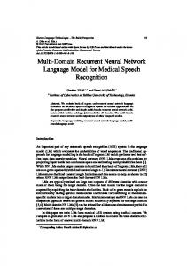
Figure 1. Truncated ESR1 amplifications in two metastatic endometrial carcinomas. Dot plots of ESR1 copy-numbers (y-axis) determined by GeneChip measurements (grey dots) of two metastatic endometrial carcinomas (above: #4, below #2) are shown on the left. Horizontal red lines indicate the segmented copynumber level of chromosomal positions (mega base pairs) on chromosome 6 (x-axis). Position of full length ESR1 (vertical green lines) as well as ESR1 exons 1-4 and 5-8 are indicated as green rectangles (see also Figure 2). Regarding FISH signals of ESR1 (green) and centromere 6 (orange) within a tumor nucleus (blue) are shown on the right. FISH and regarding GeneChip copy-number data of 28 metastatic endometrial carcinoma are summarized in Appendix A. FISH analyses of these tumors are documented in Supplementary Optical Dataset S1.
that alter the hormone-binding domain have been shown to generate hormone independence or resistance to anti-estrogen therapy in breast and endometrial cancers22–31, related genetic alterations could play a role for therapy outcome in primary endometrial carcinoma. Recent studies identified mutations of ESR1 in breast cancer that alter their hormone binding domain coding sequence, to be linked to endocrine therapy resistance in a metastatic setting26–28. One study by Li et al. even demonstrates an ESR1 fusion in endocrine treatment resistant breast cancer, truncating the hormone-binding domain coding exons28, while a later study by Veeraraghavan et al. identified evidence for another type of recurrent ERα -altering gene fusions in this tumor type32. However, structural genetic alterations of ESR1 have not been suggested to play a role in endometrial cancer carcinogenesis. Due to the potential importance of such ESR1 alterations in endometrial cancer, we analyzed an tumor test subset of 29 primary endometrial cancers for somatic gene copy-number alterations (SCNA) and explored The Cancer Genome Atlas (TCGA)33 for concerning SCNA and mRNA expression data of endometrial carcinoma.
Results
Across a cancer study subset of 29 primary endometrial carcinomas that had gone on to metastasize, we characterized the copy-number changes by GeneChips and validated amplifications of ESR1 in these cancers by fluorescence in-situ hybridization (FISH). The Pearson correlation of ESR1 GeneChip copy numbers with FISH determined absolute average ESR1 copy numbers per nucleus and average ESR1 to centromere 6 (CEN6) ratios were r = 0.743 (p