Regular Article
ANALYTICAL AND BIOANALYTICAL CHEMISTRY RESEARCH Published by the Iranian Chemical Society
1 2
3
Anal. Bioanal. Chem. Res., Vol. 4, No. 1, , June 2017.
5
4 7
6 8 9
Immunoassay for Human Chorionic Gonadotropin Based on Glassy Carbon Electrode Modified with an Epitaxial Nanocomposite
10 11
Akram Valipour and Mahmoud Roushani*
12
Department of Chemistry, Ilam University, Ilam, Iran
13
(Received 1 June 2016, Accepted 22 December 2016)
14 15
A highly sensitive electrochemical immunosensor was developed to detect hCG based on immobilization of hCG-antibody (anti-hCG)
16
onto robust nanocomposite containing Gr, Chit, 1-methyl-3-octyl imidazolium tetra fluoro borate ionic liquid (IL) (Gr-IL-Chit). AuNPs
17
were used to immobilize hCG antibody on the modified electrode. The amine groups of the antibody are covalently attached to AuNPs/Gr-
18
IL-Chit nanocomposite. CV, EIS and SEM were employed to characterize the assembly process and the performance of the immunosensor.
19
DPV and EIS studies demonstrated that the formation of antibody-antigen complexes decreased peak current and increased Rct of
20
[Fe(CN)6]3−/4− redox pair at the AuNPs/Gr-IL-Chit/GCE. The optimization of the pH of supporting electrolyte and the incubation time were
21
studied in details. Because of the synergistic effect of IL, Chit and Gr and the unique properties of AuNPs, the obtained immunosensor
22
exhibited a wide linear response to hCG in two ranges from 0.005-1.484 and 1.484-411.28 (mIU ml-1). A relatively low detection limit of
23
0.0016 mIU ml-1 (S/N = 3) was calculated from DPV. Satisfactory results were obtained for determination of hCG in human serum
24
samples.
25 26
Keywords: Electrochemical immunosensor, Human chorionic gonadotropin, Nanocomposite, Impedance spectroscopy
27 28
47
INTRODUCTION
29
Human chorionic gonadotropin (hCG) is a 37 kDa glycoprotein hormone. This is the first glycoprotein 32 produced by trophoblasts of the placenta during pregnancy 33 and is secreted by trophoblastic neoplasms and a variety of 34 non-trophoblastic tumors [1]. The hCG molecule consists of 35 two combined, dissimilar subunits designated alpha and 36 beta. The beta subunit confers biological and 37 immunological specificity to the entire hCG molecule by 38 virtue of its unique amino acid sequence and content. The 39 alpha subunit is essentially identical to the alpha subunit of 40 the pituitary glycoprotein hormones: luteinizing hormone 41 (LH), follicle-stimulating hormone (FSH), and thyroid42 stimulating hormone (TSH) [2,3]. The appearance of hCG 43 in urine or serum soon after conception and its rapid rise in 44 concentration makes it an ideal indicator for the detection 30 31
45 46
*Corresponding author. E-mail:
[email protected]
and confirmation of pregnancy. Thus, exact determination 49 of the concentration of hCG in urine or serum plays an 50 important role in monitoring of trophoblastic diseases in all 51 modern immunological pregnancy tests [4]. Electrochemical 52 immunosensors based on the specificity recognition of 53 antigen and antibody is of great interest in clinical 54 diagnosis. Electrochemical immunosensor is one of the 55 most promising methods for detecting pathogenic biological 56 species of clinical interest, for their low cost, fast response, 57 simplicity, short response time, simple fabrication, small 58 size, high sensitivity and a relatively low detection limit [5]. 59 In order to increase sensitivity and selectivity of 60 electrochemical immunosensing strategies, investigation of 61 new composite materials has attracted widespread attention. 62 Advances in nanotechnology have greatly influenced the 63 field of electrochemical biosensors over the past few years. 64 Much attention has been paid to the development of 65 biocompatible and highly conductive nanomaterials for 66 biosensing and biomedical applications, such as carbon 48
Valipour & Roushani/Anal. Bioanal. Chem. Res., Vol. 4, No. 1, , June 2017. 67
113
68
114
nanotube, gold nanoparticles (AuNPs), graphene (Gr) and quantum dot. Among these nanomaterials, recently, Gr has 71 been utilized in a number of forms for sensor and biosensor 72 applications because of its interesting properties such as, 73 large specific area, good conductivity and biocompatibility 74 [6]. For immobilization of immunoreagent onto the 75 electrode surface an effective and simple immobilization 76 method is very important. Recently, organic-inorganic 77 composite (or hybrid) materials have been one of the key 78 research fields for investigation in today’s material science. 79 They combine the physicochemical attributes of 80 components and improve their features. The use of ionic 81 liquids (ILs) as binders with formation of IL-Gr paste due to 82 the cation-cation-interactions of ILs with Gr can prevent 83 the aggregation of Gr [7]. Chitosan (poly-β-(1-4)-D84 glucosamine), is a polysaccharide derived from 85 deacetylation of chitin. It possesses many advantages such 86 as excellent membrane-forming ability, high permeability 87 towards water, good adhesion, biocompatibility, and high 88 mechanical strength. Also, it has abundant reactive amino 89 and hydroxyl functional groups; so, it has been widely used 90 as an immobilization matrix for biofabrication [8]. Recently, 91 some immunosensors based on various nanoparticles such 92 as AuNPs, Ag nanoparticles and TiO2 nanoparticles, etc., 93 have been reported. AuNPs can firmly adsorb antibody 94 because of their large specific surface areas, good 95 biocompatibility and high surface free energies. 96 Herein, we design an electrochemical immunoassay 97 using AuNPs/Gr-IL-Chit composite modified electrode, 98 which constructs an effective antibody immobilization 99 matrix and makes the immobilized immunocomponents 100 possessing high stability and bioactivity. The performance 101 of the developed hCG immunosensor is superior to that of 102 other methods reported in the literature, especially in 103 comparison with the reported LODs (Table 1). The 104 immunosensor showed high sensitivity, and wide linear 105 range owing to high surface to volume ratio and electronic 106 structure of Gr, large-surface area of AuNPs causing large 107 amounts of anti-hCGs immobilized on the electrode surface 108 and covalent attachment of AuNPs with amin groups of Chit 109 and anti-hCG. The resulting immunosensor is evaluated 110 using Differential pulse voltammetry (DPV) and impedance 111 spectroscopy (EIS), and utilized in the detection of hCG in 112 biological samples. 69
115
70
116 117
MATERIALS AND METHODS Reagents and Apparatus
Anti-hCG, hCG, HAuCl4, sodium citrate (Na3C6H5O7.2H2O), Chit, Gr, Bovine serum albumin (BSA), 120 Progesterone and IL (1-methyl-3-octyl imidazolium tetra 121 fluoro borate) were purchased from Sigma-Aldrich Co. LLC 122 (USA). All other reagents with analytical grade such as 123 glucose, ascorbic acid and NaOH were obtained from 124 Merck or Fluka and used without further purification. All 125 experiments were carried out at room temperature. 126 Phosphate buffer solution (PBS, 0.1 M, pH = 7.4) 3-/4127 containing 2.5 mM [Fe (CN)6] and 0.1 M KCl was used 128 as a working solution. All aqueous solutions were prepared 129 with deionized water. The real samples (human blood 130 serum) were provided by a local clinical laboratory; a stock 131 solution of human blood serum was diluted 50 times with 132 PBS buffer (0.1 M), and then analyzed. 133 Cyclic voltammetry (CV), DPV and EIS experiments 134 were performed with a µ-AUTOLAB electrochemical 135 system type III and FRA board computer controlled 136 Potentiostate/Galvanostate (Eco-Chemie, Switzerland) 137 driven with NOVA software in conjunction with a 138 conventional three electrode system with glassy carbon 139 electrode (GCE) modified and unmodified as the working 140 electrode, a platinum wire as the counter electrode, and an 141 Ag/AgCl (satd 3.0 M KCl) as the reference electrode. The 142 DPV measurements were performed by scanning the 143 potential from -0.1 to 0.7 V with modulation time of 50 ms 144 and modulation amplitude of 25 mV. CV measurements 145 were carried out from -0.2 to 0.6 V as initial and stop 146 potential. EIS analysis was carried out with a bias potential 147 of 0.2 V and a frequency range between 0.1 Hz and 100 148 kHz with signal amplitude of 5 mV. Nanocomposite and 149 AuNPs were characterized by scanning electron microscopy 150 (SEM). SEM images were recorded using a Vega-Tesacn 151 electron microscope. The morphology of the Au 152 nanoparticles was determined by a Hitachi H-800 153 transmission electron microscopy (TEM) at an operating 154 voltage of 200 kV. A Metrohm model 780 pH/mV meters 155 was used to measure the pH. 118 119
156 157 158
Synthesis of Gold Nanoparticle Gold nanoparticles were synthesized according to the
Immunoassay for Human Chorionic Gonadotropin/Anal. Bioanal. Chem. Res., Vol. 4, No. 1, , June 2017. 159
205
160 161
206
Table 1. Comparison of Different Immunosensors for Detection of hCG 207
162
208
Modified Electrode
163 164
209 Method 210
c
Anti-hCG/nano-Au/MB /GCE
165
CV211
Anti-hCG/nano-gold and CS hybrid film/GCE
166 167
AMP
Anti-hCG/GNPsd/pPAe/MWCNTsf/GCE
170
CV
0.2-1000
Ref. -1
[11]
-1
[12]
0.3 (mIU ml ) 0.1 (mIU ml ) 0.3 (mIU ml-1)
[5]
25-400
12 (mIU ml-1)
[13]
0.1-100
0.08 (mIU ml-1)
[14]
0.01-12
7.50 (pg ml-1)
[15]
0.5-40.00
0.034 (ng ml-1)
[16]
1-10, 10-160
Anti-hCG/Pt–Au alloy nanotube array/GCE
AMP215
Anti-hCG/gold nanotubesarray/GCE
AMP
216
171
217
Pt@MSN g/HRPh/Ab 2i/hCG Atigene/Ab1/
172
AMP218
THj/Graphene/GCE
173
219
174
220
Anti hCG/NPGk-Gsl/GCE
175
AMP221
Anti-hCG/Pd@SBA-15/TH/HSO3-GS/GCE
176
AMP222
177
0.01-16.00
223
Anti-hCG/Au@SiC–CS/GCE
DPV
178
-1
8.60 (pg ml )
[17]
-1
0.1-5,5-1000
0.042 (mIU ml )
[18]
0.005-1.484,
0.0016 (mIU ml-1)
This work
224
179
Anti-hCG/AuNPs/Gr-IL-Chit/GCE
225
DPV
180
226
181
227
a
184
1-1000
LODb
214
169
183
m 212 213
168
182
LRa
b
c
1.484-411.28
d
Linear rang. Limit of Detection. Methyleneblue. Gold228nanoparticles. ePoly-(2,6-pyridinediamine). fMultiwalled carbon nanotubes. gMesoporous silica nanoparticles. hHorseradish peroxidase. iAnti body. jThionine. kNanoporous 229 l m gold. Graphene nanosheets. Amperometry. 230
185
231
186
232
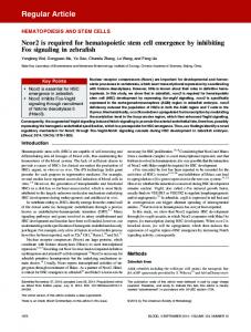
references [9,10]. For a short time, at first, 500 ml HAuCl4 188 0.01% W/V solution was poured into a round-bottom flask 189 equipped with a condenser and was heated and stirred until 190 it reached boiling temperature. While stirring, 7.5 ml 191 sodium citrate 1% was added to the solution. After 30 s, the 192 solution turned into blue and after 70 s it turned into red. 193 Boiling lasted for 10 min. Then, the heating was stopped 194 and the solution was stirred for 15 min. The obtained 195 solution was red and its particles were about 10 nm. After 196 cooling, the solution was kept in refrigerator. The 197 synthesized AuNPs were characterized by TEM (Fig. 1A).
Gr. The nanocomposite modified GCE was fabricated by 234 casting nanocomposite onto the surface of a GCE and 235 letting it stay at room temperature (for 6 h). The GCE 236 modified with Gr-IL-Chit was washed with deionized water 237 and then 8 µl of AuNPs solution was pipetted onto the 238 surface of the modified electrode and after drying, the 239 modified electrode was washed with distilled water. In this 240 time the modified electrode can be used for electrochemical 241 experiments. The composites of Gr with Chit and IL 242 exhibited improved robustness and facilitated 243 immobilization of antibodies. Gr was applied because of its 198 244 particular properties, such as large surface area and high 199 Preparation of Gr -IL- Chit Nanocomposite 245 electrical conductivity. IL was used due to its ability in 200 Modified Electrode 246 preventing the aggregation of Gr and improving properties 201 The GCE was polished with alumina powder, and then 247 of nanocomposite and Chit have been used as 202 washed with deionized water and sonicated in ethanol, 248 immobilization matrices for the immobilization of 203 deionized water, respectively. The nanocomposite was 249 nanoparticle onto the surface of electrodes, since it has 204 prepared by mixing 1 µl of IL, 0.2 mg of Chit and 2 mg of 250 abundant reactive amino and hydroxyl functional groups. 187
233
Valipour & Roushani/Anal. Bioanal. Chem. Res., Vol. 4, No. 1, , June 2017. 251
297
252
298
253
299
254
300
255
301
256
302
257
303
258
304
259
305
260
306
261
307
262
308
263
309
264
310
265
311
266
312
267
313
268
314
269
315
270
316
271
317
272
318
273
319
274
320
275
321
276
322
277
323
278
324
279
325
280
326
281
327 Gr-IL-Chit B) SEM images of AuNPs/Gr-IL-Chit. Fig. 1. A) TEM image of AuNPs A) SEM images of
282
328
283
329
An interaction between AuNPs and amine group of chitosan 285 resulted in a covalent immobilization of antibody molecules 286 on the surface of the electrode. 284
287 288
290
in use.
331 332
Electrochemical Measurements
The formation of antigen and antibody complexes was performed by immersing the BSA/anti-hCG/AuNPs/Gr-IL335 Chit/GCE into hCG solution for 50 min. After the specific 336 reaction of antibody-antigen, the formed antigen-antibody 337 immunocomplex on the electrode surface hindered the 338 electron transfer toward the electrode surface, resulting in a 339 decrease of electrochemical signal. The electrochemical 340 measurements of the modified GCE were performed in 10 3−/4− 341 mM PBS (2.5 mM Fe(CN)6 (the concentrations of 4− 3− 342 Fe(CN)6 and Fe(CN)6 was 2.5 mM) + 0.1 M KCl, pH 333
Fabrication of the Electrochemical Immunosensor
The GCE modified with AuNPs/Gr-IL-Chit was washed with water and immersed in PBS containing anti-hCG (1 mg -1 291 ml ) at 4 °C for 12 h. At last, the resulting electrode was 292 incubated in BSA solution (0.25% w/w) about 1 h in order 293 to block possible remaining active sites and eliminate the 294 risk of non-specific binding. The assemble process steps for 295 the preparation of the immunosensor are shown in (Scheme 296 1). The prepared immunosensor was stored at 4 °C when not 289
330
334
Immunoassay for Human Chorionic Gonadotropin/Anal. Bioanal. Chem. Res., Vol. 4, No. 1, , June 2017. 343
389
344
390
345
391
346
392
347
393
348
394
349
395
350
396
351
397
352
398
353
399
354
400
355
401
356
402
357
403
358
404
359
405
360
406
361
407
362
408
363
409
364
410
365
411
366
412
367
413
368
414
369
415
370
416
371
417
372
418
Scheme 1. The schematic illustration of the 419 stepwise immunosensor fabrication process
373 374 375
420
7.4)
376 377
380
covalent bond between Au and amine groups of the anti423 hCG) and accelerating electron transfer. 422
RESULT AND DISCUSSION
378 379
421
424
Characterization of AuNPs/Gr-IL-Chit Modified GCE
The expected SEM images of GCE modified with, GrIL-Chits and AuNPs/Gr-IL-Chit is shown in (Fig. 1). A 383 direct evidence for the attachment of AuNPs to the Gr-IL384 Chit surface is given by the SEM tests. Compared with Fig. 385 1 C, Fig. 1B clearly shows that the AuNPs have decorated 386 uniformly on the walls of Gr-IL-Chit. Furthermore, this 387 uniform nanostructure provides an efficient electrode 388 surface for loading anti-hCG (through the formation of
425 426
Electrochemical Immunosensor
Characterization
of
In this work Gr-IL-Chit nanocomposite was used for modification of the electrode and AuNPs were used to 429 immobilize anti-hCG because it can be attached to NH2 430 group of anti-hCG from one head, and to NH2 group of 431 Chitosan from another head existing in nanocomposite. 432 Investigation of immunosensor was carried out by CV and 433 EIS spectroscopy. 434 Cyclic voltammetry is an effective technique for probing
381
427
382
428
Valipour & Roushani/Anal. Bioanal. Chem. Res., Vol. 4, No. 1, , June 2017. 435
481
436
482
437
483
438
484
439
485
440
486
441
487
442
488
443
489
444
490
445
491
446
492
447
493
448
494
449
495
450
496
451
497
452
498
453
499
454
500
455
501
456
502
457
503
458
504
459
505
460
506
461
507
462
508
463
509
464
510
465
511
466
512
467
513
468
514
469
515
470
516
471
517
472 473 474 475 476 477 478 479 480
518
Fig. 2. A) Cyclic voltammograms of different electrodes in pH = 7.4 PBS solution containing 2.5 mM 519 3−/4− -1 [Fe(CN)6] + 0.1 M KCl; scan rate, 50 mV 520 s . a) Bare GCE, b) Gr-IL-Chit/GCE, c) AuNPs/ Gr-IL-Chit/GCE, d) Anti-hCG/AuNPs/Gr-IL-Chit/GCE, e) BSA-Anti hCG-AuNPs/Gr-IL521 Chit/GCE. B) Nyquist plots for different electrodes in pH 7.4 PBS solution containing 2.5 mM 522 3−/4− [Fe(CN)6] + 0.1 M KCl. a) Bare GCE, b)523Gr-IL-Chit/GCE, c) AuNPs/Gr-IL-Chit/GCE, d) Anti-hCG/AuNPs/Gr-IL-Chit/GCE e) BSA-Anti hCG-AuNPs/Gr-IL-Chit/GCE. The inset is the 524 equivalent circuit of the immunosensor. 525 526
Immunoassay for Human Chorionic Gonadotropin/Anal. Bioanal. Chem. Res., Vol. 4, No. 1, , June 2017. 527
573
528
574
the feature of the modified electrode surface. The CV was carried out to investigate electrochemical behaviors after 531 each assembly step. The CVs of the different modified 3−/4− 532 electrodes in [Fe(CN)6] solution are presented in Fig. 3-/4533 2A. At the bare GCE, the Fe(CN)6 redox label revealed 534 reversible cyclicvoltammogram (voltammogram‘‘a’’). After 535 modification of GCE with Gr-IL-Chit nanocomposite, the 536 peak current increased greatly due to increase the effective 537 surface area in the presence of Gr (voltammogram‘‘b’’). 538 When AuNPs were immobilized on the electrode surface, 539 the peak current decreased (voltammogram‘‘c’’). The 540 AuNPs with negative charge on the electrode surface repel 4/3541 the negatively charged [Fe(CN)6] anions; so, the response 542 of redox probe was reduced and thereby led to enhanced 543 electron-transfer resistance. 544 When anti-hCG was immobilized on the electrode 545 surface the peak current clearly decreased. This result 546 indicates the immobilization of anti-hCG on the electrode 547 surface reducing the effective surface area and available 548 active sites for electron transfer process 549 (voltammogram‘‘d’’). Peak current decreased in the same 550 way (voltammogram‘‘e’’) after BSA was used to block non551 specific sites. 552 The immunosensor fabrication processes were also 553 characterized by electrochemical impedance spectroscopy 554 (EIS). Figure 2B shows the Nyquist plots during stepwise 555 construction of immunosensor. The impedance spectra 556 include a semicircle portion and a linear portion. The 557 semicircle diameter at higher frequencies corresponds to the 558 electron-transfer resistance (Rct) which controls the electron 559 transfer kinetics of the redox probe at the electrode 560 interface. These results indicate very low resistance of 561 nanocomposite modified electrode for redox probe. In this 562 graph, we see that the EIS of the bare GCE displays a small 563 semicircle at high frequencies and linear part at low 564 frequencies (curve a, Rct = 429 Ω). After the bare electrode 565 was modified with Gr-IL-Chit composite film, the 566 resistance for the redox probe decreased (curve b, Rct = 78.2 567 Ω), indicating that Gr is an excellent electric conducting 568 material accelerating the electron transfer. After dropping 569 AuNPs on the surface of electrode, Rct increased because of 570 its negative charge surface (curve c, Rct = 146.2 Ω). 571 Subsequently, when the anti-hCG was placed on the surface 572 of AuNPs, Rct increased dramatically (curve d, Rct = 333.5
Ω). This indicates that the antibody is immobilized on the electrode surface and further prevents the redox probe to the 577 electrode surface. After incubating of immonosensor in 578 BSA solution Rct increased further more in the same way 579 (curve e, Rct = 427.3 Ω). 580 The CVs of the obtained immunosensor in PBS at 581 different scan rates were investigated. Useful information 582 involving electrochemical mechanism can be acquired from 583 this investigation. As shown in Fig. 3, both anodic peak 584 currents (Ipa) and cathodic peak currents (Ipc) increased with 585 the increase of scan rate. In addition, the peak currents of 586 the immunosensor were directly proportional to the square 587 root of the scan rate (insert in Fig. 3), suggesting that the 588 electrode reaction is a diffusion-controlled electrochemical 589 process.
529
575
530
576
590 591
Optimization of Experimental Conditions
pH, incubation time in the hCG solution and also urea solution showed an important influence on the analytical 594 performance of hCG immunosensor. In what follows, we 595 investigated the importance of these factors. 592 593
596 597
pH Optimizing
Since activities of biomolecules are pH dependent, we 599 investigated the effect of this factor on the immunosensor 600 response. The reduction peak current increased with 601 increasing pH from 5 to 7.4 and then decreased as pH 602 increased further. Therefore, a pH = 7.4 of the working 603 buffer was applied for further experiments. This result is 604 accordance with the fact that biological agents are more 605 effective in an environment similar to that of the human 606 body. The results are indicated in Fig. 4A. 598
607 608
Optimization of Incubation Time
An important parameter for immuno-complex formation between antibody and antigen is the incubation time. The 611 effect of incubation time on the immunoreaction is shown in 612 Fig. 4, curves (B) and (C) showed respectively the change 613 of current response with the increase in incubation time in 614 hCG solution and urea solution. Based on the study of the 615 incubation time, we found that current response of the -1 616 immunosensor for 0.07 mIU ml hCG decreased with the 617 increase of incubation time before 50 min and leveled off 618 after 50 min. With the increase of hCG concentration, 609 610
Valipour & Roushani/Anal. Bioanal. Chem. Res., Vol. 4, No. 1, , June 2017. 619
665
620
666
621
667
622
668
623
669
624
670
625
671
626
672
627
673
628
674
629
675
630
676
631
677
632
678
633
679
634
680
635
681
636
682
637
683
638
684
639
685
640
686
641 642 643
687
Fig. 3. CV studies of BSA/anti-hCG/Au/Gr-IL-Chit/GCE immunosensor at different scan rates (from a to f): 688 20, 40, 60, 80, 100 and 120 mV s-1 in 2.5 mM Fe(CN)6 3−/4−, 0.1 M KCl + 0.1 M PBS solution (pH = 689 7.4.), inset shows the plot of peak currents vs. v1/2.
644
690
645
691
current response decreased because of the formed hCG/anti 647 hCG complex acting as inert block layer and hindered the 648 transfer of electrons toward the surface of the modified 649 electrode. In addition, we examined incubation time in urea 650 solution (4 M) and we found that increasing of incubation 651 time in urea to 20 min caused an increase in current 652 response (For breaking the link between hCG and anti-hCG) 653 and for upper time, it remained stable. 646
654 655
Calibration Curve of Immunosensor
Step by step, the standard solution of hCG at a known 657 concentration was added into the incubation solution, and 658 under optimal conditions the DPV of the immunosensor in 659 the presence of different concentrations of hCG was 660 performed in 0.1 M PBS solution containing 2.5 mM Fe 3−/4− 661 (CN)6 . The hCG in solution interaction with anti-hCG 662 immobilized on the surface of biosensor. The DPV peak 663 current decreases with increasing the hCG concentration in 664 the incubation solution (Fig. 5). The decrease of peak 656
current was proportional to the concentration of hCG in two linear ranges from 0.005 to 1.484 mIU ml-1, with a linear -1 694 slope of 1.1331 µA/( mIU ml ) and correlation coefficient -1 695 of 0.999 (n = 3) and 1.484 to 411.28 mIU ml with a linear -1 696 slope of 0.0037 µA/( mIU ml ) and correlation coefficient 697 of 0.992 (n = 8), inset in Fig. 5. A relatively low detection -1 698 limit of 0.0016 mIU ml (S/N = 3) was calculated by DPV. 699 In this study, we also investigated the effect of hCG 700 concentration on the immunosensor with EIS (LOD = -1 701 0.0009 mIU ml ). As illustrated in Fig. 6, increasing of 702 hCG concentration leads to an increase in semi-circle 703 diameter which indicates hCG interaction with anti-hCG. 704 With increasing in concentration, more hCG will be placed 705 on the surface, and this will consequently increase the 706 resistance (Rct). In other words, the impedance is influenced 707 with the changes in amount, growth and morphological 708 behavior of adherent substance. Therefore, this method can 709 be proposed as an efficient way to monitor the formation of 710 antigen-antibody interaction. 692 693
Immunoassay for Human Chorionic Gonadotropin/Anal. Bioanal. Chem. Res., Vol. 4, No. 1, , June 2017. 711
757
712
758
713
759
714
760
715
761
716
762
717
763
718
764
719
765
720
766
721
767
722
768
723
769
724
770
725
771
726
772
727
773
728
774
729
775
730
776
731
777
732
778
733
779
734
780
735
781
736
782
737 738 739 740
783
Fig. 4. Investigated three different parameters on the response of the immunosensor incubated with 0.07 mIU ml-1 hCG 784
in 2.5 mM Fe(CN)64−/3− solution containing 0.1 M KCl. A) Influence of the pH of the solution B) Dependence of 785 peak currents on incubation time of immunoreaction786and C) incubation time in Urea solution (4.0 M).
741
787
742
788
The analytical performance of the proposed 744 immunoassay has been compared with other sensors 745 reported previously (summarized in Table 1). The low 746 detection limit and wide linear response to hCG for the 747 proposed immunosensor is satisfactory. Acceptable factors 748 affecting detection limit, sensitivity and linear range of the 749 proposed immunosensor are: 1) High surface to volume 750 ratio and electronic structure of Gr and unique features of IL 751 such as cation-intraction with Gr, 2) Large surface area of 752 AuNPs causes the large amounts of anti-hCG immobilized 753 on the electrode surface, 3) Covalent attachment of AuNPs 754 with amin groups of Chit and anti-hCG lead to the more 755 stability and repeatability in comparison with the adsorption 756 method. 743
789
Selectivity and Regeneration of Immunosensor
Selectivity is assessed by evaluating the effect of 791 compounds present in the test solution, other than the target 792 analyte (hCG), on the analytical response of a sensor device. 793 Within the investigation of the selectivity of the 794 immunosensors, it is found that the response of the sensor to -1 795 hCG (0.6 mIU ml ) is significantly larger than its signal to 796 some potentially interfering species such as ascorbic acid 797 (AA), glucose (Gl), BSA, CEA and progesterone (PR) (60 -1 798 mIU ml ) (Fig. 7). 799 An important factor in studies of antibody-antigen is 800 regeneration of immunosensor. After using the 801 immunosensor to detect hCG, the modified electrode was 802 treated with 4 M urea solution for 20 min to dissociate 790
Valipour & Roushani/Anal. Bioanal. Chem. Res., Vol. 4, No. 1, , June 2017. 803
849
804
850
805
851
806
852
807
853
808
854
809
855
810
856
811
857
812
858
813
859
814
860
815
861
816
862
817
863
818
864
819
865
820
866 Fig. 5. Calibration curve for hCG determination (mIU ml-1), from up to down: 0, 0.005, 0.0742, 1.484, 2.5, 867 14.84, 63.6, 127.2, 190.8, 262.88, 318 and 411.28 in 0.1 M pH = 7.4 PBS containing 2.5 mM [Fe(CN)6]3-/4- and 0.1 M KCl. Pulse width: 0.05868S; amplitude: 0.025 V, Inset: Calibration plots of the reduction current vs. concentration of hCG. 869
821 822 823 824
870
825
871
826
872
827
873
828
874
829
875
830
876
831
877
832
878
833
879
834
880
835
881
836
882
837
883
838
884
839
885
840
886 Fig. 6. Nyquist plots of the BSA/anti-hCG/AuNPs/Gr-IL-Chit/GCE Immonosensor, obtained in PBS pH = 7.4 3-/4887 containing 2.5 mM of Fe(CN)6 previously incubated in increasing concentrations of hCG, from a to 888 k (0, 0.005, 0.0742, 1.484, 2.5, 14.84, 63.6, 127.2, 190.8, 262.88 and 318 mIU ml-1). Inset shows the 889 plot of ∆Rct vs. log[hCG].
841 842 843 844
890
845
891
Antigen-antibody linkage. The regenerated immunosensors were used to detect the same hCG concentration. The 848 reproducibility of the electrode regeneration was examined
by repeating the use-regeneration cycle six times. The RSD of 1.95% was obtained at the hCG concentration of 0.07 -1 894 mIU ml . This result indicates that the immunosensor
846
892
847
893
Immunoassay for Human Chorionic Gonadotropin/Anal. Bioanal. Chem. Res., Vol. 4, No. 1, , June 2017. 895
941
896
942
897
943
898
944
899
945
900
946
901
947
902
948
903
949
904
950
905
951
906
952
907
953
908
954
909
955
910
956 Fig. 7. Amperometric response of the immunosensor to interferents: glucose (Glu), carcinoembryonic antigen 957 (CEA), ascorbic acid (AA), BSA and Progestron (Pr) (60 mIU ml-1).
911 912
958
913 914
959
-1
960 Table 2. hCG Detection (mIU ml ) in Serum Samples with a Developed Immunosensor
915
961
916
962
Sample
hCG in serum
917
0 A
0
920
0
922
19
964 965
19.5 ± 0.2
102.6
96
95.5 ± 0.1
99.4
328 967
326.5 ± 0.2
99.7
968
923
969
924
B
0
0.35 970
0.30 ± 0.02
95
0
971 1.5
1.45 ± 0.11
95
3.4 ± 0.1
90
1.55 ± 0.12
99.22
2.28 ± 0.14
99.56
926
972
0
927 928
3.5 973 974
929
0.53
930 931
Recovery (%)
966
921
925
Founded
963
918 919
Added
975
1
976
C
932
0.29
2
977 978
933
979
85% of the initial response. The repeatability was evaluated via assaying 10 mIU ml-1 hCG solution using the obtained 936 Stability and Repeatability of Immunosensor 982 immunosensor for 5 times and the relative standard 937 Stability of immunosensors is an important factor that 983 deviation (RSD) was 1.22%. This result indicates 938 shows their performance. In order to investigate this factor, 984 satisfactory stability and repeatability of the immunosensor. -1 939 during three weeks, the DPV of 10 mIU ml hCG solution 985 Intra-electrode and inter-electrode coefficients of variation 940 was recorded, in every two days. After 21 days, it retained 986 were used to investigate the reproducibility. The relative 934 935
could be regenerated and used again.
980 981
Valipour & Roushani/Anal. Bioanal. Chem. Res., Vol. 4, No. 1, , June 2017. 987
1032
988
1033
standard deviation (RSD) of reproducibility was 3.3% for 5 measurements of hCG with the different immunosensors 991 (Inter-electrode). Also for five times, the reproducibility of 992 the immunosensor was estimated by determining hCG with 993 one immunosensor (Intra-electrode) and RSD was 994 calculated at 4.1%. 989
1034
990
1035
995 996
[1]
1037
[2] 1039 [3] 1038
1040
Real Sample Analysis
Recovery experiments were performed by standard 998 addition methods in spiked blood serum to evaluate the 999 feasibility of the proposed immunosensor for real sample 1000 analysis. To this end, the human serum samples were 1001 ordered from a local clinical laboratory and subjected to an 1002 ultrafiltration by loading into a centrifugal filtration tube at 1003 3000 rpm (30 min). Afterwards, the serum samples were 1004 diluted with PBS (0.1 M) and different concentrations of 1005 hCG were spiked to these samples. Finally, the serum hCG 1006 concentrations were detected with the calibration curve of 1007 the hCG immunosensors. The experimental results are 1008 shown in Table 2. They show an acceptable recovery. These 1009 results indicate that the system presented in this study can 1010 be valid for the analysis of hCG in biological fluids. 997
1011 1012
1036
REFERENCES
1041 1042
[4]
1043 1044 1045
[5]
1046 1047
[6]
1048 1049 1050
[7]
1051 1052
[8]
1053
[9] 1055 [10] 1054
1056
CONCLUSIONS
1057
[11]
1013 1058
In this work, we have designed a novel electrochemical 1015 immunosensor based on AuNPs/Gr-IL-Chit composite 1016 modified electrode for a rapid and sensitive immunoassay 1017 The results indicated that the AuNPs/Gr-IL-Chit based 1018 immunosensor can be used for detection of hCG at low 1019 detection limit. The detection limit, calculated from DPV, 1020 was 0.0016 mIU ml-1 based on (S/N = 3). The 1021 immunosensor exhibited a wide linear response to hCG in -1 1022 two ranges from 0.005-1.484 mIU ml and 1.484-411.28 -1 1023 mIU ml . In addition, the proposed sensitive approach holds 1024 great promise for the extended application in the fields of 1025 clinical diagnosis, bioaffinity assays and environmental 1026 monitoring. 1014
1059
[12]
1060 1061 1062
[13]
1063 1064
[14]
1065 1066 1067
[15]
1068 1069 1070
[16]
1071 1027 1028
ACKNOWLEDGMENTS
1072
[17]
1073 1029
The financial suppot of Ilam University is gratefully 1031 acknowledged.
1074
1030
1075
[18]
T. Fang, Y. Feng, J. Huangxian, Biosens. Bioelectron. 22 (2007) 2945. F.J. Morgan, Endocrinology 88 (1971) 1045. O.M. Bahl, R.B. Carlsen, R. Bellisario, N. Swaminathan, Biochem. Biophys. Res. Commun. 48 (1972) 416. H. Lund, S.B. Torsetnes, E. Paus, K. Nustad, L. Reubsaet, T.G. Halvorsen, J. Protein. Res. 8 (2009) 5241. J. Wang, R. Yuan, Y. Chai, S. Cao, S. Guan, P. Fu, Li. Min, Biochem. Eng. J. 51 (2010) 95. Q. Wei, R. Li, B. Du, D. Wu, Y. Han, Y. Cai, Y. Zhao, X. Xin, H. Li, M. Yang, Sens. Actuators, B 153 (2011) 256. J.Y. Sun, K.J. Huang, S.F. Zhao, Y. Fan, Z.W. Wu, Bioelectrochemistry 82 (2011) 125. K.J. Huang, D.J. Niu, W.Z. Xie, W. Wang, Anal. Chim. Acta 659 (2010) 102. G. Frens, Nature Phys. Sci. 241 (1973) 20. W.S. Sutherland, J.D. Winefordner, J. Colloid Interface Sci. 148 (1992) 129. R. Chai, R. Yuan, Y.Q. Chai, C.F. Ou, S.R. Cao, X.L. Li, Talanta 74 (2008) 1330. G.M. Yang, Y.B. Chang, H. Yang, L. Tan, Z.S. Wu, X.X. Lu, Y.H. Yang, Anal. Chim. Acta 644 (2009) 72. M. Tao, X.F. Li, Z.S. Wu, M Wang, H. Mei, Y.H. Yang, Clin. Chim. Acta 412 (2011) 550. G.M. Yang, X.Y. Yang, C.Y. Yang, Y.H. Yang, Colloids Surf. A: Physico Chem. Eng. Asp, 389 (2011) 195. Q. Wei, R. Li, B. Du, D. Wu, Y. Han, Y. Cai, Y. Zhao, X. Xin, H. Li, M. Yang, Sens. Actuators, B 153 (2011) 256. R. Li, D. Wu, H. Li, C. Xu, H. Wang, Y. Zhao, Y. Cai, Q. Wei, B. Du, Anal. Biochem. 414 (2011) 196. D. Wu, Y. Zhang, L. Shi, Y. Cai, H. Ma, B. Du, Q. Wei, Electroanalysis 25 (2013) 427. L. Yang, H. Zhao, Sh. Fan, Sh. Deng, Q. Lv, J. Lin, C.P. Li, Biosens. Bioelectron. 57 (2014) 1996.