Carcinogenesis vol.19 no.3 pp.485–491, 1998
Relationships between the synthesis of N-nitrosodimethylamine and immune responses to chronic infection with the carcinogenic parasite, Opisthorchis viverrini, in men
Soisungwan Satarug1,11, Melissa R.Haswell-Elkins1, Paiboon Sithithaworn2, Helmut Bartsch6, Hiroshi Ohshima7, Mitsuhiro Tsuda8, Pisaln Mairiang3, Eimorn Mairiang4, Puangrat Yongvanit5, Hiroyasu Esumi9 and David B.Elkins10 1National
Research Centre for Environmental Toxicology, 39 Kessels Road, Coopers Plains, Queensland 4108, Australia, 2Departments of Parasitology, Medicine, 4Radiology, 5Biochemistry, Faculty of Medicine, Khon Kaen University, Khon Kaen, Thailand, 6Division of Toxicology and Cancer Risk Factors, German Cancer Research Center, Im Neuenheimer Feld 280, D-69120 Heidelberg, Germany, 7Unit of Endogenous Risk Factors, International Agency for Research on Cancer, 150 cours Albert-Thomas, 69372 Lyon Cedex 08, France, 8Division of Pharmacology, National Institute of Health Sciences, 1-18-1 Kamiyoga, Setagaya-ku, Tokyo 158, Japan, 9Division of Investigative Treatment, National Cancer Centre Research Institute East, 6-5-1 Kashiwanoha, Chiba, Japan and 10Tropical Health Program, Queensland Institute of Medical Research, Bancroft Centre, Herston Road, Brisbane, Australia
3Internal
11To
whom correspondence should be addressed. Email:
[email protected]
This study investigated the relationship between immune responses to infection with the liver fluke, Opisthorchis viverrini, and the synthesis of the carcinogen, N-nitrosodimethylamine (NDMA) in humans. It also examined associations between synthesis of nitric oxide (NO) and nitrosation of amines, in vivo. Antibody and T cell responses to fluke antigens and post-alcohol urinary NDMA excretion were assessed among three groups of 40–50 men with no, moderate and heavy liver fluke infection. Markers of NO synthesis (nitrate, nitrite) and nitrosation (nitrosamino acids) were also measured in biological fluids. Assessments were carried out under controlled conditions which minimised intake of exogenous nitrate and nitrite and were carried out at two time points, namely before and 4 months after elimination of the infection with praziquantel treatment. No statistically significant variation was observed in the amount of NDMA excreted between the 3 groups. However, during active infection, a strong negative association was observed between in vitro lymphoproliferative responses to some liver fluke antigens and NDMA excretion. After treatment this association was reduced. Multivariate statistical models revealed a highly significant relationship between NDMA levels and urinary nitrate, stimulation indices for two T cell responses to two parasite antigens (MW 37 kDa and 110 kDa) and gall bladder dimensions. NDMA levels after treatment were best described by the ratio between parasite-specific IgG2 and IgE, background levels of T cell proliferation, a urinary marker of nitrosation (N-nitrosothioproline) and usual level of alcohol consumption. These results suggest that individual background immunologic activity, parasite-specific responses and/or parasite products and NO synthesis are important determinants *Abbreviations: NO, nitric oxide; NDMA, N-nitrosodimethylamine; cpm, counts per minute; SI, stimulation index; GC/TEA, gas chromatography/ thermal energy analysis; DMA, dimethylamine. © Oxford University Press
of endogenous generation of nitrosamines in O.viverriniinfected humans.
Introduction The synthesis of nitric oxide (NO*) by inducible nitric oxide synthase in hepatocytes and inflammatory cells is now considered one of the most important host defence mechanisms against some murine bacterial, protozoal and helminth infections (1–3). This pathway is not without cost, however, since NO is mutagenic, cytotoxic and, through nitrosation of available amines, can give rise to potentially carcinogenic Nnitroso compounds (4–9). While infection with susceptible organisms may be cleared by nitric oxide-generating immune responses, those which are refractory may elicit long-standing exposure to tissue damaging agents generated by the host’s own immune response. Heterogeneity in immune responses is a characteristic of parasitic infection, and tissue damage is often immunopathological, rather than directly parasite related (10–12). Thus pathology only partly depends on worm burden, since individuals with equivalent burdens may vary in the degree of tissue damage as a result of different immune responses mounted against parasite antigens. Opisthorchis viverrini-associated cholangiocarcinoma is increasingly being studied as a model system to understand the role of immune responses to chronic infection in carcinogenesis (13–22). These parasites establish long-standing infection within the intrahepatic bile ducts and do not appear to be susceptible to host inflammatory responses. The risk of cholangiocarcinoma is strongly associated with intensity of infection (21). In Northeast Thailand where ~30% of the population is infected, the age-standardised incidence of this normally rare cancer is extremely high (84.6 and 36.8 per 100 000 males and females, respectively) (22). Liver fluke infected hamsters express nitric oxide synthase in macrophages, mast cells and eosinophils in the inflamed biliary tissue (17) and develop bile duct cancer after exposure to normally sub-carcinogenic doses of N-nitrosodimethylamine (NDMA) (18–20). Human studies have detected higher levels of urinary nitrate, nitrosoproline and nitrosothioproline among infected people (13–16). However, it remains unclear whether NDMA is also endogenously generated and is the relevant mutagen in human cholangiocarcinoma. Due to its extensive and rapid metabolism in the body, NDMA is not normally excreted in the urine (23,24). However, Spiegelhalder and Preussman demonstrated that NDMA can be detected after consumption of a small amount of alcohol (‘ethanol effect’) which inhibits first-pass (hepatic) clearance of NDMA via cytochrome P450-mediated catabolism (25). This method has received less attention than proline loading to estimate the potential formation of mutagenic and carcinogenic nitrosamines by endogenous nitrosation. Although increased levels of circulating and excreted NDMA occurs in some diseases, e.g. renal failure, bacterial urinary-tract and Schisto485
S.Satarug et al.
soma haematobium infections (26–30), the determinants of NDMA synthesis and excretion during infection and inflammation have not been well studied. The amount of NDMA excreted in the urine after alcohol consumption may be a better indicator of extra-gastric nitrosation than the amount of nitrosoproline in the urine after dosing with proline. Nitrosoproline is produced predominantly in the stomach with relatively small amounts in the tissue, while a comparatively small amount of NDMA is formed in the stomach (6,31–34). Because of the basicity of dimethylamine (DMA, pKb 5 3.25), DMA is mostly present in a chemical form that is not reactive with nitrosating agents at low pH (32–34). Thus only small amounts of NDMA (,30 µg or ,0.5 µmol) are formed in the human stomach, even when large amounts of precursors are co-ingested (33,35). In contrast, at neutral pH of the tissue, DMA readily reacts with nitrosating agents, such as N2O3/N2O4, which are generated during biosynthesis of NO from arginine (6). This study was undertaken to quantify the link between the synthesis of NO and NDMA in human subjects using alcohol to allow urinary excretion of NDMA. It also tested the hypothesis that immune responses to Opisthorchis viverrini antigens are important determinants of endogenous synthesis of NDMA during infection. Materials and methods The rationale, study design and methodology have been described in detail (13) and were approved by official Ethics committees in Thailand and Australia. Briefly, a total of 148 men, aged 30–50 years, were selected from groups stratified on the basis of egg counts from a cross-sectional study of cholangiocarcinoma in villages in Northeast Thailand (21). The range of egg counts of the three intensity groups were: 0 (uninfected), 1000–6000 (moderate infection) and over 6000 (heavy infection) eggs per g of faeces. Those who wished to participate provided written informed consent and stayed together for 1 week in 13 groups of 12–15 men of mixed infection status, eating only the supervised low nitrate diet provided and refraining from smoking. Subjects were examined by abdominal ultrasound as previously described to measure gall bladder dimensions and detect hepatobiliary and renal abnormalities. After initial sample collection, the men were treated with the anthelmintic drug, praziquantel (40 mg/kg), and 134 (90%) attended identical post-treatment assessments 4 months later. Potentially important determinants of NDMA generation (Table I) were measured as described (13,16). These included markers of NO synthesis (salivary nitrite, plasma and urinary nitrate) and endogenous nitrosation (salivary thiocyanate, nitrosoproline, nitrosothioproline), urinary tract infection and hepatitis B surface antigen carriage. Usual smoking frequency (cigarettes per day) and alcohol intake (frequency per month3amount usually consumed) were estimated by interview. Humoral and cellular immune responses to somatic O.viverrini antigens were measured by ELISA and T cell Western blotting, respectively. Relative levels of parasite-specific antibody isotypes were determined by ELISA essentially as described (36) with the additional application of isotype-specific monoclonal antibodies (Dako-Patts, Copenhagen) (for IgG1, IgG2, IgG4, IgE) and avidin-biotin amplification procedures (ABC complex, Dako-Patts) (for IgG4, IgE). T cell proliferation assays were performed as previously described (37) on 97 participants by isolating mononuclear cells on Ficoll-Hypaque (Pharmacia, Sweden), washing and culturing them for 7 days at 106 cells per ml with one of 28 mol. wt fractions of somatic O.viverrini antigen (manuscript in prep.). The antigens were separated on SDS-polyacrylamide gel electrophoresis, transferred to nitrocellulose paper by electroblotting, cut into individual pieces, then each fraction was solubilized with DMSO. [3H]Thymidine was added for the final 12 h and counts per min (cpm) on harvested filters were determined by scintillation counting. Stimulation indices (SI) were calculated as cpm of [3H]thymidine incorporated by cells exposed to the fraction with solubilized fluke antigens on nitrocellulose paper divided by cpm of [3H]thymidine incorporated by cells exposed to solubilized nitrocellulose paper alone. On the final day all subjects voluntarily consumed 355 ml of beer containing 5% ethanol. Urine samples potentially containing NDMA were collected for the next 8 h and stored at 4°C until the collection was complete. The total
486
volume was then recorded, the samples made alkaline (pH .10) by addition of NaOH and then stored at –20°C prior to analysis. NDMA concentrations in urine were determined by gas chromatography/thermal energy analysis (GC/TEA) following extraction with dichloromethane using the Extube column (ToxElute) as described by Spiegelhalder and Preussman (25). NNitrosomethylpentylamine (0.4 µg, MPNA) was added to the urine prior to extraction to serve as an internal standard. The amount of NDMA was calculated based on the ratio between NDMA and MPNA obtained from the GC/TEA of a mixture of various standard volatile nitrosamines including the NDMA and MPNA. The detection limit was 0.05 µg per litre. The data were analysed statistically using SPSS for Windows (Version 6.1) after transformation to normality. For NDMA, this required taking the square root of µg of NDMA excreted in 8 h, while most other variables were logtransformed. This also stabilized variances across the infection and T cell response groups. Differences in excreted levels of NDMA between the three intensity groups before and after praziquantel treatment were assessed by one way analysis of variance (ANOVA), while correlations between NDMA and individual continuous variables are described by Pearson’s (r) correlation coefficient. Variables were then tested in stepwise multiple regression models to determine which factors were the strongest determinants of NDMA excretion. The adjusted beta, T value and significance of each variable are given to indicate the relative strengths of association. Missing values in some measurements account for the differences in sample size.
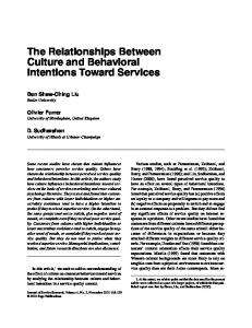
Results Average levels of various biochemical measurements, including markers of NO generation and nitrosation, among the three liver fluke intensity groups for the sample population are shown in Table I. Significantly higher levels of plasma nitrate, salivary nitrite, nitrosoproline and nitrosothioproline were observed among the infected groups prior to treatment, as previously reported in these studies (13,16). NDMA was detected in urine of 70% and 66% of men before and after treatment, and very high amounts (maximum 7.73 µg) were recorded in some individuals. The mean backtransformed square root amounts of NDMA excreted within 8 h after alcohol intake were 0.271 µg (standard deviation: 0.264, 95% CI: 0.192–0.364) among 152 men tested before treatment and 0.326 µg (standard deviation: 0.310, 95% CI: 0.223–0.447) among 126 men tested after praziquantel treatment. Differences between pre- and post-treatment levels are not statistically significant (paired t-test, t 5 0.91, P . 0.05). Figure 1 shows the square root transformed average amounts of NDMA (µg) excreted in 8 h post-alcohol urine which did not differ significantly between the three infection groups before (ANOVA; F 5 0.79, df 2,149, P . 0.05) or after treatment (F 5 0.27, df 2,123, P . 0.05). A significant correlation (r 5 0.30, P , 0.001) existed between the amount of NDMA excretion within individuals before and after treatment. No association was observed between NDMA excretion and urinary tract infection or hepatitis B infection. Table II records correlations observed between urinary excreted amounts of NDMA and other measured variables. NDMA amounts were only weakly associated with plasma nitrate, urinary nitrate and salivary nitrite in samples collected on preceding days (ranged from 0.10–0.19), but were more strongly associated with urinary nitrate levels in the 8 h postalcohol urine sample (r 5 0.30, P , 0.001). Urinary Nnitrosoproline, N-nitrosothioproline and creatinine levels during the previous 24 h with proline loading correlated more strongly with post-treatment than with pre-treatment NDMA excretion levels. Salivary thiocyanate concentration (a catalyst of gastric nitrosation) was not associated with urinary NDMA at either time point. A number of immunological responses correlated with NDMA excretion (Table II). While parasite-specific IgE levels
Immune responses and N-nitrosodimethylamine excretion
Table I. Characterization of the sample group with mean values for various demographic and biochemical measurements potentially related to NDMA synthesis Intensity group Variable
Pre-treatment
Egg count category (eggs/g faeces) Mean EPG Mean worm burden Number of men Age (years) Body wt (kg) Alcohol index Creatinine (mg/kg/day)
1 0 0 0 42 39.7 57.5 0.35 25.6
Markers of nitric oxide Plasma nitrate (µM) Urinary nitrate (µmol/kg/day) Salivary nitrite (µM)
38 14.8 74.1
Markers of nitrosation Salivary thiocyanate (µM) Nitrosamino acidsa: NPRO (µg/day) NTPRO (µg/day)
1046 0.87 1.19
Post-treatment 2 1000–6000 2720 6.9 56 38 57.4 0.45 24.8 46.4 17.0 124.6 1083 1.38 1.79
3 .6000 8129 125.9 54 37.6 56.9 0.43 25.9 46.0* 16.8 109.2* 962 1.16* 1.961
1 0 0 –
2 0 0 –
3 0 0 –
38 39 57.4 0.35 25.9
48 37.8 57.4 0.5 24.8
40 37 57.7 0.55 25.3
41.5 17.4 66.2
43.1 16.2 88.4
46 16.2 91.6
767 1.39 2.03
945 1.48 2.50
848 1.90 2.76
aNPRO
and NTPRO were measured in 24 h urine after dosing with 300 mg of proline. Symbols placed under intensity group 3 indicate statistically significant variation in the log-transformed means of the three intensity groups determined by one-way ANOVA at the level of: 1P ,0.05; *P ,0.01.
Fig. 1. Average amounts of NDMA excreted in post-alcohol urine stratified by liver fluke infection status before and after elimination of the infection. Square root transformed mean and SE values of µg NDMA excreted in the 8 h post-alcohol urine specimens collected from individuals with no infection (leftmost boxes), moderate infection (1000–6000 eggs per g of faeces) and heavy infection (.6000 eggs per g) with the liver fluke, Opisthorchis viverrini. Pre-treatment indicates the results of assessments done prior to elimination of the infection, while post-treatment results are from urines collected from the same subjects 4 months later. Sample sizes for these groups are: uninfected (n 5 42, 38), moderate infection (n 5 56, 48) and heavy infection (n 5 54, 40), for pre- and post-treatment assessments, respectively. Differences in NDMA level between intensity groups were not statistically significant before or after treatment (P .0.05).
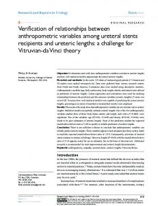
showed negative trends, IgG2 levels correlated positively with both pre- and post-treatment NDMA levels. Background levels of T cells proliferation (log-transformed cpm of [3H]thymidine incorporated by cells exposed to media alone) correlated positively with NDMA levels, both before and after treatment. In contrast, the stimulation indices of most T cell proliferative responses to different O.viverrini antigen fractions showed significant negative associations with NDMA excretion, with the 37 kDa antigen fraction showing the strongest associations (r 5 –0.43, P , 0.001, Table II). As shown in Figure 2, men whose T cells exhibited marked suppression of proliferation in vitro (indicated by an SI of ,0.50) following exposure to
the 37 kDa antigen excreted the highest levels of NDMA during infection; these trends are highly significant (ANOVA, F 5 7.31, df 2,95, P 5 0.001). These relationships between excreted NDMA and T cell proliferative responses were not apparent in post treatment assessment (ANOVA, F 5 1.87, df 2, 75, P 5 0.16). T cell responses (assessed either pre- or post-treatment) showed weaker correlations with post-treatment NDMA excretion, although six remained statistically significant (r 5 –0.20 to –0.27, Table II). Plasma and urinary nitrate, urinary Nnitrosoproline and N-nitrosothioproline excreted after proline loading showed significant associations with post-treatment NDMA, as did the ratio between parasite-specific IgG2 and IgE. Multivariate regression models of NDMA excretion Pretreatment. A systematically developed multivariate model incorporating five variables accounted for 52.8% of the total variation in pre-treatment NDMA excretion levels (Table III). During infection, T cell proliferative responses to two liver fluke antigen fractions (37 kDa, 110 kDa), parasite-specific IgG2 in serum (positive association), nitrate in 8 h post-alcohol urine (positive) and gall bladder length explained highly significant variation in NDMA. Although background levels of T cell proliferation were shown to be strongly correlated with NDMA levels in univariate tests, this variation was explained by its close correlation with nitrate levels (r 5 0.39, P , 0.001). Therefore it did not enter the multivariate model after entry of nitrate, suggesting that its influence on NDMA synthesis may be through NO synthesis. Post-treatment. Modelling the post-treatment data was successful in explaining 17.5% of the variation in NDMA excretion levels. Four variables were incorporated, namely the ratio between parasite-specific IgG2 and IgE (positive association), the amount of N-nitrosothioproline excreted during a previous day with proline loading (positive), 8 h nitrate levels and usual alcohol intake. The immunoglobulin ratio controlled for all 487
S.Satarug et al.
NDMA (µg/kg body wt/8 h post-alcohol urine)
variation associated with T cell proliferation to the crude parasite extract and the T cell responses to six antigens found to maintain statistical significance in univariate correlations (Table II).
Variable
Pre-treatment
Post-treatment
Discussion
Body wt Age Smoking index Alcohol index Intensity group
–0.09 –0.12 0.05 –0.07 0.10
–0.11 –0.11 0.04 0.24** 0.01
Our data provide strong evidence of a link between individual immunological activity and endogenous synthesis of nitrosamines that could play a role as aetiological agents in fluke-associated cholangiocarcinoma in humans. In this study, heterogeneity in lymphoproliferative responses after in vitro exposure to parasite antigens was strongly linked to urinary
Table II. Correlation coefficients (Pearson’s r) describing the single variable relationships between NDMA excretion and other measured variables
Pre-treatment ultrasound observations Ratio GB length/width –0.15* Portal radicle echoes 0.01 Biochemical measurements Markers of nitric oxide Nitrate in plasma Nitrate in 24 h urine Nitrate in post-alcohol urine Salivary nitrite Markers of nitrosation Salivary thiocyanate N-nitrosothioproline N-nitrosoproline Urinary creatinine (24 h)
0.06 –0.12
0.18* 0.19* 0.30***
0.20* 0.20* 0.17*
0.10
0.12
0.04 0.06 0.11 0.00
0.07 0.22* 0.18* 0.20
Parasite-specific immunological measurements Serum IgG2 0.17* Serum IgE –0.02 Ratio IgG2/IgE 0.14
0.13 –0.15 0.23**
T cell proliferation to: Background (cpm) Purified protein derivative 37 kDa parasite protein 145 kDa parasite protein 28 kDa parasite protein 22 kDa parasite protein 110 kDa parasite protein
0.33*** –0.08 –0.23* –0.16 –0.02 –0.21* –0.08
0.30*** –0.18* –0.43*** –0.40*** –0.40*** –0.41*** –0.09
Fig. 2. Average amounts of NDMA excreted in post-alcohol urine stratified by stimulation index (SI) of T-cell response following in vitro exposure to a 37 kDa parasite antigen. Square root transformed mean and SE values of µg NDMA excreted in the 8 h post-alcohol urine specimens collected from individuals who showed a suppressed proliferation (stimulation index , 0.5) relative to the control cells exposed to media alone (the leftmost bars). The middle boxes represent mean values of NDMA excretion of those with a SI between 0.5 and 1.5, while the rightmost boxes represent those with a SI over 1.5. Sample sizes for these groups are: suppressed group (n 5 24,17), SI 0.5–1.5 (n 5 43,34) and SI .1.5 (n 5 31,25), for pre- and post-treatment assessments, respectively. Differences in NDMA levels between these immune response groups were highly significant in the presence of infection (P ,0.001).
*P ,0.05; **P ,0.01; ***P ,0.001.
Table III. Multiple regression model of NDMA excretion before and after treatment with praziquantel including T cell immune responses. Pre-treatment
Post-treatment
Variable
Adjusted beta
t-test
P value
Adjusted beta
t-test
P value
37 kDa response 110 kDa response Urinary nitrate NTPRO Creatinine Alcohol index
–0.603* 0.285* 0.475* –0.107 –0.056 –0.128
–5.6 2.7 6.4 –1.1 –0.6 –1.5
.000 .010 .000 .296 .521 .142
–0.139 0.077 0.180* 0.267* 0.111 0.229*
–1.1 0.6 2.0 2.9 1.2 2.5
.262 .533 .051 .004 .224 .013
Gall bladder: Length/width Length Width
–0.185* –0.153 0.164
–2.4 –2.1 1.9
.016 .041 .058
0.013 0.053 0.036
0.1 0.5 0.3
.890 .620 .762
Parasite-specific: IgG2/IgE IgG2 IgE No infection Moderate flukes Heavy flukes
0.140 0.167* –0.043 –0.060 –0.015 0.014
1.8 2.2 –0.6 –0.8 –0.2 0.2
.075 .033 .571 .455 .830 .869
0.274* 0.143 –0.171 0.034 0.096 –0.170
3.0 1.6 –1.9 0.4 1.0 –1.8
.003 .119 .064 .709 .305 .06
TOTAL
r 5 0.744; adjusted r2 5 0.528; df 5,87
Stars in the adjusted beta column indicate those variables which incorporated into the final model.
488
r 5 0.455; adjusted r2 5 0.175; df 4,97
Immune responses and N-nitrosodimethylamine excretion
NDMA excretion prior to, but not after, eliminating infection with praziquantel. We offer two possible explanations of this finding. In one immunological pathway, CD41and CD81T cells produce γ-interferon and tumour necrosis factor which stimulate hepatocytes, macrophages (at least in the murine system) and other inflammatory cells to express NO synthase which generates NO via oxidation of arginine (38,39). Nitrogen oxides (N2O3/N2O4) that are formed from NO after oxidation can be incorporated in surrounding secondary and tertiary amines, most likely dimethylamine (DMA) which is most abundant in the tissue fluid. As a result, NDMA and other nitrosamines are formed (6–9). Excessive NO is in turn a potent inhibitor of cell proliferation and can mediate immunosuppression (reduced in vitro responsiveness to mitogens) in experimental malaria and trypanosome infections (40–42). The marked negative association between amounts of NDMA and in vitro T cell proliferation following exposure to some parasite antigen suggests that people whose immunological response to liver fluke infection is characterized by suppressed proliferation in vitro, particularly after exposure to a 37 kDa parasite antigen, may be those whose T cells respond to fluke antigens with a so-called T helper 1 phenotype (gamma interferon and tumour necrosis factor). These cytokines stimulate macrophages (and monocytes) to generate NO which may inhibit cell proliferation in a dose-dependent manner under in vitro conditions (43). Positive relationship between NDMA and parasite-specific IgG2 may reflect the tendency for B cells exposed to gamma interferon to undergo an isotype switch to produce IgG2, while IgE switching may be favoured by cytokines of the ‘T helper 2’ phenotype (39,44). An alternative explanation of the findings is that the negative associations with parasite antigens reflect a combination of γinterferon release (by both spontaneously activated T cells and those activated by the parasite antigen), together with a parasite-derived molecule with LPS-like activity which could itself provide the ‘second signal’ for NO generation. Studies by Sternberg and Mabbott (45) have suggested a similar interaction between γ-interferon and blood stream-forms of Trypanosoma brucei. If this is correct, both specific and nonspecific responses would contribute to the relationship between in vivo NDMA synthesis and the measured in vitro proliferative responses. This may explain why associations are observed in both infected and non-infected individuals. At present it is unclear whether we have demonstrated a close relationship between types of T cell responses to specific parasite antigens and NDMA synthesis or perhaps more broadly, the ability of a parasite to direct the type of immune response which is mounted against it. By possessing and secreting molecules with LPS-like activity, parasite could play a direct role in the enhancement of NO generation. In the case of the liver fluke, NO could be advantageous to the parasite by mediating vasodilation and increased leakiness of the biliary epithelium allowing serum components to enter the bile for consumption by the fluke (46). Liu et al. (47) detected increased generation of NDMA in woodchucks infected with woodchuck hepatitis B virus, while others have directly measured NDMA in urine from humans with bacterial and Schistosoma hematobium infections of the urinary tract (28–30). The elevation in urinary tract infection results in part from bacterial enzymatic catalysis of nitrosation. In our study, we were not able to demonstrate a significant increase in NDMA excretion associated with infection status.
This may be because the observed trends are complicated by small amounts of NDMA (2% of total body burden) excreted in post-alcohol urine samples and by dual effects of liver fluke infection on the synthesis and degradation of NDMA. In both experimental animals and in humans, liver fluke infection induces the expression of CYP2A6 which converts NDMA to genotoxic metabolites (48,49). Concurrent increases in synthesis of NDMA and expression of CYP2A6 would result in more DNA damages although no net increase in the amount of NDMA was observed. It is possible that increased generation of NDMA occurs in all infected individuals, but is obscured by increased activity of CYP 2A6 which catalyses the conversion of NDMA to metabolites not excreted in urine. The increase may become detectable above background only where the generation of NDMA is markedly increased, e.g. among those with particular types of immune responses to infection. Previous studies using the ‘ethanol effect’ to inhibit first pass clearance of NDMA by P450-mediated catabolism in healthy people detected increased urinary NDMA following the consumption of nitrate and amine precursors (25,35). Under these conditions, the amount of NDMA excreted was very small and probably generated intragastrically. In contrast, the present study involved tight control of dietary and tobaccorelated intake of nitrate, nitrite and nitrosamines, which would have reduced gastric production and facilitated detection of NDMA coupled to NO synthesis and inflammation. The strong correlations between NDMA levels, liver fluke-specific and background T cell responses and urinary nitrate excretion indicate that nitrosating agents are produced along with the synthesis of NO at the site of infection (13,16). This study provides the first evidence in humans of the close link between endogenous nitrate and NDMA synthesis. In summary, it is likely that nitrosamines generated in situ play a very important role in inflammation-associated carcinogenesis, especially when their production is both chronic and located in close proximity to cells containing P450 enzymes which can metabolize the nitrosamine to DNA methylating agents. NDMA produced during inflammatory responses to liver fluke antigens may diffuse to the biliary epithelium and become activated by hepatic CYP 2E1 and possibly CYP 2A6. The nearby biliary epithelium may be highly susceptible to malignant transformation due to chronic proliferation which is another pathologic response to infection. This combination of events could explain the very high risk of cholangiocarcinoma associated with liver fluke infection. Acknowledgements We would like to thank Drs T.Sugimura, R.Maclennan, A.Green, and especially M.Good and P.O’Rourke for advice, the community leaders and study subjects for enthusiastic cooperation and Auomporn Mongwongkolroj and Rati Boonmak, whose excellent work made this project possible. We also gratefully acknowledge the Foundation for Promotion of Cancer Research (Tokyo) for providing a Foreign Research Scholarship for Dr S.Satarug and the International Agency for Research on Cancer for supporting laboratory expenses for the measurements of NDMA. This study was supported by the National Health and Medical Research Council of Australia.
References 1. James,S.L. and Hibbs,J.B.(1990) The role of nitrogen oxides as effector molecules of parasite killing. Parasitol. Today, 6, 303–305. 2. Boockvar,K.S., Granger,D.L., Poston.R.M., Maybodi,M., Washington,M.K., Hibbs,J.B. and Kurlander,R.L. (1994) Nitric oxide during murine listeriosis is protective. Infect. Immun., 62, 1089–1100.
489
S.Satarug et al. 3. Liew,F.Y., Millot,S., Parkinson,C., Palmer,R.M.J. and Moncada,S. (1990) Macrophage killing of Leishmania parasite in vivo is mediated by nitric oxide from L-arginine. J. Immunol., 144, 4794–4797. 4. Wink,D.A., Kasprzak,K.S., Maragos,C.M., Elespuru,R.K., Misra,M., Dunams,T.M., Cebula,T.A., Koch,W.H., Andrews,A.W., Allen,J.S. and Keefer,L.K. (1991) DNA deaminating ability and genotoxicity of nitric oxide and its progenitors. Science, 254, 1001–1003. 5. Nguyen,T., Brunson,D., Crespi,C.L., Penman,B.W., Wishnok,J.S. and Tannenbaum,S.R.(1992) DNA damage and mutation in human cells exposed to nitric oxide in vitro. Proc. Natl Acad. Sci. USA, 89, 3030–3034. 6. Miwa,M., Stuehr,D.J., Marletta,M.A., Wishnok,J.S. and Tannenbaum,S.R. (1987) Nitrosation of amines by stimulated macrophages. Carcinogenesis, 8, 955–958. 7. Leaf,C.D., Wishnok,J.S. and Tannenbaum,S.R. (1989). Mechanisms of endogenous nitrosation. Cancer Surv., 8, 323–334. 8. Leaf,C.D., Wishnok,J.S. and Tannenbaum,S.R. (1991) Endogenous incorporation of nitric oxide from L-arginine into N-nitrosomorpholine stimulated by Escherichia coli lipopolysaccharide in the rat. Carcinogenesis, 12, 537–539. 9. Ohshima,H., Tsuda,M., Adachi,H., Ogura,T., Sugimura,T. and Esumi,H. (1991) L- arginine-dependent formation of N-nitrosamines by the cytosol of macrophages activated with lipopolysaccharide and interferon. Carcinogenesis, 12, 1217–1220. 10. Rocklin,R.S., Brown,A.P., Warren,K.S., Pelley,R.P., Houba,V., Siongok,T.K.A., Ouma,J., Sturrock,R.F. and Butterworth,A.E. (1980) Factors that modify the cellular immune response in patients infected with Schistosoma mansoni. J Immunol., 125, 1916–1923. 11. Clark,I.A. (1987) Cell-mediated immunity in protection and pathology of malaria. Parasitol. Today, 3, 300–305. 12. Flavell,D.J. and Flavell,S.U. (1986) Opisthorchis viverrini: pathogenesis of infection in immunodeprived hamsters. Parasite Immunol., 8, 455–466. 13. Haswell-Elkins,M.R., Satarug,S., Tsuda,M., Mairiang,E., Esumi,H., Sithithaworn,P., Mairiang,P., Saitoh,M., Yongvanit,P. and Elkins,D.B. (1994) Liver fluke infection and cholangiocarcinoma: model of endogenous nitric oxide and extra gastric nitrosation in human carcinogenesis. Mutat. Res., 305, 241–252. 14. Srianujata,S., Tonbuth,S., Bunyaratvej,S., Valyasevi,A., Promvanit,T.N. and Chaivatsagul,W. (1987) High urinary excretion of nitrate and Nnitrosoproline in opisthorchiasis subjects. In Bartsch,H., O’Neill,I.K. and Schulte-Hermann,R. (eds) The Relevance of N-Nitrosocompounds in Human Cancer: Exposures and Mechanisms. IARC Scientific Pubications, Lyon, no. 84, pp. 544–546. 15. Srivatanakul,P., Ohshima,H., Khlat,M., Parkin,M., Sukarayodhin,S., Brouet,I. and Bartsch,H. (1991) Endogenous nitrosamines and liver fluke as risk factors for cholangiocarcinoma in Thailand. Int. J. Cancer, 48, 821–825. 16. Satarug,S., Haswell-Elkins,M.R., Tsuda,M., Mairiang,P., Sithithaworn,P., Mairiang,E., Esumi,H., Sukprasert,S., Yongvanit,P. and Elkins,D.B. (1996) Thiocyanate-independent nitrosation in humans with carcinogenic parasite infection. Carcinogenesis, 17, 1075–1081. 17. Ohshima,H., Bandaletova,T.Y., Brouet,I., Bartsch,H., Kirby,G., Ogunbiyi,F., Vatanasapt,V. and Pipitgool,V. (1994) Increased nitrosamine and nitrate biosynthesis mediated by nitric oxide synthase induced in hamsters infected with liver fluke (Opisthorchis viverrini). Carcinogenesis, 15, 271–275. 18. Flavell,D.J. and Lucas,S.B. (1983) Promotion of N-nitrosodimethylamineinitiated bile duct carcinogenesis in the hamster by the human liver fluke, Opisthorchis viverrini. Carcinogenesis, 4, 927–930. 19. Thamavit,W., Bhamarapravati,N., Sahaphong,S., Vajrasthira,S. and Angsubhakorn,S. (1978) Effects of dimethylnitrosamine on induction of cholangiocarcinoma in Opisthorchis viverrini infected Syrian golden hamsters. Cancer Res., 38, 4634–4639. 20. Lee,J.H., Yang,H.M., Bak,U.B. and Rim,H.J. (1994) Promoting role of Clonorchis sinensis infection on induction of cholangiocarcinoma during two-step carcinogenesis. Korean J. Parasitol., 32, 13–18. 21. Haswell-Elkins,M.R., Mairiang,E., Mairiang,P., Chaiyakum,J., Chamadol,N., Loapaiboon,V., Sithithaworn,P. and Elkins,D.B. (1994) Cross-sectional studies of Opisthorchis viverrini infection and cholangiocarcinoma in communities within a high-risk area in Northeast Thailand. Int. J. Cancer, 58, 505–509. 22. Working Group (1994) IARC Monographs on the Evaluation of carcinogenic risks to humans: Volume 61. Schistosomes, liver flukes and Helicobacter pylori. IARC, Lyon, pp. 121–175. 23. Gombar,C.T., Polypiw,H.M. and Harrington,W. (1987) Pharmacokinetics of N-nitrosodimethylamine in beagles. Cancer Res., 47, 343–347.
490
24. Gombar,C.T., Harrington,G.W., Polypiw,H.M., Bevill,R.F., Thurmon,J.C., Nelson,D.R. and Magee,P.N. (1988) Pharmacokinetics of Nnitrosodimethylamine in swine. Carcinogenesis, 9, 1351–1354. 25. Spiegelhalder,B. and Preussman,R. (1985) In vivo nitrosation of amidopyrine in humans: use of ‘ethanol effect’ for biological monitoring of N-nitrosodimethylamine in urine. Carcinogenesis, 6, 545–548. 26. Dunn,S.R., Simenhoff,M.L., Lele,P.S., Goyal,S., Pensabene,J.W. and Fiddler,W.(1990) N-nitrosodimethylamine blood levels in patients with chronic renal failure: modulation of levels by ethanol and ascorbic acid. J. Natl. Cancer Inst., 82, 783–787. 27. Tricker,A.R., Stickler,D.J., Chawla,J.C. and Preussman,R. (1991) Increased urinary nitrosamine excretion in paraplegic patients. Carcinogenesis, 12, 943–946. 28. Ohshima,H., Calmels,S., Pignatelli,B., Vincent,P. and Bartsch,H. (1987) N-nitrosamine formation in urinary tract infections. In Bartsch,H., O’Neill,I.K. and Schulte-Hermann,R. (eds) The Relevance of NNitrosocompounds in Human Cancer: Exposure and Mechanisms. IARC Scientific Publications, Lyon, no. 84, pp. 292–296. 29. Tricker,A.R., Mostafa,M.H., Spiegelhalder,B. and Preussmann, R. (1989) Urinary excretion of nitrate, nitrite and N-nitroso compounds in schistosomiasis and bilharzia bladder cancer patients. Carcinogenesis, 10, 547–552. 30. Bartsch,H., Ohshima,H., Shuker,D.E.G., Pignatelli,B. and Calmels,S. (1990) Exposure of humans to endogenous N-nitroso compounds: implications in cancer etiology. Mutat. Res., 238, 255–267. 31. Ohshima,H. and Bartsch,H. (1981) Quantitative estimation of endogenous nitrosation in humans by monitoring N-nitrosoproline excretion in the urine. Cancer Res., 41, 3658–3666. 32. Challis,B.C. and Kyrtopoulos,S.A. (1977) Rapid formation of carcinogenic N-nitrosamines in aqueous alkaline solutions. Br. J. Cancer, 35, 693–696. 33. Mirvish,S.S. (1970) Kinetics of dimethylamine nitrosation in relation to nitrosamine carcinogenesis, J. Natl Cancer Inst., 44, 633–639. 34. Licht,W.R. and Dean,W.M. (1988) Theoretical model for predicting rates of nitrosamine and nitrosamide formation in the human stomach. Carcinogenesis, 9, 2227–2237. 35. Milligan,J.R., Zucker,P.F., Swann,P.F. and Archer,M.C. (1986) Alcohol consumption does not lead to urinary excretion of N-nitrosodimethylamine in the fasting human. Carcinogenesis, 7, 1401–1402. 36. Haswell-Elkins,M.R., Sithithaworn,P., Mairiang,E., Elkins,D.B., Wongratanacheewin,S., Kaewkes,S. and Mairiang,P. (1991) Immune responsiveness and parasite-specific antibody levels in human hepatobiliary disease associated with Opisthorchis viverrini infection. Clin. Exp. Immunol., 84, 213–218. 37. Lamb,J.R., O’Hehir,R.E. and Young,D.B. (1988) The use of nitrocellulose immunoblots for the analysis of antigen recognition by T lymphocytes. J. Immunol. Method, 110, 1–10. 38. Salgame,P., Abrams,J.S., Clayberger,C., Goldstein,H., Convit,J., Modlin,R.L. and Bloom,B.R. (1991) Differing lymphokine profiles of functional subsets of human CD4 and CD8 T cells clones. Science, 254, 279–282. 39. Scott,P. and Kaufmann,S.H.E. (1991) The role of T-cell subsets and cytokines in the regulation of infection. Immunol. Today, 12, 346–348. 40. Wei,X.-Q., Charles,I.G., Smith,A., Ure,J., Feng,G.-J., Huang,F.-P., Xu,D., Muller,W., Moncada,S. and Liew,F.Y. (1995) Altered immune responses in mice lacking inducible nitric oxide synthase. Nature, 375, 408–411. 41. Taylor-Robinson,A.W., Liew,F.Y., Severn,A., Xu,D., McSorley,S.J., Garside,P., Padron,J. and Phillips,R.S. (1994) Regulation of the immune response by nitric oxide differentially produced by T helper type 1 and T helper type 2 cells. Eur. J. Immunol., 24, 980–984. 42. Rockett,K.A., Awburn,M.M., Rockett,E.J., Cowden,W.B. and Clark,I.A. (1994) Possible role of nitric oxide in malarial immunosuppression. Parasite Immunol., 16, 243–249. 43. Liew,F.Y. (1995) Regulation of lymphocyte functions by nitric oxide. Curr. Opin. Immunol., 7, 396–399. 44. King,C.L., Gallin,J.I., Malech,H.L., Abramson,S.L. and Nutman,T.B. (1995) Regulation of immunoglobulin production in hyper immunoglobulin E recurrent infection syndrome by interferon gamma. Proc. Natl Acad. Sci. USA, 86, 10085–10089. 45. Sternberg,M.J. and Mabbott,N.A. (1996) Nitric oxide-mediated suppression of T cell responses during Trypanosoma brucei infection: soluble trypanosome products and interferon-gamma are synergistic inducers of nitric oxide synthase. Eur. J. Immunol., 26, 539–543. 46. Sithithaworn,P., Haswell-Elkins,M.R., Mairiang,P., Satarug,S., Mairiang,E., Vatanasapt,V. and Elkins,D.B. (1994). Parasite-associated morbidity: liver fluke infection and bile duct cancer in Northeast Thailand. Int. J. Parasitol., 24, 833–843.
Immune responses and N-nitrosodimethylamine excretion 47. Liu,R.H., Baldwin,B., Tennant,B.C. and Hotchkiss,J.H. (1991) Elevated formation of N-nitrosodimethylamine in woodchucks (Marmota monax) associated with chronic woodchuck hepatitis virus infection. Cancer Res., 51, 3925–3929. 48. Kirby,G.M., Pelkonen,P., Vatanasapt,V., Camus,A.-M., Wild,C.P. and Lang,M.A. (1994) Association of liver fluke (Opisthorchis viverrini) infestation with increased expression of cytochrome P450 and carcinogen metabolism in male hamster liver. Mol. Carcinogenesis, 11, 81–89. 49. Satarug,S., Lang,M.A., Yongvanit,P., Sithithaworn,P., Mairiang,E., Mairiang,P., Pelkonen,P., Bartsch,H. and Haswell-Elkins,M.R. (1996) Induction of cytochrome P450 2A6 expression in humans by the carcinogenic parasite infection, opisthorchiasis viverrini. Cancer Epidemiol. Biomarkers Prev., 5, 795–800. Received on February 6, 1997; revised on September 2, 1997; accepted on October 31, 1997
491