Resting State EEG-Based Biometrics for Individual Identification Using Convolutional Neural Networks Lan Ma, James W. Minett, Thierry Blu, Fellow, IEEE and William S-Y. Wang Abstract— Biometrics is a growing field, which permits identification of individuals by means of unique physical features. Electroencephalography (EEG)-based biometrics utilizes the small intra-personal differences and large inter-personal differences between individuals’ brainwave patterns. In the past, such methods have used features derived from manually-designed procedures for this purpose. Another possibility is to use convolutional neural networks (CNN) to automatically extract an individual’s best and most unique neural features and conduct classification, using EEG data derived from both Resting State with Open Eyes (REO) and Resting State with Closed Eyes (REC). Results indicate that this CNN-based joint-optimized EEG-based Biometric System yields a high degree of accuracy of identification (88%) for 10-class classification. Furthermore, rich inter-personal difference can be found using a very low frequency band (0-2Hz). Additionally, results suggest that the temporal portions over which subjects can be individualized is less than 200 ms.
I. INTRODUCTION Biometrics is a method used to identify individuals for purposes of surveillance or security. It utilizes specific and unique individual human characteristics, scanning and ”matching” these characteristics against a database to provide positive identification. Common and widespread applications of biometrics include fingerprints, retinal scans, and voice or facial recognition [1]. These methods are utilized by various organizations including police departments, banks, and other secure facilities (such as high-security government installations), and provide varying degrees of accuracy and precision. In the modern era, however, some unique and modern methods of biometrics may become necessary. One possible method is to use electroencephalography (EEG), which identifies unique electrical brainwave patterns non-invasively measured along the scalp. EEG possesses many special properties that would make it useful in this context, such as a high time resolution which opens a window to see the dynamics of the brain [2]. Previous studies have observed that EEG provides important information about differences between individuals, as pertains to the anatomical and functional traits of their brains [3]. An individual person’s EEG, furthermore, is both stable and specific [4]. That is to say, EEG provides small intra-personal differentiation and large inter-personal differentiation, which is why it is ideal for biometrics. L. Ma, J. Minett, T. Blu, and W. Wang are with Department of Electronic Engineering, The Chinese University of Hong Kong, Hong Kong, China. ∗ Corresponding author: Lan Ma (
[email protected])
978-1-4244-9270-1/15/$31.00 ©2015 IEEE
With the right application, this technology can provide a kind of ”brain fingerprint”, impossible to fake or replicate, thus ensuring the highest level of biometric security [5]. Furthermore, the EEG might be adapted to detect high levels of distress in the subject, ensuring a higher level of security where necessary (removing the possibility of access to the secure area in the case of, e.g., a hostage situation). EEG-based biometric systems can be basically organized into two different states: task-related state and resting state. In this paper we are interested in using resting state EEG. There are two reasons for this. First, evidence indicates that electrical activities’ resting state organizes and coordinates neuronal functions [6]. Second, certain tasks cannot be performed by certain group of people, e.g., Attention Deficit Disorder (ADD) or handicapped patients. Resting state EEG includes Resting-state EEG with open eyes (REO) and Resting-state EEG with closed eyes (REC). In this paper, we will consider both conditions. The common procedure of EEG-based biometrics involves data collection, preprocessing feature extraction, and pattern recognition [7]. However, resting state EEG lacks a taskrelated feature, thus making it difficult to manually design the best feature to extract. In this paper, we use convolutional neural networks (CNN) for the purpose of automatically extracting features and conducting classification [8].The whole procedure is then joint-optimized based on gradient descent. The paper is organized as follows: The first part consists of an introduction to biometrics. The second part gives a general overview of EEG-Based Biometrics, including the criteria, the underlying basis, and the framework. The third part, ”Materials and Methods”, describes the subjects and the experiment, as well as the topology of the CNN. Finally, the results, discussion and conclusion are detailed in Sections 4 and 5. II. EEG-BASED BIOMETRICS A. Criteria of Biometric Systems Biometric systems must fit several criteria, as follows: Universality (presence of the identifying feature in every user accessing the system); Uniqueness (distinctiveness of the identifying feature among all users); Collectability (data can be collected rapidly); Stability (the identifying feature is consistent); etc. [9].
2848
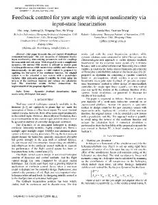
Fig. 2. Common framework of the EEG-based Biometric Systems (a) and the framework of our joint-optimized CNN-based EEG Biometric Systems (b).
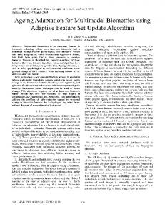
Fig. 1. Two subjects’ three 10-Hz spectrum-topographies under REO and REC conditions. This illustrates small intra-personal differences and large inter-personal differences. Each spectrum-topography is analyzed in 10-second duration.
B. The Base for EEG-Based Biometrics The base for EEG-Based Biometrics is based on two things: the small intra-personal differences (differences within an individual’s brain) and large inter-personal differences (differences between one individual’s brain and another’s). As an illustration of this, Fig.1 shows three 10-Hz spectrum-topographies taken from two subjects under REO and REC conditions. Each of the six spectrum-topographies is analyzed for duration of 10 seconds. From this graph we can see that, under the REO condition, the distribution of the spectrum-topography at a certain frequency is relatively stable over time in individual subjects. However, between the two subjects there is distinction. Under the REC condition, both subjects have large amplitude of energy around the occipital region at 10Hz. However, we can still see the pattern of each subject is stable, while between subjects they are different. This figure is only for illustration. For resting state EEG, we will avoid designing hand-craft features in favor of automatic extraction and classification by the system. C. The Framework of EEG Based Biometric Systems The framework of the commonly-used EEG-based Biometric Systems and the framework of the CNN-based jointoptimized EEG-based Biometric Systems which we propose are shown in Fig.2. Instead of manually designing the feature, we let the CNN extract the most discriminant feature though the optimization and propagation steps. III. METHODS AND MATERIALS A. Data Set In this study, EEG signals came from a publicly available dataset [10], [11] . EEG recordings were originally acquired using the 64-channel BCI2000 system with a sampling rate of 160 Hz. Our analysis was performed on 10 subjects under both REO and REC conditions using 55 seconds for each single condition in time. The 55-second dataset was divided into 55 1-second sub-datasets; 50 of these sub-datasets were used for training the networks and 5 of them for testing.
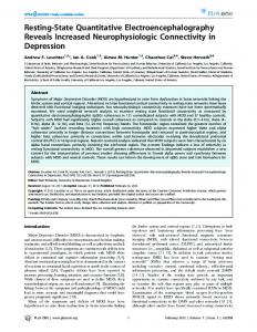
Thus, for each condition (REO, REC, and REO + REC), there were 500 training samples and 50 testing samples in total. Before entering the CNN, each subjects EEG signals were normalized. The input matrix of CNN takes this form: Nelec ∗ Nt , where Nelec is the number of the electrodes (64), and Nt is the number of time points for each sample, which is Nt = T ime ∗ sampling rate. In our case, Nt is 160. B. Convolutional Neural Network (CNN) CNN is a neural network which consists of a multilayer perceptron (MLP), and which possesses a special topology containing multiple hidden layers. It is used for feature extraction and classification, and takes its inspiration from biological systems, in which cells are sensitive to a small sub-region of the input space. Its most successful early application has been for recognition of speech and handwritten characters. Raw information is preserved as input without pre-processing procedures, apart from scaling and centering the input vector. This models advantages are most apparent when the input data contains inner structure (i.e., for images), permitting discovery of invariant features. However, the networks topology setting is empirical and requires repeated validation [12]. In 2011, Cecotti et.al. used CNN on EEG data for P300 detection with the application of braincomputer interface, and obtained a high degree of accuracy [13]. C. Network Topology Our CNN models topology is shown in Fig.3. It accomplishes the tasks of both feature extraction and classification. This CNN has five layers, including two convolutional layers, two pooling layers and one fully connected layer [14]: - The input data matrix has a structure of 64 ∗ 160 (as described above). The convolutional layer convolves this input data matrix via six 5 ∗ 5 filters and outputs the filtered data map. (The structures of these filters are learned by back propagation during the training procedure.) - The filtered data maps are then entered into the averagepooling layer, which divides the input-filtered data maps into sets of 2 ∗ 2 rectangles. For each of these sub-regions, the average-pooling layer outputs the average value. The filtered data maps are then down-sampled and feature maps
2849
TABLE I (10-C LASS ) I NDIVIDUAL I DENTIFICATION U SING R ESTING S TATE EEG UNDER D IFFERENT C ONDITIONS Individual Identification (10-class)
Accuracy
REO
REC
REO+REC
88%
86%
82%
TABLE II (10-C LASS ) I NDIVIDUAL I DENTIFICATION U SING BAND - PASS F ILTERED R ESTING S TATE EEG UNDER D IFFERENT C ONDITIONS Fig. 3.
The topology of five-layer CNN.
are obtained. During this step the computational complexity for upper layers is reduced. - The second convolutional layer uses the feature maps as input data. Through this and the second average-pooling layer, the first two steps are repeated. As a result, the features become more abstract and the computational complexity is further reduced. - Finally, the features obtained by the second averagepooling layer are transferred to the fully connected layer. Ultimate classification is based on these features. Softmax function is used as the activation function. The objective function is mean squared error (MSE).
IV. RESULTS AND DISCUSSION
REO
REC
0Hz-2Hz
68%
64%
0Hz-4Hz
72%
64%
0Hz-6Hz
72%
70%
0Hz-8Hz
74%
72%
0Hz-10Hz
74%
74%
0Hz-20Hz
78%
76%
0Hz-30Hz
82%
78%
0Hz-40Hz
84%
80%
0Hz-50Hz
84%
82%
0Hz-60Hz
84%
86%
0Hz-70Hz
88%
86%
0Hz-80Hz
88%
86%
C. The Accuracy of the Divided Temporal-Structure Data Set
A. The Accuracy of the Full Data Set The classification results are shown in Table I. Accuracy rate is the percentage of test set samples that are correctly classified by the CNN. Of the individuals whose data were input, the highest rate of identification accuracy was derived from REO data. Generally, REO data yielded a higher rate of accuracy than REC data. There was no significant difference among these three dataset about accuracy. The accuracy reached by REO+REC data shows that it is possible to use REO and REC data in tandem; integration of this data might suggest the CNN can find the underline structure of the data. B. The Accuracy of the Filtered Data Set Additionally, the REO and REC datasets were filtered into different frequency bands to evaluate which frequency band is important for identification. The REO and REC datasets were filtered in the following ways (classification results shown in Table II): The frequency bands started from a very low range (0-2 Hz) and become progressively wider, ultimately reaching 080Hz. The CNN trained by the narrow-band EEG data would be used for the initial setup of the wide-band data’s network. The purpose was to avoid the two networks falling into different local minimums. Generally speaking, as the upper limit of the frequency band went wider, the accuracy increased. Another important point which should be noted is that the very low-frequency band (0-2Hz) is highly informative.
Before, we used a 1-s time period as the time base to do the identification, and obtained good classification results. In this section, we would like to find the minimum temporal length of EEG signal which is still able to preserve the identification information. We divided each 1-s REO EEG signal into various smaller portions (from 6.25ms-1000ms) and then performed random permutations. Because the sampling rate is 160, the minimum length is 6.25ms. Using this procedure, the temporal structure of the original EEG signal is damaged to varying degrees. The classification results are shown in Table III. From the table we can see that, as the duration of divided 1-s EEG portions became longer, the accuracy increased. At the point that we maintained a 62.5ms temporal structure, the classification accuracy already reached 76%. The results may suggest that the temporal portions over which subjects can be individualized is less than 200 ms.
2850
TABLE III (10-C LASS ) I NDIVIDUAL I DENTIFICATION U SING D IVIDED T EMPORAL -S TRUCTURE DATA S ET
REO
REO
6.25ms
12.5ms
25ms
50ms
62.5ms
34%
44%
54%
64%
76%
100ms
200ms
500ms
800ms
1000ms
78%
88%
88%
88%
88%
V. CONCLUSIONS The proposed method, based on resting state EEG using CNN, may represent an appropriate technique to develop EEG-based biometric systems which supply good classification performance. In the future, this CNN-based system should be tested on a larger group of subjects, providing further confirmation of the robustness and realworld applicability of the system. This form of biometrics, in addition to providing a higher level of security, would have other useful applications, e.g., in the case of a disabled individual unable to use fingerprints, retinal scans, or other such methods. This could have farreaching applications to various fields, particularly those of security and law enforcement. R EFERENCES [1] Jain A K, Ross A, Prabhakar S. An introduction to biometric recognition[J]. Circuits and Systems for Video Technology, IEEE Transactions on, 2004, 14(1): 4-20. [2] Nunez P L. Toward a quantitative description of large-scale neocortical dynamic function and EEG[J]. Behavioral and Brain Sciences, 2000, 23(03): 371-398. [3] Berkhout J, Walter D O. Temporal stability and individual differences in the human EEG: an analysis of variance of spectral values[J]. Biomedical Engineering, IEEE Transactions on, 1968 (3): 165-168. [4] Van Dis H, Corner M, Dapper R, et al. Individual differences in the human electroencephalogram during quiet wakefulness[J]. Electroencephalography and clinical neurophysiology, 1979, 47(1): 87-94. [5] Pham T, Ma W, Tran D, et al. A study on the feasibility of using eeg signals for authentication purpose[C]//Neural Information Processing. Springer Berlin Heidelberg, 2013: 562-569. [6] Salinas E, Sejnowski T J. Correlated neuronal activity and the flow of neural information[J]. Nature reviews neuroscience, 2001, 2(8): 539550. [7] Palaniappan R, Mandic D P. EEG based biometric framework for automatic identity verification[J]. The Journal of VLSI Signal Processing Systems for Signal, Image, and Video Technology, 2007, 49(2): 243250 [8] Ciresan D C, Meier U, Masci J, et al. Flexible, high performance convolutional neural networks for image classification[C]//IJCAI Proceedings-International Joint Conference on Artificial Intelligence. 2011, 22(1): 1237. [9] Delac K, Grgic M. A survey of biometric recognition methods[C]//Electronics in Marine, 2004. Proceedings Elmar 2004. 46th International Symposium. IEEE, 2004: 184-193. [10] Schalk G, McFarland D J, Hinterberger T, et al. BCI2000: a generalpurpose brain-computer interface (BCI) system[J]. Biomedical Engineering, IEEE Transactions on, 2004, 51(6): 1034-1043. [11] Goldberger A L, Amaral L A N, Glass L, et al. Physiobank, physiotoolkit, and physionet components of a new research resource for complex physiologic signals[J]. Circulation, 2000, 101(23): e215e220. URL:http://www.physionet.org/physiobank/database/eegmmidb/ [12] Bengio Y. Learning deep architectures for AI[J]. Foundations and trends in Machine Learning, 2009, 2(1): 1-127. [13] Cecotti H, Graser A. Convolutional neural networks for P300 detection with application to brain-computer interfaces[J]. Pattern Analysis and Machine Intelligence, IEEE Transactions on, 2011, 33(3): 433-445. [14] Palm R B. Prediction as a candidate for learning deep hierarchical models of data[J]. Technical University of Denmark, Palm, 2012, 25.
2851