oncology reports 26: 523-531, 2011
Restored expression of the tumor suppressor gene RUNX3 reduces cancer stem cells in hepatocellular carcinoma by suppressing Jagged1-Notch signaling Shin-ichi Nishina1, Hidenori Shiraha1, Yutaka Nakanishi1, Shigetomi Tanaka1, Minoru Matsubara1, Nobuyuki Takaoka1, Masayuki Uemura1, Shigeru Horiguchi1, Junro Kataoka1, Masaya Iwamuro1, Takahito Yagi2 and Kazuhide Yamamoto1 Departments of 1Gastroenterology and Hepatology, and 2Gastroenterological Surgery, Transplant, and Surgical Oncology, Okayama University Graduate School of Medicine and Dentistry, 2-5-1 Shikata-cho, Kita-ku, Okayama 700-8558, Japan Received December 1, 2010; Accepted February 28, 2011 DOI: 10.3892/or.2011.1336 Abstract. Runt-related transcription factor 3 (RUNX3) is a candidate tumor suppressor gene that is downregulated in various cancers. In the present study, we analyzed the regulatory function of RUNX3 on Jagged-1 (JAG1) expression and cancer stem cell (CSC) signaling in hepatocellular carcinoma (HCC). Eleven HCC cell lines and 30 human HCC tissues were used. RUNX3 and JAG1 expression levels were analyzed by immunoblotting and immunohistochemistry. Ectopic RUNX3 expression was induced by introducing RUNX3 cDNA into the RUNX3-negative HCC cell line Hep3B and Huh7 cells. Furthermore endogenous RUNX3 expression was knocked down by RUNX3 siRNA in SK-Hep-1 cells. In order to analyze JAG1 transcriptional regulation, we conducted reporter assays, chromatin immunoprecipitation (ChIP) assays and electrophoretic mobility shift assays (EMSAs). Tumorigenicity was analyzed using a SCID mouse liver injection model. An inverse correlation was observed between RUNX3 expression and JAG1 expression in most HCC cell lines and tissues. Restoring RUNX3 expression decreased the expression of JAG1 in Hep3B and Huh7 cells, whereas JAG1 expression was upregulated in RUNX3 siRNA-treated SK-Hep-1 cells. Reporter assays, ChIP assays and EMSAs revealed that RUNX3 directly bound to the transcriptional regulatory region of JAG1 and suppressed JAG1 transcription. Moreover, RUNX3 restoration downregulated CSCs by suppressing JAG1-mediated Notch signaling. The tumorigenic capacity of RUNX3-expressing Hep3B cells was lower compared to that of control Hep3B cells. RUNX3 expression suppressed JAG1 expression and
Correspondence to: Dr Shin-Ichi Nishina, Department of Gastroenterology and Hepatology, Okayama University, 2-5-1 Shikata-cho, Kita-ku, Okayama 700-8558, Japan E-mail:
[email protected]
Key words: runt-related transcription factor 3, Jagged-1, Notch signaling, cancer stem cell, hepatocellular carcinoma
resulted in downregulation of tumorigenesis by suppression of JAG1-mediated CSCs. Introduction Hepatocellular carcinoma (HCC) is the third most frequent cause of cancer-related death worldwide, and its incidence rate is increasing (1-3). Patients diagnosed with HCC have a poor prognosis because of the aggressive nature of the disease (1,4). Although various therapeutic strategies have been developed, such as operation, ablation and chemotherapy, therapies to treat patients with advanced HCC are lacking. There is an urgent need to elucidate the pathologic mechanism underlying molecular-targeted therapies in HCC. Many studies have considered that the dysfunction of oncogenes and tumor suppressor genes, reactivation of developmental pathways, and activation of growth factors and their receptors play major roles in the development and progression of HCC (1,5). Further, the expression of a number of tumor suppressor genes is attenuated by a combination of genetic and epigenetic events, including loss of an allele, mutation, or promoter methylation (6,7). The loss of heterozygosity (LOH) on chromosome 1p36 was reported as one of the most frequent LOH in HCC (7,8). LOH on chromosome 1p36 suggests the presence of important tumor suppressor genes on that chromosome. It was reported that Runt-related transcription factor 3 (RUNX3), located on chromosome 1p36, correlates with tumorigenesis and progression of gastric cancer (9). RUNX3 is an apoptotic factor located downstream from TGF- β and the cell differentiation mediator of conditions such as intestinal metaplasia of the gastric mucosa (10,11). Decreased RUNX3 expression accounts for ~45-60% of human gastric cancers and has been shown in various human malignancies such as those of the colon, lung, pancreas and bile duct (12-16). It has been reported that RUNX3 gene expression is decreased in 30-80% of HCC tissues due to LOH and methylation of its promoter lesion (6,17). RUNX3 is reported to be a multifunction transcription factor; therefore, the functions of RUNX3 in HCC need to be elucidated. In the
524
nishina et al: RUNX3 suppresses cancer stem cells in HCC
present study, we demonstrated that the restoration of RUNX3 expression repressed Jagged-1 (JAG1)-Notch signaling. The Notch signaling pathway is an evolutionarily conserved intercellular signaling mechanism that affects not only many differentiation processes and cell-fate determination during embryonic and postnatal development, but also the regulation of self-renewing tissues and stem cell maintenance (18). Four Notch receptors (Notch-1 to Notch-4) and 5 ligands (JAG1 and -2, and δ-like-1 -3 and -4) have been described in mammals (19). The activation of Notch signaling is mediated by the interaction of bordering cells via cell-to-cell contact of the membrane-associated Notch receptor and ligand. Although Notch signaling has been associated mostly with oncogenic and growth-promoting roles, depending on the tissue type, it can also function as a tumor suppressor (20,21). It remains controversial whether Notch functions in HCC as an oncogene or tumor suppressor. On the other hand, JAG1-dependent Notch signaling is dispensable for hematopoietic stem cell self-renewal and differentiation (22). Others have reported that Notch signaling regulates cancer stem cells (CSCs) in breast cancer, colon cancer and melanoma (23-25). The CSC hypothesis has received enormous attention from researchers. Initial studies on leukemia provided a paradigm for the general CSC model, suggesting that a similar model existed for solid tumors with CSCs at the top of a hierarchy (26,27). The dysregulated self-renewal of hepatic stem cells may cause hepatocarcinogenesis. In the current study, we show that loss of RUNX3 causes aberrant JAG1 expression and induces CSCs in HCC. Our investigation provides a novel molecular mechanism in HCC and suggests that the aberrant expression of JAG1 influences stem cell dysfunction and hepatocarcinogenesis. Materials and methods HCC tissues and immunohistochemistry. Thirty patients, consisting of 23 men aged 18-76 years (average, 58.3 years) and 7 women aged 54-82 years (average, 64.7 years) at the time of hepatic resection, were included in this study. HCC tissues along with adjacent liver tissues were used for analysis. Human HCC tissues were obtained through a written informed consent process that adhered to the stringent ethical criteria of the Okayama University Graduate School of Medicine, Dentistry and Pharmaceutical Sciences. Immunohistochemistry was performed on formalin-fixed, paraffin-embedded tissue sections that were dewaxed and dehydrated. After rehydration, endogenous peroxidase activity was blocked for 10 min in a methanol solution containing 0.3% hydrogen peroxide. After antigen retrieval in a citrate buffer, the sections were blocked for 30 min at 4˚C. The sections were probed with 1:100 mouse monoclonal anti-RUNX3 antibody (ab49117; Abcam, Cambridge, MA) and 1:250 polyclonal anti-JAG1 antibody (sc-34473; Santa Cruz Biotechnology, Santa Cruz, CA). The primary antibody was detected using a biotinylated antibody (Dako Japan, Tokyo, Japan). The signal was amplified by avidin-biotin complex formation and developed with diamino-benzidine followed by counterstaining with hematoxylin, dehydration in alcohol and xylene, and mounting. The sections were scored for RUNX3 and JAG1 expression with a 4-titer scale as follows: 0, negative; 1, weak signal; 2, interme-
diate signal; and 3, strong signal (28). All sections were scored independently by 2 blinded observers. All discrepancies in scoring were reviewed, and a consensus was reached. Cell lines and cell culture. Eleven human HCC cell lines were utilized. HCC cell lines HepG2, Hep3B, PLC/PRF/5 (PLC) and Sk-Hep-1 were obtained from the American Type Culture Collection (Manassas, VA). HCC cell lines Huh1, Huh7, JHH1, JHH2, JHH4, HLE and HLF were obtained from the Health Science Research Resources Bank (Osaka, Japan). HepG2, Hep3B, Huh1, Huh7, JHH4, HLE, HLF, PLC and Sk-Hep-1 cells were maintained in Dulbecco's modified Eagle's medium (DMEM) (Invitrogen, Carlsbad, CA). JHH1 and JHH2 were cultured in William's medium E (Invitrogen). DMEM and William's medium E were supplemented with 10% heatinactivated fetal bovine serum (FBS) (Sigma, St. Louis, MO), 1% non-essential amino acids (Sigma), 1% sodium pyruvate (Sigma), and 1% penicillin/streptomycin solution (Sigma). Cells were cultured at 37˚C in a humidified atmosphere of 5% CO2 and 95% air. In some experiments, 25 µM γ-secretase inhibitor (N-[N-(3,5-difluorophenacetyl-l-alanyl)]-S-phenylglycine t-butyl ester [DAPT]) (Calbiochem, San Diego, CA) was added every 3 to 4 days to the culture medium, and the controls consisted of cultures treated with the same concentration of vehicle (DMSO). Immunoblot analysis. Cells were plated in 6-well tissue culture plastic dishes and grown to confluence. The cells were washed twice with cold phosphate-buffered saline (PBS) and lysed in 150 µl of sample buffer [100 mM Tris-HCl pH 6.8, 10% glycerol, 4% sodium dodecyl sulfate (SDS), 1% bromophenol blue, 10% β-mercaptoethanol]. The samples were resolved by SDS-polyacrylamide gel electrophoresis (PAGE) and transferred to an Immobilon-P TM polyvinylidene difluoride membrane (Millipore, Bedford, MA). The membranes were blocked using PVDF Blocking Reagent for Can Get SignalTM Solution (Toyobo Life Science, Osaka, Japan) for 1 h. The membranes were incubated with anti-RUNX3 antibody (ab40278, Abcam), anti-JAG1 antibody (no. 2620, Cell Signaling Technology, Beverly, MA), anti-Notch1 antibody (no. 2495, Cell Signaling), anti-cleaved Notch1 antibody (no. 2421, Cell Signaling), anti-HES1 antibody (AB5702, Millipore), anti-HEY1 antibody (ab22614, Abcam) and antiactin antibody (A2066, Sigma) dissolved in Can Get Signal solution A overnight at 4˚C. Target proteins were probed with peroxidase-conjugated secondary antibodies dissolved in Can Get Signal solution B for 1 h at room temperature. The blotted proteins were detected by using an ECL plusTM kit (GE Healthcare Biosciences, Piscataway, NJ). RUNX3 transfection. The human RUNX3 or chloramphenicol acetyltransferase (CAT) (control) constructs were cloned into the pCEP4 (Invitrogen) expression vector. The RUNX3 or CAT constructs were transfected into Hep3B cells by using FuGENE6TM transfection reagent (Roche Diagnostics, Basel, Switzerland). These transfected cells were designated as Hep3BRUNX3 and Hep3B-CAT cells, respectively. Forty-eight hours after transfection, cells were selected in the complete medium containing 250 µg/ml hygromycin (Roche). Polyclonal lines consisting of >20 colonies were established. At least 2 indepen-
oncology reports 26: 523-531, 2011
dent transfections and stably transfected lines were established for each construct. Gene silencing of RUNX3 with small interfering RNA. The RUNX3 gene was knocked down by using the siRNA expression vector pSilencer2.1-U6 hygro (Applied Biosystems, Foster City, CA). Sense (5'-GATCCGTCGGAACTGAACCC ATTCTTTCAAGAGAAGAATGGGTTCAGTTCCGAGGT TTTTTGGAAA-3') and antisense (5'-AGCTTTTCCAAAAA ACCTCGGAACTGAACCCATTCTTCTCTTGAAAGAAT GGGTTCAGTTCCGACG-3') siRNA template oligonucleotides were annealed with annealing solution at 90˚C for 3 min according to the manufacturer's instructions. The annealed oligonucleotides were inserted into the psilencer2.1-U6 hygro vector. RUNX3 siRNA and control siRNA expression vectors were transfected into Sk-Hep-1 cells by using FuGENE6 transfection reagent in 6-well tissue culture plastic dishes. At 48 h after transfection, cells were used for the following experiments. Luciferase reporter assay. A 574-bp transcriptional regulatory region of JAG1 [transcriptional regulatory element database (TRED) promoter ID no. 113024 from -574 to -1] was cloned by polymerase chain reaction (PCR) from genomic DNA of normal human primary hepatocytes (hNHeps; Sanko Junyaku, Tokyo, Japan) (29). The 574-bp region contains the RUNX3 binding site (5'-ACCACA-3') at -145 to -140. A transcriptional regulatory region of a different length (from -100 to -1) without the RUNX3 binding site was also cloned. These transcriptional regulatory regions of JAG1 were subcloned into the firefly luciferase reporter vector pGL4.23 (Promega, Madison, WI). A luciferase reporter vector including the mutant transcriptional regulatory region of JAG1 was also generated. The RUNX3 binding site in the transcriptional regulatory region of JAG1 was mutated to 5'-GTGTAC-3' by using the Site-Directed Mutagenesis kit (Stratagene, La Jolla, CA) according to the manufacturer's instructions. These constructs were designated as pGL4-574, pGL4-100 and pGL4-574m, respectively. All constructs were verified by sequencing. The reporter plasmids, 8 µg of the indicated pGL4.23 vector containing the transcriptional regulatory region of JAG1, 1 µg of the pGL4.74 vector (Promega), which contains the Renilla luciferase gene as a transfection efficiency control, and 8 µg of RUNX3/control construct per 1x10⁵ cells were cotransfected using FuGENE6 transfection reagent. After 48 h, cells were washed with PBS, and lysates prepared by adding 500 µl of passive lysis buffer (Dual LuciferaseTM Reporter Assay System, Promega). The activities of firefly and Renilla luciferase were measured sequentially with a luminometer (Berthold Technologies, Bad Wildbad, Germany). These experiments were performed independently 3 times with similar results. Statistical analysis was performed on luciferase activity by using JMP software (SAS Institute, Cary, NC). The significance of the data was determined using the unequal variance t-test (Student's t-test). Electrophoresis mobility shift assay. An electrophoresis mobility shift assay (EMSA) was performed using nuclear extract prepared from Hep3B-CAT and Hep3B-RUNX3 cells plated into 10-cm plates at 80% confluence. Nuclear
525
extract was prepared using a nuclear extraction kit (Panomics, Redwood, CA) and stored at -80˚C until use. Complementary oligonucleotides corresponding to the RUNX3-binding site (5'-GGCTTTTCAACCACAAAACTCATC-3') of the transcriptional regulatory region of JAG1 were labeled with digoxigenin at the 3' end and used as probes. EMSA was performed using a DIG Gel Shift kit 2nd generation (Roche) according to the manufacturer's instructions. For a supershift assay, nuclear extracts were incubated with an anti-RUNX3 antibody (Abcam) for 30 min before electrophoresis. Binding reactions were resolved by running extracts on a 10% polyacrylamide gel for 80 min at 100 V, followed by transfer onto a nylon membrane for 40 min at 0.4 A. After blotting, nylon membranes were cross-linked with ultraviolet light using a StratalinkerTM UV crosslinker (Stratagene). Digoxigenin signals were visualized by chemiluminescent detection by using the DIG DNA detection kit (Roche). Chromatin immunoprecipitation assay. Chromatin immunoprecipitation (ChIP) assays were performed using the ChIP assay kit (17-295, Upstate Biotechnology, Lake Placid, NY) according to the manufacturer's protocol. Briefly, 2x10⁶ cells were cross-linked by formaldehyde, lysed and fragmented by sonication. Prior to crosslinking, an aliquot of the cells was removed to analyze input chromatin DNA. Cell extracts were pretreated with agarose beads before incubation with an anti-RUNX3 monoclonal antibody (ab40278, Abcam) or a non-specific antibody control overnight. After incubation with antibodies, the complex was collected with agarose beads, and crosslinking between protein and DNA was reversed. Immunoprecipitated DNA was analyzed by PCR by using JAG1 ChIP primers 5'-AGTCTGTTCTGGTAATCGGGGTAT-3' and 5'-CAGTGGTGTTTATTCAAGCAGTAT-3'. Flow cytometry analysis. For flow cytometry, cells were detached with AccutaseTM (Chemicon International, San Diego, CA) and washed twice in the staining solution containing Ca2+and Mg2+-free PBS with 1 mM EDTA, 25 mM HEPES (pH 7.0; Sigma), and 1% FBS. Cells were stained live in staining solution containing anti-human CD44-fluorescein isothiocyanate (FITC; Beckman Coulter, Miami, FL), CD90-FITC (Beckman Coulter), CD133-allophycocyanin (APC; Miltenyi Biotech, Bergisch Gladbach, Germany), and epithelial cell adhesion molecule (EpCAM)-APC (Miltenyi Biotech) for 30 min at 4˚C. Isotype-matched mouse immunoglobulins served as controls. Labeled cells were analyzed on a FACSAria (BectonDickinson, San Jose, CA). Dead cells were eliminated using 0.25 µg/ml propidium iodide (PI) (Dojindo Laboratories, Kumamoto, Japan). A minimum of 500,000 viable cell events were recorded per sample. The data were analyzed using FlowJo software (Treestar, San Carlos, CA). HCC xenografts in severe combined immunodeficiency mice. The care and treatment of mice were in accordance with the guidelines of the IACUC at the Okayama University Faculty of Medicine. Severe combined immunodeficiency (SCID) mice (8 weeks of age) were purchased from Japan SLC (Hamamatsu, Japan). Then, 2x10⁶ of either Hep3B-CAT or Hep3B-RUNX3 cells were resuspended in 100 µl of culture medium and injected into the livers of SCID mice. Eight weeks after the injection,
526
nishina et al: RUNX3 suppresses cancer stem cells in HCC
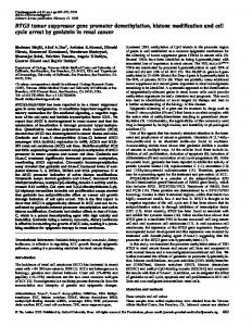
Figure 1. RUNX3 and JAG1 expression in HCC. (A) RUNX3, JAG1 and actin protein expression levels were analyzed by immunoblotting in HCC cell lines and normal liver tissue. Shown here are representative gels from 3 independent studies. (B) Representative images of RUNX3 and JAG1 staining in HCC tissues by immunohistochemistry. Representative images are from immunohistochemical analysis for RUNX3 (upper) and JAG1 (middle) expression and histology with hematoxylin and eosin staining (lower) in HCC tissues. Three sets of consecutive tissue sections represent negative (left upper) and strong (right upper) RUNX3 expression. (C) RUNX3 and JAG1 expression levels were analyzed by immunohistochemistry in human HCC tissues. The sections were scored for RUNX3 and JAG1 expression with a 4-titer scale as follows: 0, negative; 1, weak signal; 2, intermediate signal; and 3, strong signal. The inverse correlation between RUNX3 expression and JAG1 expression was assessed by Spearman correlation coefficient analysis. p