the fully convolutional neural networks (CNNs) to generate a vessel probability ... features for pixel representation, and classified each pixel by using a neural ...
RETINAL VESSEL SEGMENTATION VIA DEEP LEARNING NETWORK AND FULLY-CONNECTED CONDITIONAL RANDOM FIELDS Huazhu Fu, Yanwu Xu, Damon Wing Kee Wong, Jiang Liu Ocular Imaging Department, Institute for Infocomm Research Agency for Science, Technology and Research (A*STAR), Singapore. {fuhz, yaxu, wkwong, jliu}@i2r.a-star.edu.sg ABSTRACT Vessel segmentation is a key step for various medical applications. This paper introduces the deep learning architecture to improve the performance of retinal vessel segmentation. Deep learning architecture has been demonstrated having the powerful ability in automatically learning the rich hierarchical representations. In this paper, we formulate the vessel segmentation to a boundary detection problem, and utilize the fully convolutional neural networks (CNNs) to generate a vessel probability map. Our vessel probability map distinguishes the vessels and background in the inadequate contrast region, and has robustness to the pathological regions in the fundus image. Moreover, a fully-connected Conditional Random Fields (CRFs) is also employed to combine the discriminative vessel probability map and long-range interactions between pixels. Finally, a binary vessel segmentation result is obtained by our method. We show that our proposed method achieve a state-of-the-art vessel segmentation performance on the DRIVE and STARE datasets. Index Terms— Vessel segmentation, Conditional Random Fields, Convolutional Neural Networks. 1. INTRODUCTION Retinal vessel has diagnostic significance, it is widely used in monitoring the disease progression, and evaluation of various ophthalmologic diseases [1]. However, manually vessel segmentation by trained specialists delineate is a repetitious and time-consuming task. In the last two decades, many approaches have been introduced to segment the retinal vessel automatically. For example, Marin et al. [2] extracted a 7-D vector composed of gray-level and moment invariants-based features for pixel representation, and classified each pixel by using a neural network scheme. Nguyen et al. [3] utilized a multi-scale line detection to produce the final vessel segmentation. Orlando et al. [4] proposed an automatic method for vessel segmentation based on fully-connected Conditional Random Fields (CRFs), which learned its configuration by using a structured output SVM. However, most of the existing methods are based on manual designed features, which
lack the efficient discriminative and are easily affected by the pathological regions in the fundus images. Deep learning (DL) architecture is formed by the composition of multiple linear and non-linear transformations of the data, with the goal of yielding more abstract and ultimately more useful representations, which has been demonstrated having the powerful ability in many computer vision tasks (e.g., image classification and object detection). Convolutional Neural Networks (CNNs) are DL architectures, which are not only improving for whole-image classification [5], but also making progress on local tasks with structured output [6]. Long et al. [7] showed that a fully CNNs trained end-to-end, pixels-to-pixels on semantic segmentation exceeded the most state-of-the-art methods without further machinery. Based on fully CNNs, Xie et al. [8] further proposed a holisticallynested edge detection (HED) method to automatically learn the rich hierarchical representations with deep supervision, and resolved the challenging ambiguity in edge and object boundary detection. In this paper, we formulate the retinal vessel segmentation to a boundary detection problem, and utilize the fully CNNs architecture to learn the discriminative features that better characterize the important hidden patterns related to vessels and backgrounds in the fundus images. A similar DL-based vessel segmentation work is proposed by Melinscaket al. [9], which addressed the retinal vessel segmentation into a pixel-level binary classification task. They used a deep neural network [5] to classify each pixel in the fundus image. However, it has two disadvantages: first, the classification for each individual pixel is less the global smoothness correlation, which makes the method failed for some local pathological regions; second, the pixel-to-pixel classification strategy significantly spends plenty of running times on both training and testing phases. By contrast, our method is based on fully CNNs architecture [7, 8], which is an image-to-image training system and provides a multi-scale and multi-level visual responses. A vessel probability map is generated as the output of our fully CNNs architecture. And then a fully-connected CRFs is utilized further to take into account the global pixel correlation and output a binary vessel segmentation results.
Training image
Testing image
Conv3-64, Conv3-64
Side-output layer
MaxPooling Conv3-128, Conv3-128, Conv3-128 MaxPooling
Input image
Side-ouput 1
Side-ouput 2
Side-ouput 3
Side-ouput 4
Fusion Result
Conv3-256, Conv3-256, Conv3-256 MaxPooling Conv3-512, Conv3-512, Conv3-512
Vessel Probability map Ground-truth
CRFs
Fig. 2. The map for each side-output in our architecture.
Result
the transformation rule:
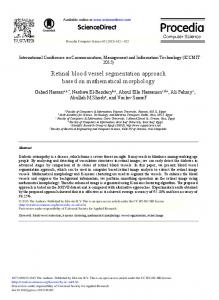
Fig. 1. The architecture of our retinal vessel segmentation method, which contains one fully CNNs network and one fully-connected CRFs. The fully CNNs network is a fourstages HED-like architecture, where side-output layers are inserted after the last convolutional layers in each stage (marked in Bold). The convolutional layer parameters are denoted as “Conv-”. The ReLU activation function is not shown for brevity. 2. PROPOSED METHOD Fig. 1 give the illustration of our architecture for retinal vessel segmentation. Our proposed method includes two parts: one fully CNNs network used to learn the discriminative features and generate the vessel probability map; one fully-connected CRFs used to produce the binary vessel segmentation result with the dense global pixel correlation. 2.1. Vessel probability map via deep learning networks Different from the existing DL-based method [9], which employed a pixel-to-pixel classification, our method treats vessel segmentation as a counter detection problem. To capture the inherent scales of vessel maps, we build our architecture on the top of HED system [8], which is based on the ideas of fully fully CNNs [7] and deeply-supervised net [10]. Fully convolutional networks: Suppose xij is the data vector at location (i, j) in a particular layer, and yij is for the following layer. The function of output yij is defined as: yij = fks ({xsi+δi,sj+δj }0≤δi,δj≤k ),
(1)
where k and s are the kernel size and the stride factor, respectively. fks denotes the layer manipulation (e.g., convolution, pooling, or activation function). This functional form is maintained under composition, with kernel size and stride obeying
fks ◦ gk0 s0 = (f ◦ g)k0 +(k−1)s0 ,ss0 .
(2)
A fully CNNs is that the whole networks are composed by this form layers to compute the nonlinear filter. A major advantage is that the fully CNNs can take input of arbitrary size and produce correspondingly-sized output with efficient inference and learning. Our architecture: The original HED has five stages. Each stage includes multiple convolutional and ReLU layers, and the side-output layers are connected to the last convolutional layer in each stage to suppose the deep layer supervision. In our architecture, we employ four stages to generate vessel probability map, as shown in Fig. 1. The main reason is that the retinal vessels in fundus images are different from the general object boundaries in natural images. The object boundary separates two regions with different appearances, which makes the boundary available in the small plane outputted from deeper layer. By contrast, the retinal vessel is a line-shape object, which is too-thin to output a meaningful response in the higher stride layer. Thus, we only employ a four-stage architecture. The generated map for each sideoutput in our architecture is shown in Fig. 2. Implementation details: We implement our framework using the Caffe library[11] and build on top of implementation of HED [8]. The model parameters follow the configuration used in [8]. For the fine-tuning data, we employ the ARIA dataset [12], which is the largest vessel segmentation dataset containing 143 fundus images with 768 × 576 resolution. We a) rotate the images to 4 different angles and crop the largest rectangle in the rotated image; b) flip the image at each angle. Moreover, due to the fully convolutional network can take input of arbitrary size, we also add the dataset by scaling the images to 50% and 75% of its original size. The fine-tuning phase takes about two days on a single NVIDIA K20 GPU (10, 000 iterations). For a 565 × 584 image, it takes about 200 ms to generate a vessel probability map.
2.2. Fully-connected CRFs segmentation:
Table 1. Performances on the two datasets.
Although the fully CNNs networks could produce the satisfactory vessel probability maps, they still have some problems. Firstly, traditional CNNs have convolutional filters with large receptive fields and hence produce coarse maps when pixel-level vessel segmentation (e.g., non-sharp boundaries and blob-like shapes). Secondly, fully CNNs lack smoothness constraints that may result in small spurious regions in segmentation output. Thus, we introduce a CRFs to obtain the binary vessel segmentation result. CRFs models pixel labels as random variables that form a Markov Random Field (MRF) when conditioned upon a global correlation. In the fullyconnected CRFs, each node is a neighbor of every other [13]. Following this approach, the method is able to take into account long-range interactions in the whole image. We denote v = {vi } as a labeling over all pixels of the image. The energy of a label assignment v is given by: X X E(v) = ψu (vi ) + ψp (vi , vj ), (3) i
i