lauf and Nancy Papalopulu for very valuable comments and discussions. This work was ... N. G., JENKINS, N. A., GRAHAM, E. AND. DAVIDSON, D. R. (1989).
75
Development 109, 75-80 (1990) Printed in Great Britain © The Company of Biologists Limited 1990
Retinoid-binding protein distribution in the developing mammalian nervous system
M. MADEN 1 '*, D. E. ONG 2 and F. CHYTIL2 'Anatomy and Human Biology Group, King's College London, Strand, London WC2R 2LS, UK Department of Biochemistry, University of Vanderbilt Medical School, Nashville, Tennessee, 37323 USA
2
• Author for correspondence
Summary We have analysed the distribution of cellular retinolbinding protein (CRBP) and cellular retinoic acidbinding protein (CRABP) in the day 8.5-day 12 mouse and rat embryo. CRBP is localised in the heart, gut epithelium, notochord, otic vesicle, sympathetic ganglia, lamina terminalis of the brain, and, most strikingly, in a ventral stripe across the developing neural tube in the future motor neuron region. This immunoreactivity remains in motor neurons and, at later stages, motor axons are labelled in contrast to unlabelled sensory axons. CRABP is localised to the neural crest cells, which are particularly noticeable streaming into the branchial arches. At later stages, neural crest derivatives such as Schwann cells, cells in the gut wall and sympathetic ganglia are immunoreactive. An additional
area of CRABP-positive cells are neuroblasts in the mantle layer of the neural tube, which subsequently appear to be the axons and cell bodies of the commissural system. Since retinol and retinoic acid are the endogenous ligands for these binding proteins, we propose that retinoids may play a role in the development and differentiation of the mammalian nervous system and may interact with certain homoeobox genes whose transcripts have also been localised within the nervous system.
Introduction
correspondingly exaggerated (Durston et al. 1989). Endogenous RA is present in whole Xenopus embryos at this stage. RA also promotes neurite outgrowth in explanted amphibian spinal cord and is present endogenously in this tissue (Hunter etai, unpublished). These results suggest that RA may be involved in various aspects of the developing nervous system as well as the limb. It is therefore a matter of considerable interest to uncover the mechanism of action of RA within the cells of the embryo. It has been known for some time that, at least in embryonal carcinoma cells, RA causes a change in the pattern of gene activity and thus can act on the nucleus (Roberts and Sporn, 1984). Two types of protein are thought to mediate its action between the cell membrane and nucleus, namely binding proteins and receptors. Cellular retinoic acid-binding protein (CRABP) is a 15.6X10³ MT protein with a high degree of ligand specificity for RA (Chytil and Ong, 1984). CRABP is found in the cytoplasm of a variety of cell types in the adult rat, particularly those that require RA for their normal function, such as skin and testis (Ong et al. 1982). The distribution of this protein in mammalian embryonic tissues has not previously been described,
It is becoming increasingly apparent that retinoic acid (RA), the most biologically active naturally occurring form of vitamin A, may play an important role in the morphogenesis of the embryo. In particular, the limb and the nervous system seem to employ RA during crucial phases of their development. When it is administered to the developing chick limb bud (Tickle et al. 1982; Summerbell, 1983) or the regenerating amphibian limb (Maden, 1982) it has the remarkable ability to stimulate the production of extra limbs. What is more, endogenous RA has been identified in the chick limb bud (Thaller and Eichele, 1987) and the regenerating amphibian limb (Maden, Summerbell and Waterson, unpublished results). In the chick, endogenous RA is differentially distributed across the anteroposterior axis of the limb bud, whereas its metabolic precursor, retinol, is not. This is precisely the behaviour one would expect of RA if it was acting as a morphogen. In the developing nervous system of Xenopus, the administration of RA causes anteroposterior transformations such that the forebrain and midbrain are reduced or absent and the hindbrain and spinal cord are
Key words: retinoic acid, retinol, cellular retinoic acidbinding protein, cellular retinol-binding protein, neural tube, notochord, motor nerves, sensory nerves, lamina terminalis, neural crest, commissural neurons.
76
M. Maden, D. E. Ong and F. Chytil
although it is now known that the CRABP gene is transcribed in the limb bud (Dolle et al. 1989) and in discrete populations of neuroblasts in the brain and spinal cord of early mouse embryos (Vaessen etal. 1989; Perez-Castro etal. 1989). Thus there may be an association between developmental systems that are affected by RA and the presence of CRABP and in the work reported here we confirm this by looking in detail at the distribution of the protein in the mouse embryo. A closely related protein, cellular retinol-binding protein (CRBP), has a ligand specificity for retinol (Chytil and Ong, 1984), the metabolic precursor of RA, and a wide distribution in the cytoplasm of the cells of the adult rat (Ong et al. 1982). Again, its distribution in the mammalian embryo has not yet been investigated, although the CRBP gene is transcribed in the brain and spinal cord amongst other organs such as the gut, liver and heart (Perez-Castro etal. 1989). In the chick embryo CRABP is indeed concentrated in precisely those systems whose development is under the influence of RA. It is expressed at highest levels in the limb buds, followed by the spinal cord and brain and absent in the rest of the trunk (Momoi et al. 1988). In the limb bud, CRABP was localised by immunocytochemistry to the rapidly dividing, RA-responsive cells at the distal tip of the bud (Maden etal. 1988). In the nervous system, CRABP is found in the neural crest cells, sensory axons, and the axons and cell bodies of the commissural system (Maden etal. 1989a). Thus, there is a clear association in the chick embryo between the presence of CRABP in a developing organ and the involvement of RA in its development. The receptors for RA have recently been discovered (Petkovich etal. 1987; Giguere etal. 1987) and these have a structure, including ligand-binding and DNAbinding domains similar to steroid receptors. There are at least three types of receptor in the mouse, a, fi and y (Zelent etal. 1989), and an additional one in the newt limb, termed 8 (Ragsdale etal. 1989). The a and 0 receptors seem to have a widespread distribution in tissues of the adult mouse whereas the y receptor is predominantly expressed in skin (Zelent et al. 1989). In the mouse limb bud, the a and y receptors are expressed uniformly and the (3 receptor is not expressed at all at early stages, in contrast to the graded distribution of CRABP transcripts (Dolle et al. 1989). The receptors identified in the newt are expressed in the regenerating limb (Giguere etal. 1989; Ragsdale etal. 1989), which, as described above, is a developing system responsive to the profound pattern changes induced by RA. Thus in the mechanism of action of RA the receptors are the component that interacts with the DNA to alter the pattern of gene activity in the cell, but the function of the binding proteins is not clear. They may be responsible for regulating the intercellular level of free RA that is available for binding to the receptors (Maden etal. 1988); they may define populations of cells that are responsive to RA (Maden et al. 19896) or they may be involved in the metabolism of retinoids. Another possible function involving homoeobox transcripts is considered in the Discussion. To help resolve
these issues, we have investigated by immunocytochemistry the distribution of CRBP and CRABP at various stages of mouse and rat embryogenesis. We find that CRBP is localised in several differentiating organs of the early embryo, but, most interestingly, as a stripe across the ventral neural tube where motoneurons form. Later staining is found in the motor axons, the sympathetic ganglia and in discrete regions of the brain. CRABP, on the other hand, seems to be specific for the neural crest and the axons and cell bodies of the commissural system. The possible relationships between CRBP, CRABP, their ligands and the localisations of some relevant homoeobox genes is discussed. Materials and methods Crossbred strains of mice and rats were used and their embryos staged according to Rugh (1968). Embryos were fixed in Perfix (Fisher Scientific, New Jersey) for 3h, dehydrated, cleared in xylene and embedded in wax. 7^/m sections were cut. CRBP was immunolocalised with an affinitypurified anti-rat CRBP rabbit lgG and CRABP was immunolocalised with an affinity-purified anti-rat CRABP rabbit IgG. The technique has been described previously (Porter et al. 1985) and was used with only one modification, namely that the dilution buffer for all immunochemical reagents was phosphate-buffered saline containing 1 % normal goat serum and 0.1 % crystalline bovine serum albumin. The anti-CRBP IgG was diluted to an absorbance at 280 nin of 0.35 and the anti-CRABP IgG was diluted to an absorbance at 280 nM of 0.01. Colour was developed by the avidin-biotinylated peroxidase complex with a kit from Vector Laboratories (Burlingame, California). For the Western blot, several day 10 mouse embryos were homogenised in solubilisation buffer for SDS-polyacrylamide gel electrophoresis according to Laemmli (1970) and centrifuged to remove debris. Aliquots of the extract (100-200/.ig), pure rat CRBP (100 ng) and pure rat CRABP (80 ng) were electrophoresed on an 11 % SDS-polyacrylamide gel and the proteins transferred to nitrocellulose paper. Immunoreactivity was detected using affinity-purified IgG fractions from rabbit antisera to rat CRBP and rat CRABP and radioiodinated protein A as previously described (Porter et al. 1985). Results
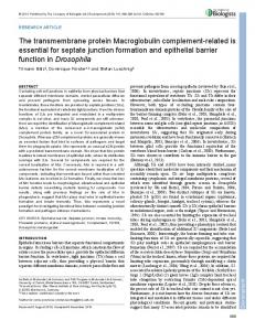
The experiments described here were conducted both on rat and mouse embryos with identical results. Originally rat embryos were used because we knew that our affinity-purified antibodies reacted with a single protein of the correct molecular weight on a Western blot of rat tissues (Porter etal. 1985). We then found that sections of mouse embryos gave the same staining patterns as sections of rat embryos and the descriptions below apply equally to each species. To confirm that the affinity-purified antibodies only reacted with a single band of the appropriate molecular weight in mouse embryos, homogenates of day 10 mouse embryos were blotted. In Fig. 1, it can be seen that both CRBP and CRABP antibodies identify such a band. In addition to these Western blots, we have also shown that sections
CRBP and CRABP in mammalian embryo
I Fig. 1. Lanes a and b - Western blot of pure rat CRBP (lane a) and day 10 mouse embryo (lane b) reacted with affinity-purified antibody to CRBP demonstrating the presence of a single band at the appropriate molecular weight in mouse embryos. Lanes c and d - blot of pure rat CRABP (lane c) and day 10 mouse embryo (lane d) reacted with affinity-purified antibody to CRABP demonstrating the presence of a single band of the appropriate molecular weight in mouse embryos. treated with antibody that had been preincubated with purified CRBP or CRABP gave no staining as do sections treated with preimmune serum (Fig. 7). These controls confirm the specificity of the immunoreactivity described below.
CRBP On day 8.5 of development, several areas of the embryo showed immunoreactivity to CRBP. The strongest and most obvious staining was found in the myocardium of the heart. The epithelium of the gut was also intensely stained, especially in the mid- and hindgut region (Fig. 2) and less so in the foregut. A weaker, but significant, level of reactivity was detected in the notochord (Fig. 3) and this showed a greater intensity at more rostral levels suggesting that CRBP is associated with some differentiative event appearing in an anterior-to-posterior sequence during development. The only other structure showing any reactivity in these sections was the otic vesicle, which develops adjacent to the neural tube of the hindbrain. Sections at this level showed a clear contrast between positive reactivity of the otic vesicle and the absence of reactivity in the hindbrain. On subsequent days, certain staining patterns such as the myocardium and gut lining remained a constant feature of the embryos (e.g. Fig. 6). The notochord increased its immunoreactivity and by day 12 was physically well-separated from the ventral floor plate of the neural tube (cf. Fig. 3) to remain an island of intense staining in the unstained sclerotome (Fig. 5). In other organ systems, however, new patterns of immunoreactivity emerged and this was most striking in the nervous system. On day 10, the neural tube showed a clear band of staining in the future motor neuron
77
region (Fig. 4). The band extended from the lumen to the periphery of the neural tube thus including both proliferating and differentiating cells. As a stripe it did not extend ventrally all the way to the floor plate on first appearance (Fig. 4), but did so as motor neuron differentiation was established (Figs 5, 6, 8). This staining had begun very faintly on day 9.5 in the forelimb region (later in the hindlimb region) and, by day 10.5 when motor neurons had extended axons, these were also immunoreactive in clear contrast to the sensory axons. This differentiation between CRBP positive motor axons and CRBP negative sensory axons had become so clear by day 12 that, at the brachial plexus where the two types of nerve mix (Fig. 8), stripes of labelled and unlabelled axons could be seen (Fig. 9). One other component of the nervous system that became intensely labelled upon differentiation was the sympathetic ganglia, which is in contrast to the lack of reactivity of the dorsal root ganglia (Fig. 8). The developing brain also showed localisations in two regions. Neuroblasts in the floor of the hindbrain were heavily labelled as the ventrally located immunoreactivity of the motor neurons in the spinal cord continued anteriorly into the hindbrain. No areas of labelling were obvious in the mid-brain, but in the forebrain the lamina terminalis was an area of intensity surrounded by unlabelled regions (Fig. 10). CRABP There were far fewer cell types that were immunoreactive to CRABP than to CRBP. In fact, only two subsets of neural cells and their descendents were labelled to any great extent and, furthermore, they were a different class of neural cells from those described above. In transverse sections through the trunk region of day 8.5 embryos, individual CRABP-positive cells could easily be identified, although there were not many of them. They were outside the neural tube and in two tracts, one underneath the epidermis and the other more medial and ventral around the dorsal aorta. In sections through the hindbrain, by contrast, there were large numbers of immunoreactive cells concentrated in the newly formed cephalic ganglia and streaming out from them into the branchial arches (Fig. 11). At this stage, no cells within the neural tube were labelled although in some sections individual cells that had just detached or were in the process of detaching from the dorsal neural tube were labelled. This distribution and behaviour suggested that these CRABP-positive cells were the neural crest and that the commencement of migration was related to the first appearance of (or increase in levels of) CRABP. By day 9.5, the period of extensive neural crest migration had terminated and a second population of immunoreactive cells became apparent. These were a proportion of cells in the mantle layer of the neural tube (Fig. 12). One day later both CRABP-positive populations were clearly distinguishable in the same section (Fig. 13). Descendants of migrating neural crest cells remained immunoreactive and were located in the wall
78
M. Maden, D. E. Ong and F. Chytil
of the developing gut (Fig. 14), in the sympathetic ganglia, scattered throughout the dorsal root ganglia (Fig. 13) and distributed along the nerve fibres (Fig. 15). The cells in the gut wall are likely to be developing into enteric ganglia and the scattered cells along nerve fibres and in dorsal root ganglia are most likely Schwann cells since each of these cell types is known to be derived from neural crest, at least in the chick (Le Douarin, 1982). The tracts of immunoreactive neurons and axons in the neural tube are in the locations expected for the commissural and funicular cells, which project contralaterally and ipsilaterally as relay neurons (Altman and Bayer, 1984). In the developing brain of a day 12 embryo, CRABP was present only in the hindbrain and midbrain. In the hindbrain, the labelled neuroblasts were extensively distributed through the thickness of the hindbrain epithelium. In the midbrain, labelled neuroblasts were more restricted, being present only in the peripheral cell layers (Fig. 16). At the midbrain-forebrain boundary, CRABP immunolabelling suddenly ceased. Limb buds Using the same CRABP antibody, we have previously described a distinct pattern of immunoreactivity in the developing chick limb bud where the most intensely labelled regions are at the distal tip in rapidly dividing, undifferentiated cells and in proximal regions where dermis, connective tissue and muscle are differentiating (Maden etal. 1988). We therefore expected to see a similar localisation in mouse and rat limb buds and were surprised to observe the complete absence of any immunoreactivity in the limb buds (not shown). This is despite the demonstrated existence of high levels of CRABP in mouse limb buds by sucrose gradient centrifugation (Kwarta etal. 1985; Maden, unpublished). The following explanation of this paradox seems possible. Recently it has been shown that there are in fact two CRABPs in the chick embryo (Kitamoto etal. 1988) and the rat (Bailey and Siu, 1988) and the first 25 amino acids of the N-terminus have been sequenced in each case. Chick CRABP I has an identical sequence to rat CRABP I (Eriksson etal. 1981). Chick CRABP I differs from chick CRABP II in 3 positions whereas chick CRABP I (and rat CRABP I) differs from rat CRABP II in 7 positions. Thus it is likely that our antibody (which was made against a purified rat protein rather than a peptide sequence) does not distinguish between CRABP I and II in the chick, but does distinguish between CRABP I and II in the rat because they are more dissimilar. This suggests that the two proteins are tissue specific, CRABP I being restricted to the nervous system and CRABP II to the limb. It is known that rat CRABP I and CRABP II have a tenfold difference in dissociation constant and different ligand specificities (Bailey and Siu, 1988). Thus, even before RA gets to the multiple retinoic acid receptors (see Introduction), there may be an initial degree of tissue specificity in their interaction with CRABP.
Figs 2-10. Sections of mouse and rat embryos treated with affinity-purified anti-rat CRBP antibody. Fig. 2. Day 8.5 mouse embryo showing pale staining of the notochord (n) and more intense staining of the epithelium of the developing gut (g). nt=neural tube. Bar=25 j*m. Fig. 3. Higher power view of Fig. 2 to show reactivity in the notochord (arrow). Bar=10/