P1: DPI
April 25, 1998
16:50
Annual Reviews
AR057-13
Annu. Rev. Biochem. 1998.67:335-394. Downloaded from arjournals.annualreviews.org by CALIFORNIA INSTITUTE OF TECHNOLOGY on 09/08/05. For personal use only.
Annu. Rev. Biochem. 1998. 67:335–94 c 1998 by Annual Reviews. All rights reserved Copyright °
RNA LOCALIZATION IN DEVELOPMENT Arash Bashirullah,1, 3 Ramona L. Cooperstock,1, 2 and Howard D. Lipshitz1, 2 1Program in Developmental Biology, Research Institute, The Hospital for Sick Children, Toronto, Canada M5G 1X8; 2Department of Molecular & Medical Genetics, University of Toronto, Toronto, Canada; 3Division of Biology, California Institute of Technology, Pasadena, California; e-mail:
[email protected]
KEY WORDS: Drosophila, Xenopus, Saccharomyces, oocyte, embryo, RNA-binding protein, cytoskeleton, microtubules, microfilaments, 30 -untranslated region (30 -UTR)
ABSTRACT Cytoplasmic RNA localization is an evolutionarily ancient mechanism for producing cellular asymmetries. This review considers RNA localization in the context of animal development. Both mRNAs and non-protein-coding RNAs are localized in Drosophila, Xenopus, ascidian, zebrafish, and echinoderm oocytes and embryos, as well as in a variety of developing and differentiated polarized cells from yeast to mammals. Mechanisms used to transport and anchor RNAs in the cytoplasm include vectorial transport out of the nucleus, directed cytoplasmic transport in association with the cytoskeleton, and local entrapment at particular cytoplasmic sites. The majority of localized RNAs are targeted to particular cytoplasmic regions by cis-acting RNA elements; in mRNAs these are almost always in the 30 -untranslated region (UTR). A variety of trans-acting factors— many of them RNA-binding proteins—function in localization. Developmental functions of RNA localization have been defined in Xenopus, Drosophila, and Saccharomyces cerevisiae. In Drosophila, localized RNAs program the anteroposterior and dorso-ventral axes of the oocyte and embryo. In Xenopus, localized RNAs may function in mesoderm induction as well as in dorso-ventral axis specification. Localized RNAs also program asymmetric cell fates during Drosophila neurogenesis and yeast budding.
335 0066-4154/98/0701-0335$08.00
P1: DPI
April 25, 1998
16:50
336
Annual Reviews
AR057-13
BASHIRULLAH, COOPERSTOCK & LIPSHITZ
CONTENTS
Annu. Rev. Biochem. 1998.67:335-394. Downloaded from arjournals.annualreviews.org by CALIFORNIA INSTITUTE OF TECHNOLOGY on 09/08/05. For personal use only.
INTRODUCTION . . . . . . . . . . . . . . . . . . . . . . . . . . . . . . . . . . . . . . . . . . . . . . . . . . . . . . . . . . . 336 PATTERNS OF RNA LOCALIZATION . . . . . . . . . . . . . . . . . . . . . . . . . . . . . . . . . . . . . . . . . . 342 Drosophila Oocytes and Early Embryos . . . . . . . . . . . . . . . . . . . . . . . . . . . . . . . . . . . . . . . 342 Xenopus Oocytes . . . . . . . . . . . . . . . . . . . . . . . . . . . . . . . . . . . . . . . . . . . . . . . . . . . . . . . . . 348 Ascidian Oocytes . . . . . . . . . . . . . . . . . . . . . . . . . . . . . . . . . . . . . . . . . . . . . . . . . . . . . . . . . 352 Echinoderm Oocytes . . . . . . . . . . . . . . . . . . . . . . . . . . . . . . . . . . . . . . . . . . . . . . . . . . . . . . 353 Zebrafish Embryos . . . . . . . . . . . . . . . . . . . . . . . . . . . . . . . . . . . . . . . . . . . . . . . . . . . . . . . . 353 Polarized Somatic Cells . . . . . . . . . . . . . . . . . . . . . . . . . . . . . . . . . . . . . . . . . . . . . . . . . . . . 353 MECHANISMS OF RNA LOCALIZATION . . . . . . . . . . . . . . . . . . . . . . . . . . . . . . . . . . . . . . 355 Nucleo-Cytoplasmic Transport . . . . . . . . . . . . . . . . . . . . . . . . . . . . . . . . . . . . . . . . . . . . . . 355 Transport from One Cell Type into Another . . . . . . . . . . . . . . . . . . . . . . . . . . . . . . . . . . . . 355 Transport Out of Mitochondria . . . . . . . . . . . . . . . . . . . . . . . . . . . . . . . . . . . . . . . . . . . . . . 356 Generalized Degradation with Localized Protection . . . . . . . . . . . . . . . . . . . . . . . . . . . . . 356 Directed Cytoplasmic Transport of RNA . . . . . . . . . . . . . . . . . . . . . . . . . . . . . . . . . . . . . . . 357 Entrapment/Anchoring of RNA at the Site of Localization . . . . . . . . . . . . . . . . . . . . . . . . . 362 RNA Transport/Anchoring Particles . . . . . . . . . . . . . . . . . . . . . . . . . . . . . . . . . . . . . . . . . . 363 CIS-ACTING ELEMENTS THAT TARGET RNAs FOR LOCALIZATION . . . . . . . . . . . . . 364 Mapping of Cis-Acting Elements in Localized RNAs . . . . . . . . . . . . . . . . . . . . . . . . . . . . . 364 Cis-Acting Sequences for Localization Map to the 30-Untranslated Region of mRNAs . . . 365 Cis-Acting Sequences for Localization Map Within Non-Protein-Coding RNAs . . . . . . . . 367 Alternative Splicing Can Generate Localized vs Unlocalized RNA Isoforms . . . . . . . . . . . 367 Discrete Localization Elements . . . . . . . . . . . . . . . . . . . . . . . . . . . . . . . . . . . . . . . . . . . . . . 368 Repeated/Redundant Localization Elements . . . . . . . . . . . . . . . . . . . . . . . . . . . . . . . . . . . . 369 Dispersed/Nonredundant Localization Elements . . . . . . . . . . . . . . . . . . . . . . . . . . . . . . . . 370 Additive Function of Localization Elements . . . . . . . . . . . . . . . . . . . . . . . . . . . . . . . . . . . . 371 Elements That Function in Translational Control During or After Localization . . . . . . . . 371 Primary, Secondary, Tertiary, and Quaternary Structures . . . . . . . . . . . . . . . . . . . . . . . . . 372 TRANS-ACTING FACTORS INVOLVED IN RNA LOCALIZATION AND TRANSLATIONAL CONTROL OF LOCALIZED RNAs . . . . . . . 373 Identification of Trans-Acting Factors . . . . . . . . . . . . . . . . . . . . . . . . . . . . . . . . . . . . . . . . . 373 Factors That Interact Directly with Defined RNA Elements . . . . . . . . . . . . . . . . . . . . . . . . 374 Other Factors That Function in RNA Localization . . . . . . . . . . . . . . . . . . . . . . . . . . . . . . . 377 Summary . . . . . . . . . . . . . . . . . . . . . . . . . . . . . . . . . . . . . . . . . . . . . . . . . . . . . . . . . . . . . . . 382 DEVELOPMENTAL FUNCTIONS OF RNA LOCALIZATION . . . . . . . . . . . . . . . . . . . . . . 383 Specification of the Anterior-Posterior and Dorsal-Ventral Axes of the Drosophila Oocyte . . . . . . . . . . . . . . . . . . . . . . . . . . . . . . . . . . . . . . . 383 Specification of Anterior Cell Fates in the Drosophila Embryo . . . . . . . . . . . . . . . . . . . . . 384 Specification of Abdominal Cell Fates in the Drosophila Embryo . . . . . . . . . . . . . . . . . . . 385 Assembly of Polar Granules and Specification of Germ Cells in the Drosophila Embryo . . . . . . . . . . . . . . . . . . . . . . . . . . . . . . . . . . . . . . 386 Signaling of Dorso-Ventral Axis and Mesoderm Induction in the Xenopus Embryo . . . . . 386 Specification of Cell Fates During Asymmetric Cell Divisions . . . . . . . . . . . . . . . . . . . . . . 387 EVOLUTIONARY CONSIDERATIONS . . . . . . . . . . . . . . . . . . . . . . . . . . . . . . . . . . . . . . . . . 388
INTRODUCTION Since the early days of experimental embryology it has been suggested that the asymmetric distribution of substances in the egg cytoplasm might confer particular fates to cells that receive that cytoplasm (reviewed in 1). However, it is only in the past 13 years that specific maternally synthesized, asymmetrically distributed RNA and protein molecules have been identified in oocytes and
P1: DPI
April 25, 1998
16:50
Annual Reviews
AR057-13
Annu. Rev. Biochem. 1998.67:335-394. Downloaded from arjournals.annualreviews.org by CALIFORNIA INSTITUTE OF TECHNOLOGY on 09/08/05. For personal use only.
CYTOPLASMIC RNA LOCALIZATION
337
early embryos of Xenopus, Drosophila, ascidians, zebrafish, and echinoderms. This review focuses largely on RNAs that are localized to specific cytoplasmic regions in eggs and early embryos. It addresses both the mechanisms of cytoplasmic RNA localization and the developmental functions of this localization. Some consideration is also given to RNA localization later in development, in differentiating or differentiated cells. However, since both the mechanisms and the functions of this later localization are not well understood, the emphasis here is on RNA localization in oocytes. This review considers only RNAs that are asymmetrically distributed in the cytoplasm. Examples of RNAs that are localized to and within the nucleus— even to specific chromosomes or regions of chromosomes (2–4)—are covered elsewhere. The first maternally synthesized cytoplasmically localized RNAs were identified in Xenopus in a molecular screen for RNAs enriched in either the vegetal (Vg RNAs) or animal (An RNAs) hemisphere of the Xenopus oocyte (5). Shortly thereafter, an RNA was discovered that is localized to the anterior pole of the Drosophila oocyte and early embryo (6). This RNA is encoded by the bicoid maternal effect locus (7), which plays a crucial role in specifying cell fates in the anterior half of the early Drosophila embryo (8). The facile combination of genetics and molecular biology in Drosophila led to bicoid becoming the first case in which it was demonstrated that RNA localization per se was important for normal development. Delocalization of bicoid RNA led to defects in anterior cell fate specification (7). Over 75 cytoplasmically localized RNAs have now been identified, and many of these are localized in eggs, early embryos, or differentiating cells (Table 1). To date, it has been possible to address both the mechanisms and the developmental functions of RNA localization almost exclusively in Drosophila and Xenopus. The large size of the Xenopus oocyte has allowed mapping of sequences necessary and sufficient for RNA localization through injection of in vitro synthesized transcripts engineered to contain an exogenous reporter sequence and part or all of the localized RNA. Further, in some cases inactivation, delocalization, or degradation of specific RNAs has been induced through microinjection of antisense RNA or DNA. The ability to manipulate Xenopus oocytes and to apply various cytoskeleton-destabilizing drugs or other inhibitors has demonstrated the importance of the cytoskeleton in RNA localization. Drosophila oocytes and early embryos are also large and have also been used for drug and inhibitor studies. In contrast to Xenopus, however, the ability to generate transgenic lines that express reporter-tagged transcripts during oogenesis has obviated the need for micronjection studies, although some of these have been conducted. Finally, the ability to obtain mutations in the endogenous gene that encodes the localized RNA or in factors that function in trans in its localization or in its translational regulation, has facilitated analyses of the
P1: DPI
April 25, 1998
16:50
338
Annual Reviews
AR057-13
BASHIRULLAH, COOPERSTOCK & LIPSHITZ
Table 1 Localized RNAs
Species
Transcript name
Protein product
Localization pattern
Actin
Cytoskeletal component Auxilary protein of DNA polymerase Ribosomal component Noncoding RNA
Myoplasm and ectoplasm Ectoplasm
Cell
Reference
Oocyte
249
Oocyte
100
Myoplasm
Oocyte
102
Myoplasm
Oocyte
101
Anterior
16, 17
Anterior
Oocyte and embryo Oocyte
18
Anterior
Oocyte
19
Anterior
Oocyte and embryo Cellular blastoderm Oocyte and embryo Oocyte Cellular blastoderm Cellular blastoderm Oocyte and embryo
7, 32
Ascidians
Annu. Rev. Biochem. 1998.67:335-394. Downloaded from arjournals.annualreviews.org by CALIFORNIA INSTITUTE OF TECHNOLOGY on 09/08/05. For personal use only.
PCNA
Ribosomal protein L5 YC RNA Drosophila Add-hts Bicaudal-C
Bicaudal-D
bicoid crumbs Cyclin B egalitarian even-skipped fushi tarazu germ cell-less
gurken hairy Hsp83 inscuteable K10 mtlrRNA
Cytoskeletal component Signal transduction/ RNA-binding protein Cytoskeleton interacting protein (?) Transcription factor Transmembrane protein Cell cycle regulator Novel Transcription factor Transcription factor Nuclear pore associated protein Secreted growth factor Transcription factor Molecular chaperone Novel Novel Noncoding RNA
Apical Posterior and perinuclear Anterior Apical Apical Posterior
67 59, 62 20 69 64 57, 239
Anterior-dorsal
Oocyte
21
Apical
Cellular blastoderm Embryo
65
Posterior Apical Anterior Posterior
Neuroblast Oocyte Oocyte and embryo
63 127 22 61, 134
(Continued )
P1: DPI
April 25, 1998
16:50
Annual Reviews
AR057-13
339
CYTOPLASMIC RNA LOCALIZATION Table 1 (Continued )
Species
Transcript name
Protein product
Localization pattern
nanos
RNA binding protein RNA binding protein Novel
Posterior
Noncoding RNA Transcription factor RNA binding protein Transcription factor Transmembrane receptor Novel Secreted ligand
Posterior
orb
Annu. Rev. Biochem. 1998.67:335-394. Downloaded from arjournals.annualreviews.org by CALIFORNIA INSTITUTE OF TECHNOLOGY on 09/08/05. For personal use only.
oskar Pgc prospero pumilio runt sevenless tudor wingless
Cell
Reference 28, 43
Apical/basal
Oocyte and embryo Oocyte and embryo Oocyte and embryo Oocyte and embryo Neuroblast
Posterior
Embryo
250
Apical
Cellular blastoderm Eye imaginal Epithelial cells Oocyte Cellular blastoderm Oocyte
66
Oocyte
103
Posterior Posterior
Apical Posterior Apical
yemanuclein-α Transcription factor
Anterior
SpCOUP-TF
Hormone receptor
Lateral to animalvegetal axis
β-actin
Cytoskeletal component
Specialized periphery
Arc
Cytosketal component Noncoding RNA Noncoding RNA Signalling component PKC substrate Integral membrane receptor
23 24, 25 26 127
126 27 68 251
Echinoderms
Mammals
BC-1 BC-200 CaMKIIα F1/GAP43 InsP3 receptor
Fibroblasts, 123–125 myoblasts, and epithelial cells Somatodendritic Neurons 104
Somatodentritic Neurons and axonal Somatodendritic Neurons
107, 111
Somatodendritic Neurons
109
Somatodendritic Neurons Somatodendritic Neurons
104 110
108
(Continued )
P1: DPI
April 25, 1998
16:50
340
Annual Reviews
AR057-13
BASHIRULLAH, COOPERSTOCK & LIPSHITZ
Table 1 (Continued )
Species
Transcript name
Protein product
Localization pattern
Cell
Reference
MAP2
Cytosketal component Membrane protein
Somatodendritic
Neurons
106
Myelinating membrane
120
Peripheral
Oligodendrocyte and Schwann cells Muscle
252
Axonal
Neurons
118, 119
Axonal Axonal Somatodendritic Axon hillock
Neurons Neurons Neurons
115 116, 117 104
Neurons
112
Pre-axonal pole Specialized membrane Axonal
Neurons
113
Osteoclasts
122
Neurons
253
Periplasmic
Oocyte
254
Animal
Oocyte
5, 81
Animal
Oocyte
5, 82
Animal
Oocyte
5
Animal Animal
Oocyte Oocyte
83 83
Vegetal
Oocyte
83
Vegetal
Oocyte
83
Vegetal Vegetal Vegetal Vegetal Vegetal
Oocyte Oocyte Oocyte Oocyte Oocyte
70 70 70 70 70
Annu. Rev. Biochem. 1998.67:335-394. Downloaded from arjournals.annualreviews.org by CALIFORNIA INSTITUTE OF TECHNOLOGY on 09/08/05. For personal use only.
MBP
Myosin heavy chain OMP/odorant receptors Oxytocin Prodynorphin RC3 tau Tropomyosin-5 V-ATPase subunits Vassopressin
Cytoskeletal component Integral membrane receptor Neuropeptide Neuropeptide PKC substrate Cytoskeletal component Cytoskeletal component Membrane protein Neuropeptide
Xenopus Actin Anl (a and b)
An2 An3 An4 (a and b) βTrCP βTrCP-2 βTrCP-3 B6 B7 B9 B12 C10
Cytoskeletal component Cyoplasmic protein (ubiquitinlike) mt ATPase subunit RNA binding protein Novel Signaling molecule Signaling molecule Signaling molecule NRa NR NR NR NR
(Continued )
P1: DPI
April 25, 1998
16:50
Annual Reviews
AR057-13
341
CYTOPLASMIC RNA LOCALIZATION Table 1 (Continued )
Species
Transcript name
Protein product
Localization pattern
Cell
Reference
G-proteins
Signaling molecule Transcription factor Signaling molecule Cytoskeletal component Transcription factor Signaling molecule RNA-binding protein RNA-binding protein NR Transcription factor (?) P-rich and PEST sequences Membrane protein Noncoding RNA Secreted ligand
Animal
Oocyte
84
Animal
Oocyte
85
Animal
Oocyte
86
Periplasmic
Oocyte
254
Vegetal
Oocyte
91, 92
Vegetal
Oocyte
5
Vegetal
Oocyte
78, 79
Vegetal
Oocyte
95
Vegetal Animal
Oocyte Oocyte
70 87
Animal
Oocyte
88
Animal
Oocyte
89
Vegetal Vegetal
Oocyte Oocyte
97 98
Oct60
Annu. Rev. Biochem. 1998.67:335-394. Downloaded from arjournals.annualreviews.org by CALIFORNIA INSTITUTE OF TECHNOLOGY on 09/08/05. For personal use only.
PKCα α-tubulin VegT (Antipodean) Vgl Xcat-2 Xcat-3 Xcat-4 xl-21 Xlan4 Xlcaax-1 Xlsirt Xwnt-11 Yeast ASH1
Transcription factor
Budding site
Mother cell
128, 129
Vasa
RNA-binding protein
Cleavage plane
Early embryo
209
Zebrafish
a
NR, Not reported.
mechanisms of RNA localization, the developmental functions of these RNAs, and of their localization per se. This review begins with a description of patterns of cytoplasmic RNA localization with an emphasis on Xenopus and Drosophila. To help explain the patterns and their significance, brief descriptions of the structure and development of Xenopus and Drosophila oocytes and/or early embryos are included. After considering the patterns of RNA localization, the focus switches to mechanisms. First, the dynamics of RNA localization are considered, including the
P1: DPI
April 25, 1998
16:50
342
Annual Reviews
AR057-13
BASHIRULLAH, COOPERSTOCK & LIPSHITZ
role of the cytoskeleton in RNA transport and anchoring. Then specific components of the localization mechanism are dissected; these include cis-acting sequences and trans-acting factors that function either in localization per se or in control of RNA stability or translation during and after localization. Finally, developmental functions of RNA localization are discussed.
Annu. Rev. Biochem. 1998.67:335-394. Downloaded from arjournals.annualreviews.org by CALIFORNIA INSTITUTE OF TECHNOLOGY on 09/08/05. For personal use only.
PATTERNS OF RNA LOCALIZATION All cells are nonhomogeneous since they are compartmentalized into organelles with distinct functions and locations. These inhomogeneities can result in several forms of cellular symmetry and asymmetry. For example, positioning of the nucleus in the center of an otherwise quite homogeneous spherical cell produces spherical symmetry. In such a cell (and there are few if any examples, with the possible exception of some oocytes), certain RNAs might be localized close to the nucleus (perinuclear) while others might be positioned more peripherally. More complex cellular asymmetries result from variations in cell shape and the position of the nucleus and other subcellular organelles. Cells can be radially symmetric or even further polarized in two or three axes. In these cases, RNA localization can occur relative to one, two, or three axes (e.g. to the dorsal anterior pole). Regardless of cell shape or size, RNA distribution patterns are usually based on preexisting asymmetries and can, in turn, lead to the establishment of further asymmetries. This section describes the dynamics and patterns of subcellular distribution of cytoplasmically localized RNAs. It provides a cellular and developmental context for consideration of the mechanisms and functions of RNA localization in subsequent sections. The emphasis here is on the best understood of the examples listed in Table 1.
Drosophila Oocytes and Early Embryos STRUCTURE AND DEVELOPMENT OF THE NURSE CELL–OOCYTE COMPLEX
The two bilaterally symmetric Drosophila ovaries each consist of about 16 ovarioles. At the anterior tip of the ovariole is the germarium. Here the oogonial stem cells divide asymmetrically producing a stem cell and a commited cell, which is called a cystoblast. Each cystoblast divides four times with incomplete cytokinesis to form 16 cystocyte cells interconnected by cytoplasmic bridges that run through specialized membrane cytoskeletal structures called ring canals. Only 1 of the 16 cystocytes becomes the oocyte, and the remaining 15 become nurse cells. Each 16-cell germarial cyst becomes surrounded by somatically derived follicle cells to form a stage 1 egg chamber. The more posterior part of the ovariole comprises a connected series of progressively older egg chambers ordered such that the youngest is most anterior and the
P1: DPI
April 25, 1998
16:50
Annual Reviews
AR057-13
Annu. Rev. Biochem. 1998.67:335-394. Downloaded from arjournals.annualreviews.org by CALIFORNIA INSTITUTE OF TECHNOLOGY on 09/08/05. For personal use only.
CYTOPLASMIC RNA LOCALIZATION
343
oldest (stage 14) most posterior relative to the body axis of the female. It takes three days for an egg chamber to produce a mature egg. Except during the final six hours, the nurse cells synthesize large amounts of RNA and protein that are transported into the developing oocyte. Many of these molecules are required during the first two hours of embryonic development prior to the onset of zygotic transcription. Selection of the oocyte from among the 16 cystocytes is not random. Of the 16 cells, 2 are connected to 4 others and 1 of these always becomes the oocyte (9). A large cytoplasmic structure—called the fusome—containing several cytoskeletal proteins, runs through the ring canals that connect cystocytes and has been implicated in oocyte determination (10–13). The only microtubule organizing center (MTOC) in the 16-cell complex is localized to the pro-oocyte, and microtubule arrays connect all 16 cells through the ring canals (reviewed in 14, 15). Because the MTOC nucleates the minus ends of the microtubules, the microtubule-based cytoskeleton that connects the 16 cells is polarized. This has important consequences for RNA transport from the nurse cells into the oocyte as well as for RNA localization within the oocyte itself. RNA LOCALIZATION IN STAGE 1–6 EGG CHAMBERS Many RNAs that are later localized within the growing oocyte are first transcribed in all 16 germline cells but accumulate specifically in the pro-oocyte. These include the Adducin likehuli tai shao (Add-hts) (16, 17), Bicaudal-C (18), Bicaudal-D (19), egalitarian (20), gurken (21), K10 (22), orb (23), oskar (24, 25), Polar granule component (Pgc) (26), and tudor (27) transcripts. Other RNAs that will later be localized within the oocyte are transcribed at very low levels in the nurse cells at this early stage and so cannot be visualized easily. For example, nanos is transcribed at low levels, and oocyte accumulation can be seen only later (28). Additional transcripts, such as ovarian tumor (29) and cytoplasmic tropomyosin II (cTmII ) (30), also accumulate in the oocyte at this and later stages but are not localized. Therefore oocyte-specific accumulation is not unique to RNAs that will be localized within the oocyte during later stages of oogenesis but is a property of many RNAs synthesized in the germline of early egg chambers. The fact that many RNAs that accumulate in the early oocyte appear to do so with higher concentrations at the posterior cortex—the site of the only MTOC in the egg chamber (31)—is an early indication of the role of the polarized microtubule network in RNA transport and localization (see below). The exact stage at which transcription of different localized RNAs commences (or at least the stage at which the transcripts can first be detected) varies. For example, bicoid is first transcribed in the nurse cells of stage 5 egg chambers and then accumulates in the oocyte (32), whereas oskar (24, 25) and K10 (22) RNAs already accumulate in the oocyte in the germarium over a day
P1: DPI
April 25, 1998
16:50
344
Annual Reviews
AR057-13
BASHIRULLAH, COOPERSTOCK & LIPSHITZ
earlier. Interestingly, the dynamics of accumulation of these RNAs is identical if they are intentionally transcribed at the same time (33). Thus temporal differences in patterns of oocyte accumulation of different RNAs are a consequence of variation in time of transcription and are not indicative of a difference in underlying transport mechanism, which in fact is similar for different RNAs transcribed at distinct stages.
Annu. Rev. Biochem. 1998.67:335-394. Downloaded from arjournals.annualreviews.org by CALIFORNIA INSTITUTE OF TECHNOLOGY on 09/08/05. For personal use only.
REORGANIZATION OF THE CYTOSKELETON AND RNA LOCALIZATION DURING STAGE 7 The process of nurse cell transcription and oocyte accumulation of RNAs continues through stage 6. During this time oocyte-follicle cell interactions establish anterior-posterior polarity within the oocyte and in the surrounding follicle cells (34, 35). Reciprocal signaling between the follicle cells and the oocyte results in a reorganization of the cytoskeleton such that, by the end of stage 7, the MTOC disappears from the posterior of the oocyte and microtubules become concentrated at the anterior oocyte margin (31, 34, 35). Concomitant with this change in cytoskeletal organization, RNAs that previously accumulated at the posterior of the oocyte localize in a ring-like pattern at the anterior oocyte margin (Figure 1) (e.g. Bicaudal-C, Bicaudal-D, bicoid, egalitarian, gurken, K10, nanos, orb, oskar, and Pgc) (18–26). At this stage several proteins are also seen in an anterior ring-like pattern (e.g. Egalitarian and Bicaudal-D) (20). This redistribution of RNA and protein is likely a consequence of the reorganization of the microtubule network such that the minus ends of microtubules move from the posterior pole to the anterior. Consistent with this, a β-galactosidase fusion to the minus-end-directed microtubule motor, Nod, relocalizes from the posterior pole of the oocyte at stage 6 to a ring around the anterior margin by the end of stage 7 (36). Thus RNAs that are transported into and within the oocyte by minus-end-directed microtubule motors would be expected to accumulate at the anterior rather than the posterior pole. As expected, the transient anterior localization of transcripts (e.g. Bicaudal-D, bicoid, K10, orb) is colchicine sensitive (37), and microtubules are required for anterior Egalitarian protein localization (20). Mutations in genes involved in oocyte-follicle cell signaling during stages 6 and 7 cause defects in oocyte polarity (see below) and in the microtubule-based cytoskeleton (e.g. Delta, gurken, Notch, PKA) (34, 35, 38, 39). For example, double-anterior oocytes can form in which microtubules have their minus ends at both oocyte poles and their plus ends at its center. Mutations in homeless cause a similar disorganization of microtubules (40). Such disorganization results in bicoid RNA localization at both poles of the oocyte while oskar RNA and plus-end-directed kinesin-β-galactosidase fusion protein localize in the middle (38, 41, 42). These data emphasize that microtubule polarity directs intraoocyte transcript localization.
P1: DPI
April 25, 1998
16:50
Annual Reviews
AR057-13
Annu. Rev. Biochem. 1998.67:335-394. Downloaded from arjournals.annualreviews.org by CALIFORNIA INSTITUTE OF TECHNOLOGY on 09/08/05. For personal use only.
CYTOPLASMIC RNA LOCALIZATION
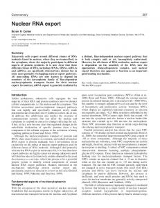
345
Figure 1 Localization of transcripts during Drosophila oogenesis. Transcript distribution patterns are shown in black or gray shading for bicoid, gurken, nanos, and oskar RNAs in stage 8 (top row), 9 (middle row), and 10B (bottom row) egg chambers. A, anterior; P, posterior; D, dorsal; V, ventral. Drawings of egg chambers are after King (9).
Further changes in RNA localization patterns occur at stages 8 and 9. Certain RNAs localize in a general anterior cortical pattern. These include Bicaudal-C (18), Bicaudal-D (19), bicoid (7), K10 (22), nanos (28, 43), orb (23), and Pgc (26). In contrast, Add-hts RNA shifts from a general cortical pattern to an anterior cortical pattern (16, 44, 45; KL Whittaker, D Ding, WW Fisher, HD Lipshitz, manuscript in preparation) whereas gurken RNA localizes in a dorso-anterior pattern around the nucleus (21). Others such as egalitarian RNA delocalize from the anterior and become
RNA LOCALIZATION AT STAGES 8–9
P1: DPI
April 25, 1998
16:50
Annu. Rev. Biochem. 1998.67:335-394. Downloaded from arjournals.annualreviews.org by CALIFORNIA INSTITUTE OF TECHNOLOGY on 09/08/05. For personal use only.
346
Annual Reviews
AR057-13
BASHIRULLAH, COOPERSTOCK & LIPSHITZ
uniformly distributed (20). Still others begin to relocalize to the posterior: Pgc RNA spreads posteriorly along the oocyte cortex (26) while oskar RNA gradually moves away from the anterior and begins to accumulate at the posterior pole of the oocyte (24, 25). During this same period a kinesin-β-galactosidase fusion protein localizes to the posterior (41). This observation suggests that, even though it has not been possible to visualize microtubules that traverse antero-posterior axis of the oocyte at this stage (31), they must be present. RNAs that enter the oocyte at this stage or that were previously localized to the anterior of the oocyte in association with minus-end-directed microtubule motors, must dissociate from these motors and associate with plus-end-directed motors in order to be translocated to the posterior pole. One such RNA is oskar, which by the end of stage 9, is present only at the posterior pole of the oocyte (24, 25). oskar RNA plays a key role in nucleating formation of the polar granules at the posterior pole of the oocyte. The polar granules, which are involved in germ-cell specification (see below), gradually assemble during stages 9–14 of oogenesis (reviewed in 47). NURSE CELL “DUMPING” AND RNA LOCALIZATION FROM STAGES 10–14 During stages 9 and 10 nurse cells increase synthesis of RNA and protein, dump their contents into the oocyte starting at stage 10B, and degenerate by the end of stage 12. This massive transfer of material is aided by contraction of the actin cortex of nurse cells. “Dumpless” mutants affect this process as well as ring canal structure. These genes, chickadee (encoding a profilin homolog), singed (encoding a fascin homolog), and quail (encoding a vilin homolog), are involved in F-actin crosslinking, indicating a major role for the actin-based cytoskeleton (48–50). Moreover, these studies demonstrate that the actin-based cytoskeleton is involved in anchoring the nurse cell nuclei so that they do not plug the ring canals during the dumping process. Interestingly, bicoid is localized apically in nurse cells during this phase (32). This apical distribution of bicoid RNA indicates a preexisting asymmetry within the nurse cells, but whether this transient bicoid localization in nurse cells serves any function is unclear. A similar apical nurse cell RNA localization pattern is observed for ectopically expressed oskar and K10 transcripts at this stage (33). During stage 10B, microtubules rearrange into subcortical parallel arrays in the oocyte, and a microtubule-based process called ooplasmic streaming begins. Capuccino and Spire proteins are required for control of ooplasmic streaming (51, 52). During stages 10B–12 the dumping of large amounts of RNA into the oocyte along with ooplasmic streaming make it difficult to distinguish delocalization of previously localized RNA on the one hand, from a transient stage during which the dumped RNA is becoming localized (i.e. is joining the
P1: DPI
April 25, 1998
16:50
Annual Reviews
AR057-13
Annu. Rev. Biochem. 1998.67:335-394. Downloaded from arjournals.annualreviews.org by CALIFORNIA INSTITUTE OF TECHNOLOGY on 09/08/05. For personal use only.
CYTOPLASMIC RNA LOCALIZATION
347
previously localized RNA at its intracellular target site) on the other. This is further complicated by the release of many transiently anteriorly localized RNAs, followed by their gradual translocation toward the posterior in some cases, or their complete delocalization in others. As a result, many RNAs appear to be generally distributed in the stage 11 oocyte. These include Bicaudal-C (18), Bicaudal-D (19), egalitarian (20), nanos (28), and orb (23). Other previously localized transcripts such as K10 and gurken disappear by stage 11 (21, 22). In contrast, the anterior localization pattern of bicoid and Add-hts transcripts is maintained throughout these stages (7, 16, 32). Maintenance of this pattern is likely the result of two factors: (a) anteriorly localized RNAs are trapped as they enter the oocyte from the nurse cells (WE Theurkauf, TI Hazelrigg, personal communication) and (b) previously anchored bicoid and Add-hts RNA is not released from the anterior pole during dumping and so does not have the opportunity to become generally distributed throughout the oocyte (16, 32). After ooplasmic streaming is completed (stage 12), subcortical microtubules are replaced by randomly oriented short cytoplasmic filaments, and F-actin reorganizes from a dense cortical filament network to an extensive deep cytoplasmic network (31, 54, 55). At this stage, several newly localized RNAs can be seen at the posterior of the oocyte. These include nanos (28, 56), germ cell-less (57), and probably orb (23). In addition, oskar and Pgc transcripts exhibit a posteriorly enriched pattern (24–26). bicoid and Add-hts transcripts remain localized at the anterior pole of late oocytes (7, 16, 32). RNA LOCALIZATION IN EARLY EMBRYOS After egg activation, the cytoskeleton reorganizes once again with actin and tubulin concentrated in the cortex and deeper filamentous networks of microtubules (31, 54, 55). Some longitudinal actin fibers may also be present in the early embryo. The Drosophila zygote undergoes 13 synchronous nuclear divisions without cytokinesis, forming a syncytial embryo containing several thousand nuclei that share the same cytoplasm (58). This syncytial state persists until the end of the 14th cell cycle when approximately 6000 nuclei reside at the cortex. Subsequently, invagination of membranes forms individual cells to give the cellular blastoderm. At this point the antero-posterior and dorso-ventral positional fates of the cells are specified. The two anteriorly localized maternal RNAs—bicoid (Figures 1 and 2) and Add-hts—persist in early cleavage embryos. Add-hts is released and diffuses posteriorly (16), while bicoid appears to remain anchored at the anterior cortex (7, 32). By the cellular blastoderm stage both RNAs are gone. Three RNAs that are posteriorly localized in the oocyte—oskar, nanos (Figures 1 and 2), and Pgc—retain posterior localization in early cleavage stage embryos. By nuclear cycle 6/7 oskar RNA is gone (24), whereas Pgc and
P1: DPI
April 25, 1998
16:50
Annu. Rev. Biochem. 1998.67:335-394. Downloaded from arjournals.annualreviews.org by CALIFORNIA INSTITUTE OF TECHNOLOGY on 09/08/05. For personal use only.
348
Annual Reviews
AR057-13
BASHIRULLAH, COOPERSTOCK & LIPSHITZ
Figure 2 Localized maternal transcripts in early Drosophila embryos. A: bicoid RNA; B: nanos RNA. Images are of whole mount RNA tissue in situ hybridizations to stage 2 embryos using digoxigenin-labeled probes. Anterior is to the left and dorsal toward the top of the page.
nanos RNAs are associated with polar granules and are taken up into pole cells together with these granules (26, 56). Some maternal RNAs do not become posteriorly localized until late in oogenesis or early embryogenesis. Examples are Cyclin B (59–62) and Hsp83 (60, 61, 63) transcripts. By the cellular blastoderm stage, maternal transcripts of Cyclin B, germ cell-less, Hsp83, nanos, orb, and Pgc can be detected only in pole cells. During the syncytial and cellular blastoderm stages, zygotic synthesis of RNA commences. Several of these zygotically synthesized transcripts, including crumbs, even-skipped, fushi tarazu, hairy, runt, and wingless are apically localized in the blastoderm (64–69), an epithelium surrounding the syncytial yolk mass of the zygote.
Xenopus Oocytes As in Drosophila, maternally synthesized gene products play a key role in the development of the Xenopus embryo (reviewed in 70). Zygotic transcription initiates at the 4000-cell mid blastula stage. Unlike in Drosophila, however, synthesis of maternal molecules occurs in the oocyte itself. Thus the issue of transport into the oocyte from interconnected nurse cells does not arise. DEVELOPMENT OF THE OOCYTE The Xenopus oocyte is initially a small spherical cell of 30 µm diameter when it is produced by mitosis of a stem cell, the oogonium (71). However, even at this stage, its nucleus and organelles are asymmetrically distributed (72, 73). Unlike Drosophila oogenesis, which lasts just over a week, Xenopus oogenesis lasts three years, although most of the
P1: DPI
April 25, 1998
16:50
Annual Reviews
AR057-13
Annu. Rev. Biochem. 1998.67:335-394. Downloaded from arjournals.annualreviews.org by CALIFORNIA INSTITUTE OF TECHNOLOGY on 09/08/05. For personal use only.
CYTOPLASMIC RNA LOCALIZATION
349
synthesis of oocyte contents occurs in the third year (71). The oocyte reaches a final diameter of approximately 1.5 mm (71). An early indicator of asymmetry is the mitochondrial cloud or Balbiani body (71). It is composed of clumps of mitochondria, rough endoplasmic reticulum, and dense granules that initially are evenly distributed around the periphery of the germinal vesicle in early stage I oocytes (∼80 µm diameter). By the end of stage I these components condense on one side of the germinal vesicle as a caplike structure that grows and assumes a spherical shape. Beginning in stage II, the mitochondrial cloud moves toward the future vegetal pole initially changing shape to become disk-like and then reorganizing into a wedge-like shape (late stage II/early stage III). Subsequently its components become localized to the vegetal cortex of the oocyte during stage III/IV. The mitochondrial cloud probably functions in the accumulation and localization of material needed for the formation of germ plasm at the vegetal pole of the early embryo (reviewed in 70, 74). In structure and function the Xenopus germ plasm is comparable to the Drosophila posterior polar plasm, and it contains germinal granules that function in germ-cell determination. However, while Drosophila polar granules are sufficient for the induction of germ cells (75, 76), Xenopus primordial germ cells are not irreversibly determined (77). Preliminary evidence indicates that certain RNA and protein components of the Drosophila and Xenopus germinal granules are evolutionarily conserved (70, 78, 79) and that in both cases RNA localization is an important mechanism used to locally assemble these structures. The oocyte cytoskeleton is symmetric early in oogenesis. Until stage II the germinal vesicle appears to serve as the only MTOC with microtubules emanating radially toward the plasma membrane (80). This array loses its symmetry as the germinal vesicle moves toward the future animal pole and the mitochondrial cloud starts condensing at the opposite (vegetal) side. At this time microtubules start to concentrate at the vegetal side of the germinal vesicle, colocalizing with the wedge-shaped mitochondrial cloud by late stage II (77). The germinal vesicle completes movement to the animal hemisphere by stage V/VI and at this time the microtubule array disappears. RNA LOCALIZATION PATTERNS The first collection of localized RNAs was reported for Xenopus oocytes. Three animal hemisphere–enriched RNAs (An1, An2, An3) and one vegetal hemisphere–localized RNA (Vg1; Figures 3 and 4) were identified in a molecular screen for cDNAs representing mRNAs differentially distributed along the animal-vegetal axis (5). The animal hemisphere (An)–enriched RNAs are not tightly localized within the animal hemisphere but are at least fourfold enriched in this hemisphere
P1: DPI
April 25, 1998
16:50
Annu. Rev. Biochem. 1998.67:335-394. Downloaded from arjournals.annualreviews.org by CALIFORNIA INSTITUTE OF TECHNOLOGY on 09/08/05. For personal use only.
350
Annual Reviews
AR057-13
BASHIRULLAH, COOPERSTOCK & LIPSHITZ
Figure 3 Localization of transcripts during Xenopus oogenesis. Transcript distribution patterns are shown in black, on the left for transcripts that show a Vg1-like pattern and on the right for transcripts that show an Xcat-2-like distribution pattern. Oocytes from stages I–IV are schematized. An, animal pole; Vg, vegetal pole. The germinal vesicle (oocyte nucleus) is shown in gray and the METRO as a white circle (stage I) or ellipsoid (stage II). Drawings of oocytes are after Kloc & Etkin (90).
relative to their vegetal concentration. Generally distributed maternal RNAs are twice as abundant in the animal than in the vegetal hemisphere (5). Thus the An RNAs are enriched in the animal hemisphere at least twofold versus other maternal RNAs. There are over a dozen known examples of An RNAs, all with similar distribution patterns: An1a (5, 81), An1b (5, 81), An2 (5, 82), An3 (5), An4a (83), An4b (83), β-TrCP (83), G protein (84), oct60 (85), PKC-α (86), xl-21 (87), Xlan-4 (88), and Xlcaax-1 (89). Unfortunately, reported in situ RNA hybridization data are lacking, making detailed comparisons of distribution patterns impossible. In contrast to the An RNAs, the vegetally localized RNAs exhibit highly restricted distribution patterns. Careful observations of the patterns of localization
P1: DPI
April 25, 1998
16:50
Annual Reviews
AR057-13
Annu. Rev. Biochem. 1998.67:335-394. Downloaded from arjournals.annualreviews.org by CALIFORNIA INSTITUTE OF TECHNOLOGY on 09/08/05. For personal use only.
CYTOPLASMIC RNA LOCALIZATION
351
Figure 4 Localized transcripts in Xenopus oocytes. A: Vg1 RNA; B: Xcat-2 RNA; C: Xlsirt RNA. All images are of whole mount RNA tissue in situ hybridizations to either late stage III (A) or early stage II (B, C ) oocytes. In A, the vegetal pole points toward the top right; in B and C the vegetal tip of the oocyte is at the center of the transcript distribution (i.e. the oocytes are viewed from the vegetal side). The small patches of Xcat-2 RNA that are radially symmetrically distributed around the vegetal ring of transcripts are in the islands of germ plasm.
in situ in whole mounts have suggested two or three distinct patterns (70, 90–92) exemplified by Vg1, Xcat-2, and, possibly, VegT/Antipodean. Four RNAs show the Vg1-like pattern of localization: Vg1 (Figures 3 and 4); and Xcat-4, B12, and B9 (70). Vg1 RNA is initially distributed uniformly in the oocyte cytoplasm and is excluded from the mitochondrial cloud during stages I and II (93). During late stage II/early stage III, Vg1 transcripts accumulate in a wedge-like pattern toward the vegetal pole before becoming restricted to the vegetal cortex by late stage III (93–95). Finally, at stage IV, Vg1 RNA is tightly localized to the cortex of the entire vegetal hemisphere (94, 96). This localization pattern correlates with the dispersion of the mitochondrial cloud (see above). The second pattern of RNA localization is exhibited by seven RNAs: Xcat-2, Xwnt-11, Xcat-3, B6, B7, C10, and the Xlsirts (Figures 3 and 4) (70, 78, 79, 95, 97, 98). This localization process includes passage through what has been called a message transport organizer (METRO) within the mitochondrial cloud (Figure 3) and occurs in three distinct steps (90): (a) movement from the germinal vesicle to the mitochondrial cloud, (b) sorting within the METRO, and (c) translocation and anchoring to vegetal cortex. Localization begins during early oocyte stage I when transcripts appear to be distributed throughout the cytoplasm at low levels with slightly higher concentrations around the germinal vesicle. As the mitochondrial cloud condenses into a sphere during mid stage I, these transcripts are transported to the METRO. For Xcat-2 this change in pattern is not the result of a reduction of RNA elsewhere, as the total amount of RNA in the oocyte remains constant or even increases (79). Within the METRO the transcripts are sorted such that, for example, Xcat-2 is localized first, followed by Xlsirts, and then Xwnt-11 (70, 79, 90, 99). By late stage I,
P1: DPI
April 25, 1998
16:50
Annu. Rev. Biochem. 1998.67:335-394. Downloaded from arjournals.annualreviews.org by CALIFORNIA INSTITUTE OF TECHNOLOGY on 09/08/05. For personal use only.
352
Annual Reviews
AR057-13
BASHIRULLAH, COOPERSTOCK & LIPSHITZ
when the METRO is disc-like, Xcat-2 RNA is in the periphery of the disc, Xwnt-11 RNA is in the center, and Xlsirts reside between these two (Figure 3). The METRO moves vegetally such that by stage IV, all three RNAs are localized at the tip of the vegetal pole (70, 79, 90, 99). At this stage, the Vg1 RNA pattern is quite distinct as it is distributed throughout the vegetal cortex (70, 79, 90, 99). The third type of localization, typified by VegT (Antipodean), appears to deviate from both of these patterns, although there is some discrepancy in the distributions described in two reports (91, 92). VegT/Apod maternal transcripts are initially uniform in the cytoplasm in stage I. Later they appear to move to the vegetal pole with a timing and pattern similar to Xlsirts, Xcat-2, and Xwnt-11 transcripts. One report describes a distribution at late stage III/early stage IV similar to that of Vg1 (92), while another describes a novel final pattern predominantly in the vegetal yolk (91). The latter distribution would imply a target of localization that is distinct from all other vegetally localized transcripts. After fertilization, the B7 transcripts disappear, the Xlsirt, Xcat-2, and Xcat-3 transcripts are associated with the germ plasm in the primordial germ cells, and the other transcripts (B9, B12, C10, Vg1, Xcat-4, and Xwnt-11) are all located in the vegetal blastomeres (70, 79, 90, 93, 98, 99; L Etkin, personal communication).
Ascidian Oocytes Several RNAs are localized in ascidian (Styela) oocytes (Table 1). These include YC RNA, PCNA mRNA, and ribosomal protein L5 mRNA. PCNA mRNA is initially uniformly distributed throughout the previtellogenic oocyte (100). During maturation, PCNA mRNA becomes concentrated in the central ectoplasm and cortical regions surrounding the myoplasm but is absent from the myoplasm per se (100). While PCNA mRNA can be observed in both the ectoplasm and the myoplasm after the first phase of ooplasmic segregation, which restricts the ectoplasm and myoplasm to the vegetal hemisphere, PCNA mRNA is absent from the myoplasm in the two-cell embryo (100). YC RNA is distributed throughout the cytoplasm of previtellogenic oocytes (101). However, during early vitellogenesis, YC transcripts are localized around the nucleus. They gradually move away from the nucleus as the oocyte increases in size, until they become restricted to the cortex of postvitellogenic oocytes (101). After the first phase of ooplasmic segregation shortly after fertilization, YC transcripts are localized in the vegetal cap of myoplasm (101). A third pattern of localization is exhibited by L5 transcripts, which are concentrated in the cortical myoplasm (except at the animal pole) during oogenesis (102).
P1: DPI
April 25, 1998
16:50
Annual Reviews
AR057-13
CYTOPLASMIC RNA LOCALIZATION
353
After fertilization, L5 transcripts become restricted to the vegetal myoplasm (102).
Annu. Rev. Biochem. 1998.67:335-394. Downloaded from arjournals.annualreviews.org by CALIFORNIA INSTITUTE OF TECHNOLOGY on 09/08/05. For personal use only.
Echinoderm Oocytes There is one example of a maternally synthesized mRNA that is localized in the echinoderm (Strongylocentrotus purpuratus) oocyte and early embryo (103). This SpCOUP-TF mRNA, which encodes an “orphan” nuclear steroid hormone receptor, is localized subcortically in one hemisphere of the sea urchin oocyte and mature egg. Since there are no markers of the animal-vegetal axis of the egg, the location of SpCOUP-TF transcripts in the egg was inferred from their distribution in cleavage stage embryos where the animal-vegetal and oral-aboral axes are evident morphologically. From this inference it was concluded that SpCOUP-TF transcripts are localized such that they are restricted to one of the two cells produced by the first cleavage (i.e. lateral to the animal-vegetal axis) and, thus, are fixed at a 45◦ angle relative to the future oral-aboral axis (103).
Zebrafish Embryos There is one example of a maternally synthesized mRNA that is localized within the cells of the zebrafish (Brachydanio rerio) embryo (209). This mRNA encodes a fish homolog of Drosophila Vasa, a DEAD-box RNA helicase that is known to function in germ plasm assembly (see below). Maternally synthesized zebrafish vasa transcripts localize to the inner (yolk-most) edges of the cleavage furrows at the first embryonic cell division (209). This localization pattern is maintained through the four-cell stage. From the 8- to the 1000-cell stage, the vasa transcripts remain in only four cells (the presumptive primordial germ cells) and are found in intracellular clumps that likely represent the assembling germ plasm. Subsequently, vasa transcripts are found in all primordial germ cells and germ cells.
Polarized Somatic Cells Many cells in addition to oocytes are polarized. Epithelial cells have an apicalbasal polarity. Differentiated neurons have dendritic arbors and an axon. Fibroblasts have specialized moving membranes (lamellopodia) at defined surfaces. These classes of cells also show asymmetric distributions of RNAs. However, in most cases, the developmental significance of RNA localization is unknown or, alternatively, transcript localization serves a function in the fully differentiated cell rather than during its development or differentiation. Several RNA localization patterns in these polarized cells are described here. Although neurons can exhibit very complex and quite varied cytoarchitectures, they are classic examples of polarized cells and generally have an axon
P1: DPI
April 25, 1998
16:50
Annu. Rev. Biochem. 1998.67:335-394. Downloaded from arjournals.annualreviews.org by CALIFORNIA INSTITUTE OF TECHNOLOGY on 09/08/05. For personal use only.
354
Annual Reviews
AR057-13
BASHIRULLAH, COOPERSTOCK & LIPSHITZ
on one side of the cell body (soma) and dendrites on the other. Most neuronal RNAs are present only in the soma and are excluded from dendrites and axons. At least 12 localized neuronal RNAs have been reported (1, 104, 105). These RNAs can be classified into two different patterns. The first is somatodendritic: MAP2 (106); BC-1 (107); BC-200 (108); CaMKII-α (109); IP3 receptor (110); and Arc, F1/GAP43, and RC3 (104) RNAs. The second is axonal: BC-1 (111), tau (112), tropomyosin-5 (113), vasopressin/oxytocin (114, 115), prodynorphin (116, 117), and odorant receptor (118, 119) RNAs. In almost all cases these RNAs are localized in differentiated neurons. The exception, tropomyosin-5 (Tm5) RNA, is localized prior to any structural polarities at the future axonal pole of differentiating neurons (113). In mature neurons Tm5 RNA is present only in the soma (113). Several types of cells have defined areas of membrane devoted to a particular function. Myelinating membranes of oligodendrocytes and Schwann cells have associated myelin basic protein (MBP) mRNA (120, 121). mRNA for vacuolar H+-ATPase subunits is localized to the bone resorption membranes of osteoclasts (122). Lamellopodia of fibroblasts (123) and myoblasts (124) contain localized cytoplasmic β-actin mRNA. Apical ends of villar epithelial cells also have high concentrations of actin mRNA (125). In these cases, the distribution of mRNA likely follows the differentiation of these cell types rather than playing a role during their differentiation. In Drosophila the location of plus-end (kinesin)- and minus-end (Nod)directed microtubule motors provides a readout of the polarities of various cell types. Localization of these motors within oocytes was mentioned earlier. In addition, these motors localize to opposite ends of polarized cells (36): epithelia (Nod is apical and kinesin is basal), mitotic spindles (Nod is at the poles), neurons (Nod is dendritic and kinesin is axonal), and muscle (Nod is at the center and kinesin at attachment sites). As mentioned previously, several mRNAs (e.g. crumbs, even-skipped, fushi tarazu, hairy, runt, and wingless) are apically localized within undifferentiated epithelia such as the embryonic blastoderm (64–69). In addition, mRNAs (e.g. sevenless) are localized within the developing epithelium of imaginal discs such as the eye disc (126). It has been reported that prospero and inscuteable mRNAs are localized within embryonic Drosophila neuroblasts (127). The inscuteable transcripts are apically localized during interphase of the neuroblast cell divisions, while prospero transcripts are apically localized at interphase but are basal from prophase to telophase. Basal prospero RNA is segregated into one daughter cell (the ganglion mother cell). The S. cerevisiae ASH1 mRNA is localized within yeast cells at the site of the future bud and is segregated into the daughter cell (128, 129).
P1: DPI
April 25, 1998
16:50
Annual Reviews
AR057-13
CYTOPLASMIC RNA LOCALIZATION
355
MECHANISMS OF RNA LOCALIZATION This section focuses on general classes of RNA localization mechanisms. Specific details of cis-acting sequences and trans-acting factors that function in RNA localization are reviewed in the following section.
Annu. Rev. Biochem. 1998.67:335-394. Downloaded from arjournals.annualreviews.org by CALIFORNIA INSTITUTE OF TECHNOLOGY on 09/08/05. For personal use only.
Nucleo-Cytoplasmic Transport An obvious way to achieve cytoplasmic RNA localization is to export transcripts vectorially from only one side of the nucleus and then to transport or anchor them in the cytoplasm on that side of the nucleus. Substantial progress has been made recently in understanding the mechanisms of nucleo-cytoplasmic transport (130); however, studies of vectorial aspects of transport from the nucleus are in their infancy. In general, it has been difficult to establish vectorial nucleo-cytoplasmic transport for particular transcripts due to experimental limitations. An exception is the case of pair-rule gene transcripts (hairy and fushi tarazu) in the cellularizing blastoderm of Drosophila (65, 131). Here it was possible to use mutations to produce two layers of nuclei (or displaced nuclei) in the cortex of the syncytial blastoderm and, thus, to show—for the inner nuclei—that transcripts are vectorially exported even in the absence of normal apical cytoskeletal structures. The fact that this is possible suggests that the nuclei themselves have a polarity independent of the cytoplasmic cytoskeleton. Moreover, this directed export depends on the 30 -UTR (65), suggesting that it is specific to these transcripts (see below for a discussion of the role of 30 -UTRs). A second example of vectorial nucleo-cytoplasmic export may be the Drosophila gurken mRNA that is localized dorso-anterior to the nucleus in stage 8 oocytes. The gurken transcripts are synthesized in the oocyte nucleus itself (R Cohen, personal communication), and the K10 and Squid proteins may function in vectorial transport of the gurken mRNA (see below).
Transport from One Cell Type into Another A second class of localization mechanism applies during Drosophila oogenesis. As outlined previously, RNAs are transported from the nurse cells into the oocyte through intercellular bridges known as ring canals. Nurse cells connect only to the presumptive anterior pole of the oocyte, so that the imported RNAs first arrive at the oocyte’s anterior pole. It is likely that anteriorly localized RNAs (e.g. bicoid ) are trapped at the anterior pole when they enter the oocyte (WE Theurkauf, TI Hazelrigg, personal communication). The fact that mutants in which nurse cells connect to the oocyte at both poles result in bipolar transport into the oocyte and trapping of bicoid RNA at both poles (42) supports this hypothesis. Recent experiments in which so-called localization particles were followed by time-lapse confocal microscopy supports this hypothesis further
P1: DPI
April 25, 1998
16:50
356
Annual Reviews
AR057-13
BASHIRULLAH, COOPERSTOCK & LIPSHITZ
(WE Theurkauf, TI Hazelrigg, personal communication). In contrast to the entrapment seen for anterior-localized RNAs, those that are localized to the posterior pole are actively transported there in association with the cytoskeleton or are localized there by other mechanisms such as degradation-protection (see below).
Annu. Rev. Biochem. 1998.67:335-394. Downloaded from arjournals.annualreviews.org by CALIFORNIA INSTITUTE OF TECHNOLOGY on 09/08/05. For personal use only.
Transport Out of Mitochondria The posterior polar plasm of Drosophila oocytes and early embryos contains large, non-membrane-bound organelles known as polar granules, which are involved in germ cell formation and specification (see below). Mitochondria are found in close association with the polar granules. One of the more remarkable examples of a localized RNA is the Drosophila 16S mitochondrial large ribosomal RNA (mtlrRNA), which is encoded by the mitochondrial genome (132). This RNA appears to be exported from the mitochondria into the cytoplasm within the posterior polar plasm and to be associated with polar granules (61, 133–137). Indeed, given the apposition of polar granules and mitochondria, the mtlrRNA may in fact be exported vectorially out of mitochondria directly into or onto the polar granules. The function of the mtlrRNA in the polar granules is unclear, although it has been implicated in pole cell formation (133). There is some disagreement, however, about whether a high local concentration of mtlrRNA is indeed necessary for pole cell formation (135–137).
Generalized Degradation with Localized Protection It was postulated several years ago that one mechanism by which a generalized RNA distribution could be converted to a restricted pattern was through degradation of the RNA throughout the cell except at the site of localization (138). Several Drosophila transcripts represent variants of this type of process. For example, while the bulk of maternally synthesized nanos and cyclin B transcripts are concentrated in the posterior polar plasm of the early embryo, a subset of these transcripts remains unlocalized (62, 139–141). The posteriorly localized transcripts are taken up into the pole cells when they bud, while the unlocalized transcripts are degraded (62, 139–141). Similarly, maternally synthesized Hsp83 transcripts are generally distributed in the early embryo (61, 63, 142; SR Halsell, A Bashirullah, RL Cooperstock, WW Fisher, A Karaiskakis, HD Lipshitz, manuscript in preparation). Hsp83 transcripts in the posterior polar plasm also are taken up into the pole cells when they bud, while the remaining transcripts are degraded (61, 63, 142; SR Halsell, A Bashirullah, RL Cooperstock, WW Fisher, A Karaiskakis, HD Lipshitz, manuscript in preparation). There is a close correlation between translational repression of unlocalized nanos transcripts and their degradation (reviewed in 144). Under normal conditions, the polar granules are necessary and sufficient for protection of nanos, cyclin B, and Hsp83 transcripts from degradation at the posterior (56, 60, 63).
P1: DPI
April 25, 1998
16:50
Annual Reviews
AR057-13
Annu. Rev. Biochem. 1998.67:335-394. Downloaded from arjournals.annualreviews.org by CALIFORNIA INSTITUTE OF TECHNOLOGY on 09/08/05. For personal use only.
CYTOPLASMIC RNA LOCALIZATION
357
Another example of transcript destabilization and localization may be maternally synthesized PCNA mRNA in the ascidian oocyte. There is sequence complementarity between a non–protein-coding RNA, YC, whose 30 end is complementary to the 30 -UTR of the PCNA RNA (100) as well as to the 50 UTR of ribosomal protein L5 RNA (ScYC26a) (102). During oogenesis the YC RNA is perinuclear, gradually moving to the cortex, and after fertilization the RNA segregates to the myoplasm and associates with the cytoskeleton (101). Uniformly expressed maternal PCNA RNA initially overlaps with YC RNA but later becomes depleted in the myoplasm (100). Investigators have suggested that the double-stranded YC-PCNA RNA hybrid in the myoplasm might somehow destabilize PCNA RNA, thus representing an example of RNA localization by degradation-protection. The hypothesis that such double-stranded RNA hybrids destabilize specific RNAs in the myoplasm is, however, confounded by the data for the L5 ribosomal protein maternal RNA. Although there is substantial sequence complementarity between YC and L5 RNAs, L5 RNA is concentrated in myoplasm along with YC RNA, rather than destabilized there (102). In this case, the YC RNA has been postulated to aid the anchoring of L5 RNA. Whether PCNA RNA localizes by a mechanism that involves hybrid-induced degradation remains an open question.
Directed Cytoplasmic Transport of RNA Asymmetries in cytoskeletal organization have been described earlier for both Xenopus and Drosophila oocytes. Further, there is colocalization of specific RNAs with either a minus-end-directed microtubule motor (Nod) or a plusend-directed motor (kinesin), in particular regions of the Drosophila oocyte’s cytoplasm (see above). There is now substantial evidence that cytoplasmic RNA transport to specific intracellular destinations is accomplished by both the microtubule- and the microfilament-based cytoskeleton. The following section reviews evidence for a role of the cytoskeleton in anchoring localized RNAs at their intracellular destinations. Here the role of the cytoskeleton in directed cytoplasmic transport is reviewed. Analysis of intracellular transport mechanisms requires the ability to systematically perturb normal cytoskeletal function. These studies have been aided in Xenopus and Drosophila by drugs that specifically perturb either the microtubule-based (colchicine, nocodazole, or taxol) or the microfilament-based (cytochalasins) cytoskeleton. In addition, mutations that affect components of the cytoskeleton have led to informative results in Drosophila and Saccharomyces. THE ROLE OF THE CYTOSKELETON IN RNA TRANSPORT DURING DROSOPHILA OOGENESIS Localized RNAs have several characteristic and sequential pat-
terns of expression during Drosophila oogenesis that correlate with particular aspects of the cytoskeleton, particularly the microtubules (see above).
P1: DPI
April 25, 1998
16:50
Annu. Rev. Biochem. 1998.67:335-394. Downloaded from arjournals.annualreviews.org by CALIFORNIA INSTITUTE OF TECHNOLOGY on 09/08/05. For personal use only.
358
Annual Reviews
AR057-13
BASHIRULLAH, COOPERSTOCK & LIPSHITZ
Over a dozen transcripts are synthesized in the nurse cells and specifically accumulate in the oocyte within early egg chambers prior to their localization [bicoid (7), nanos (28), orb (23), oskar (24, 25), Add-hts (16), Bicaudal-C (18), Bicaudal-D (19), gurken (21), Pgc (26), K10 (22), egalitarian (20), and tudor (27)]. Transport of these RNAs into the oocyte is likely to be carried out by minus-end-directed microtubule motors since the MTOC is located in the oocyte during these stages (see above). Although no specific motors have been demonstrated to be involved in this process, the kinesin-like minus-end-directed motor—Nod—localizes first to the oocyte and then to its posterior at the same stages as many of these RNAs are transported into the oocyte and then accumulate at its posterior pole (36). Dynein (a minus-end-directed motor) is also localized to the oocyte at these stages (145) but does not appear to be involved in RNA transport and localization (146). During these stages, several RNAs are present in detergent insoluble fractions (e.g. bicoid, oskar, Bicaudal-D, K10, orb) indicating association with the cytoskeleton (147). Moreover, the association of bicoid, oskar, and Bicaudal-D RNAs with the cytoskeleton is sensitive to colchicine and not to cytochalasins, indicating that microtubules but not microfilaments are involved in their transport and localization (37, 147). The phenotypes of orb, egalitarian, and Bicaudal-D mutants suggest a role in oocyte specific RNA accumulation. Egalitarian and Bicaudal-D proteins are made in nurse cells and are transported to the posterior of the oocyte presumably along minus-directed microtubules (20). The distribution of Egalitarian and Bicaudal-D proteins parallels that of the RNAs that are transported into the oocyte at these stages. Microtubule inhibitors result in delocalization of Egalitarian protein (20). Moreover, in egalitarian mutants oskar and orb RNA are no longer associated with the cytoskeleton (147). This indicates that Egalitarian and Bicaudal-D proteins may be involved—directly or indirectly— in transporting localized RNAs along microtubule networks into and to the posterior of the oocyte. Mutations in genes required for early localization also perturb oocyte polarity; egalitarian and Bicaudal-D mutations cause all 16 cells of the cyst to become polyploid nurse cells; thus oocyte-specific accumulation of transcripts cannot occur because there is no oocyte (20, 148). Orb is required for oocyte polarity, and in orb mutants, oocytes are located at ectopic positions within the egg chamber (149). In orb mutant oocytes certain RNAs (orb, oskar) are still localized—albeit at abnormal positions—whereas others are not localized at all (Add-hts, Bicaudal-D, K10) (149). It is possible that Orb protein is required to establish microtubule polarity, whereas Egalitarian and Bicaudal-D are necessary for its maintenance. As described above, the majority of RNAs transported into and localized in the oocyte have in common early transport from the nurse cells (stage 1–5),
P1: DPI
April 25, 1998
16:50
Annual Reviews
AR057-13
Annu. Rev. Biochem. 1998.67:335-394. Downloaded from arjournals.annualreviews.org by CALIFORNIA INSTITUTE OF TECHNOLOGY on 09/08/05. For personal use only.
CYTOPLASMIC RNA LOCALIZATION
359
transient localization to the posterior (stage 6), and subsequent localization to the anterior (stages 7–8). In addition, oskar and Pgc transcripts move back to the posterior pole of the oocyte at stage 9, whereas the bicoid and Add-hts RNAs never show the early posterior localization but are either always anteriorly localized (bicoid) or are initially localized throughout the cortex and subsequently localize to the anterior (Add-hts). These data suggest that a default transport and localization mechanism is carried out by minus-end-directed microtubule motors, and that certain RNAs (bicoid, Add-hts, oskar, Pgc ) initially use this mechanism to enter the oocyte but then engage a different localization machinery. Since bicoid and oskar RNA transport and localization are best understood and exemplify distinct localization mechanisms, they are discussed below. bicoid RNA is localized at the anterior pole of the oocyte by stage 8 of oogenesis and remains anterior until the late cleavage stage of embryogenesis when it is degraded. During its translocation from the nurse cells into the oocyte, it is apically localized within the nurse cells (32). Both apical nurse cell localization and anterior oocyte localization are sensitive to microtubule depolymerizing drugs (colchicine, nocodazole, tubulozole C) but not to inhibitors of F-actin polymerization (cytochalasin D and B) (150). Recent evidence suggests that transport of bicoid RNA into the oocyte actually involves several distinct steps that might be mediated by distinct localization mechanisms; for example, two distinct microtubule-dependent steps drive bicoid RNA localization particles within the nurse cell cytoplasm (WE Theurkauf, TI Hazelrigg, personal communication), but transport through the ring canals into the oocyte is resistant to both microtubule and actin filament inhibitors (WE Theurkauf, TI Hazelrigg, personal communication). The Exuperantia protein may mediate this microtubule-independent transport (151). The cytoskeletal association of bicoid transcripts is stage specific (147). During early oocyte accumulation, bicoid transcripts are associated with the cytoskeleton (i.e. the detergentinsoluble fraction). However, during stages 8–11, when bicoid transcripts are at the anterior margin of the oocyte, they are not cytoskeleton associated. This observation supports the idea that anchoring at the anterior pole at these stages is accomplished by some other structures. Later, during stage 14, bicoid RNA is localized in a tight cap at the anterior, is again associated with the microtubulebased cytoskeleton, and its localization is again sensitive to colchicine. In early embryos, bicoid RNA is no longer restricted to the cortex and is not associated with the cytoskeleton (147). A kinesin-β-galactosidase fusion protein localizes to the posterior (41) at stages 8–9, indicating that microtubules traverse the antero-posterior axis of the oocyte at these stages and are oriented with their plus ends at the posterior (41). This localization coincides with the initiation of oskar and Pgc translocation
P1: DPI
April 25, 1998
16:50
Annu. Rev. Biochem. 1998.67:335-394. Downloaded from arjournals.annualreviews.org by CALIFORNIA INSTITUTE OF TECHNOLOGY on 09/08/05. For personal use only.
360
Annual Reviews
AR057-13
BASHIRULLAH, COOPERSTOCK & LIPSHITZ
from the anterior pole of the oocyte to its posterior. Thus it is likely that oskar and Pgc switch from the use of minus-end-directed microtubule motors to the use of plus-end-directed ones in order to achieve transport to the posterior. Posterior localization of the kinesin-β-galactosidase fusion protein is lost once ooplasmic streaming begins (stage 10) and is absent in capuccino and spire mutant oocytes that undergo premature streaming (41, 52). In capuccino and spire mutants oskar RNA is not localized to the posterior during stages 8 and 9, but instead oskar RNA is uniformly distributed throughout the oocyte (24, 25). These mutants cause an early cytoplasmic streaming during stage 7 and 8 instead of 10B (52), suggesting that premature assembly of microtubules into the parallel arrays in the subcortex drives cytoplasmic streaming (52). In other words, oskar RNA does not localize to the posterior in capuccino and spire mutants because these mutants omit the stage during which antero-posterior axial organization of microtubules is used for directed transport of oskar RNA to the posterior. Evidence suggests a role for the actin-based cytoskeleton at the anterior of the oocyte in transfer of RNAs to the microtubules that run from the anterior to the posterior pole. In oocytes that are mutant for a component of the actin-based cytoskeleton—cytoplasmic (nonmuscle) tropomyosin II (cTmII)— oskar RNA remains anteriorly localized at stage 9 and never localizes posteriorly (152, 153). This observation suggests a role for cTmII—and possibly the actin-based cytoskeleton—in transfer of oskar RNA to the axial microtubules. Staufen protein similarly fails to translocate posteriorly in cTmII mutants (152), suggesting that the entire transport particle containing Staufen protein and oskar RNA fails to be transferred to the posterior translocation apparatus. THE ROLE OF THE CYTOSKELETON IN RNA TRANSPORT DURING XENOPUS OOGENESIS As discussed above, the dynamics of transcript localization to
the vegetal pole of Xenopus oocytes can largely be classified into two different patterns exemplified by Vg1 and Xcat-2 RNAs. Xcat-2 transcripts are distributed uniformly in early Xenopus oocytes (79) and are then sequestered into the METRO region of the mitochondrial cloud along with Xwnt-11, Xlsirt, and other RNAs (70, 90). This step appears to be mediated by selective entrapment of these RNAs, possibly similar to posterior polar-granule-localized RNAs in Drosophila (see below). Once localized to the METRO, Xcat-2 accompanies the mitochondrial cloud to the vegetal pole (90). Xcat-2 RNA then relocates to form a disc-like pattern at the tip of the vegetal pole (90). Vg1 RNA is initially generally distributed in the oocyte and later localizes in the wedge-shaped pattern that overlaps but differs from that of Xcat-2 RNA at the vegetal pole (90). Accumulation of Vg1 to the vegetal pole requires
P1: DPI
April 25, 1998
16:50
Annual Reviews
AR057-13
Annu. Rev. Biochem. 1998.67:335-394. Downloaded from arjournals.annualreviews.org by CALIFORNIA INSTITUTE OF TECHNOLOGY on 09/08/05. For personal use only.
CYTOPLASMIC RNA LOCALIZATION
361
functional microtubules but not actin microfilaments (94). Later Vg1 RNA is found throughout the cortex of the vegetal hemisphere, unlike Xcat-2, which is localized to a more restricted area at the vegetal pole (90). During this late stage, Vg1 RNA is enriched 30- to 50-fold in the detergent-insoluble fraction (96). Moreover, this association and cortical Vg1 RNA localization are not sensitive to microtubule-depolymerizing drugs (nocodazole and colchicine) but rather to microfilament-disrupting agents (cytochalasin B) (94). This is an indication of a two-step localization mechanism for Vg1 RNA where microtubules are required for translocation and actin filaments for anchoring (94). Recent evidence indicates that Vg1 RNA is associated with the endoplasmic reticulum (ER) and that Vg1 RNA-ER complexes move to the vegetal pole along with the mitochondrial cloud in a microtubule-dependent fashion (154). Xcat-2 RNA injected into later oocytes is able to localize cortically without prior association with the METRO (99). This localization is dependent on microtubules and cannot occur in late oocytes (stage VI) when microtubules are no longer present (99). Moreover, the cis-acting elements within the Xcat-2 30 -UTR that are required for METRO localization and cortical localization are different but overlapping (79, 99) (see below). In addition, injected Xcat-2 transcripts that localize to the vegetal cortex without METRO do so in a pattern similar to Vg1 (throughout the vegetal hemisphere) but different from that of endogenous Xcat-2 transcripts (99). Thus the differences in Vg1 and Xcat-2 localization patterns are a consequence of the fact that Xcat-2 is normally associated with the METRO, rather than in some inherent difference in their ability to associate with the microtubule-based cytoskeleton. ROLE OF THE CYTOSKELETON IN RNA TRANSPORT IN OTHER CELL TYPES Observations of localized RNAs in living neurons in culture have suggested that they are present in particles composed of several RNAs and proteins including polyribosomes (155). These particles translocate inside the cell in a microtubule- but not microfilament-dependent manner (155). Similar studies with MBP RNA injected into oligodendrocytes in culture indicate that the initially homogeneous MBP RNA becomes organized into granules that align on microtubule tracks in the peripheral processes (156). Endogenous MBP RNA is seen in granules and fractionates with the insoluble fraction in cell extracts, consistent with an association of “transport” granules with the cytoskeleton (156). Mammalian tau mRNA is localized to the proximal hillock of axons (112). This localization is mediated by the tau 30 -UTR (see below) and depends on microtubules (157). Interestingly, in vitro synthesized tau 30 -UTR injected into Xenopus oocytes localizes to the vegetal pole in a pattern identical to Vg1 RNA (158). Moreover, this localization is dependent first on microtubules and then on
P1: DPI
April 25, 1998
16:50
Annu. Rev. Biochem. 1998.67:335-394. Downloaded from arjournals.annualreviews.org by CALIFORNIA INSTITUTE OF TECHNOLOGY on 09/08/05. For personal use only.
362
Annual Reviews
AR057-13
BASHIRULLAH, COOPERSTOCK & LIPSHITZ
actin microfilaments, as for Vg1 RNA (158). This observation demonstrates that once an RNA associates with the cytoskeletal transport apparatus, it localizes according to the type of cell in which it is. This occurs even if that RNA normally would not be present in this cell type. Thus the role of the cytoskeleton in RNA localization is highly conserved across evolution (see below). RNA localization also occurs in the yeast S. cerevisiae. In this budding yeast, ASH1 mRNA is localized first to the future bud site and then to the daughter cell by a mechanism involving actin microfilaments (128, 129). The role of microfilaments was demonstrated genetically using mutants in actin, myosin, profilin, and tropomyosin (which form part of the microfilament network). In contrast, disruption of microtubules by tubulin mutants, or disruption of the process of budding with MYO2 mutants, has no effect on ASH1 transcript localization. In various somatic cells (e.g. fibroblasts and myoblasts) β-actin mRNA is localized to moving membranes (123, 124). This localization is not dependent on microtubules or intermediate filaments but on microfilaments (123). Both RNA transport and anchoring are dependent on the actin cytoskeleton (123). The data described above indicate a key role for microtubules in directed mRNA transport, especially in Drosophila and Xenopus oocytes but also in polarized cells such as neurons and glia. In several cases, microfilaments also play a crucial role in RNA localization. However, it is unclear whether the instances of microfilament-based RNA localization indicate a role in directed RNA transport versus in entrapment/anchoring of the RNA at the site of localization (see below).
Entrapment/Anchoring of RNA at the Site of Localization At stage 10B of Drosophila oogenesis, the microtubules in the oocyte assemble into subcortical arrays that direct circumferential ooplasmic streaming (see above). Thus at these stages, RNAs cannot be localized to the posterior by directed transport on axial microtubules. Localized RNAs most likely are trapped at the posterior as they circulate through the oocyte cortex along with unlocalized components of the cytoplasm. In support of this hypothesis, it has been shown that, while long-range transport of injected oskar RNA to the posterior requires microtubules (159), local injection of oskar RNA near the posterior pole of large (stage 10–11) oocytes or anywhere in smaller (stage 9) oocytes results in posterior localization in a microtubule-independent process. This observation suggests that short-range RNA transport and/or local entrapment of RNA is not dependent on microtubules. Trapping of injected oskar transcripts at the posterior pole fails in cTmII mutant oocytes (159), consistent with a role for the actin-based cytoskeleton in this process.
P1: DPI
April 25, 1998
16:50
Annual Reviews
AR057-13
Annu. Rev. Biochem. 1998.67:335-394. Downloaded from arjournals.annualreviews.org by CALIFORNIA INSTITUTE OF TECHNOLOGY on 09/08/05. For personal use only.
CYTOPLASMIC RNA LOCALIZATION
363
Egalitarian and Bicaudal-D may also be required for posterior anchoring or trapping of RNAs such as oskar. In a situation where Bicaudal-D is eliminated during late oogenesis (160), oskar RNA is initially localized normally but then is lost from the posterior pole. In these mutant oocytes kinesin-β-galactosidase protein localizes normally to the posterior; therefore, the microtubule-based cytoskeleton involved in posterior transport is not disrupted. Posterior RNA localization occurs independent of Bicaudal-D, but maintenance of localization is dependent on Bicaudal-D function. Bicaudal-D may be involved in anchoring of RNA at the posterior pole of the oocyte, where it may function in a complex with Egalitarian (20). Anchoring of RNAs at the posterior pole of the Drosophila oocyte also requires the integrity of the polar granules. These are large non-membrane-bound organelles composed of RNA and protein. Many posteriorly localized RNAs are either components of the polar granules or are associated with the granules (reviewed in 142). Mutations that disrupt the posterior polar granules (e.g. capuccino, oskar, spire, staufen, tudor, valois, and vasa) cause delocalization of posteriorly localized RNAs while ectopic assembly of polar granules at the anterior pole of the oocyte results in anterior localization of RNAs normally localized to the posterior (reviewed in 142), with the possible exception of the mtlrRNA (137). Developmental functions of the posterior polar granules and their associated localized RNAs are considered below. In Xenopus oocytes, anchoring of Vg1 RNA to the vegetal cortex of stage IV oocytes requires microfilaments (94), which as for Drosophila, implicates the actin-based cytoskeleton in anchoring of RNA. Anchoring of Vg1 RNA also involves other—non-protein-coding—localized RNAs, Xlsirts (161). Injection of oligonucleotides complementary to Xlsirt transcripts causes delocalization of anchored Vg1 RNA in a manner similar to delocalization by F-actin-disrupting drugs (161). This observation indicates a connection between Xlsirt RNA, Vg1 RNA, and F-actin; however, no direct interactions have been demonstrated. A role for the noncoding YC RNA in anchoring L5 RNA in the myoplasm of the ascidian oocyte and early embryo has also been postulated (102). However, no experimental evidence supports this hypothesis. Finally, for ASH1 RNA in yeast (128, 129) as well as for β-actin mRNA in mammalian somatic cells (123), the actin-based cytoskeleton is necessary for localization (see above). However, the mechanism of localization of these RNAs is not clear; it has not yet been determined whether localization is by selective entrapment or by directed transport.
RNA Transport/Anchoring Particles Specific trans-acting factors that interact with localized RNAs are discussed later in a separate section. Large ribonucleoprotein RNA transport particles
P1: DPI
April 25, 1998
16:50
Annu. Rev. Biochem. 1998.67:335-394. Downloaded from arjournals.annualreviews.org by CALIFORNIA INSTITUTE OF TECHNOLOGY on 09/08/05. For personal use only.
364
Annual Reviews
AR057-13
BASHIRULLAH, COOPERSTOCK & LIPSHITZ
have been visualized at the light microscope level in several systems. For example, bicoid RNA is transported from the nurse cells into the oocyte in particles that contain Exuperantia protein (162; WE Theurkauf, TI Hazelrigg, personal communication). Maintenance of bicoid transcripts at the anterior pole, as well as transport of oskar RNA to the posterior pole, likely involves particles that contain Staufen protein (163, 164). In Xenopus, RNA-containing transport particles have been visualized (90, 99). In neurons and glia, large transport particles containing tau RNA (157) and MBP RNA (156), respectively, have been identified. Particles containing ASH1 mRNA have been detected in S. cerevisiae (129). The polar granules of Drosophila and the germinal granules of Xenopus are very large (organelle-sized) ribonucleoprotein (RNP) particles that are involved in anchoring localized RNAs at the posterior and vegetal poles of Drosophila and Xenopus oocytes, respectively. Several of the particles mentioned here contain not just proteins and mRNAs, but also non-proteincoding RNAs (26, 90, 97, 134, 155).
CIS-ACTING ELEMENTS THAT TARGET RNAs FOR LOCALIZATION In principle, there are two mechanisms by which an RNA molecule could be targeted for cytoplasmic localization. An RNA element or elements could be recognized by the localization machinery. Alternatively (only in the case of mRNAs), the polypeptide product of the RNA might be recognized and the RNA translocated intracellularly along with the polypeptide. The latter mechanism—through recognition of the signal peptide by the SRP apparatus— may be used to bring mRNAs that encode secreted or transmembrane proteins into association with the endoplasmic reticulum (reviewed in 165). All other defined cytoplasmic RNA localization mechanisms involve recognition of RNA elements, particularly in the 30 -UTR of mRNAs. This section summarizes the methods used to map cis-acting sequence elements within localized RNAs. Generalities about the location of such elements within the RNAs are then drawn, and specific features of these elements are discussed. A subsequent section focuses on trans-acting factors that interact with these elements.
Mapping of Cis-Acting Elements in Localized RNAs The general method used to map cis-acting sequences that function in RNA localization is to produce hybrid RNAs that include an exogenous reporter sequence (e.g. part of the E. coli β-galactosidase RNA) and part or all of the RNA under study. These hybrid transcripts are introduced into the cell type of interest, and the reporter sequence is used as a tag to assay localization of the hybrid transcript. Initially the location of the transcripts was assayed by isolating parts of cells and carrying out RNase protection assays for the presence of the
P1: DPI
April 25, 1998
16:50
Annual Reviews
AR057-13
Annu. Rev. Biochem. 1998.67:335-394. Downloaded from arjournals.annualreviews.org by CALIFORNIA INSTITUTE OF TECHNOLOGY on 09/08/05. For personal use only.
CYTOPLASMIC RNA LOCALIZATION
365
transcripts (166). However, this method was replaced by the use of in situ hybridization with antisense reporter RNA probes (e.g. antisense β-galactosidase RNA probes) either in tissue sections or whole mounts. More recently—in the case of injected in vitro transcribed RNAs that have been fluorescently tagged— it has been possible to visualize the injected RNAs directly by fluorescence microscopy (159). Elements involved in various aspects of localization are then mapped further by testing a series of RNAs that carry deletions or mutations in the region that confers localization. Using these methods, cis-acting sequences sufficient (i.e. that are capable of conferring localization) or necessary (i.e. that when deleted result in failure of localization) can be mapped. Additional information may be gained by performing sequence alignments of the region of interest among different species. Conserved domains may represent important cis-acting elements required for localization (167). Methods available for introducing the hybrid RNA into the cell-type of interest vary depending on the system. In Drosophila transgenic flies can be obtained that synthesize the hybrid transcripts in the correct cell type and often at the correct developmental stage. This can be accomplished using an inducible promoter, a cell-type-specific promoter, or the promoter of the relevant endogenous gene. Thus there is seldom any question as to the in vivo significance of the results obtained. In many cases mutations exist that eliminate or reduce the function of the endogenous gene. This then enables one to test whether a transgene carrying all of the endogenous regulatory and protein coding sequences rescues the mutant phenotype. If so, then transgenes in which specific RNA elements are deleted can be assayed for phenotypic rescue or lack thereof; thus both the effects on RNA localization, and the phenotypic consequences of disrupting that localization, can be tested simultaneously. A second common method for introducing the hybrid transcripts into the host cytoplasm is by injection of in vitro transcribed RNA. This has been done occasionally in Drosophila (60, 159) but is most common in Xenopus oocytes where transgenic technology is still rather primitive. In both Drosophila and Xenopus, the large size of the oocyte enables the injected RNA to be introduced at a specific location within the cell. A third method, used mostly for analyses of somatic cells in culture, is to transfect expression plasmids carrying the hybrid transcription unit into the cells, to wait for transcription and possible localization to occur, and then to assay the distribution of the hybrid RNAs.
Cis-Acting Sequences for Localization Map to the 30 -Untranslated Region of mRNAs Table 2 lists localized RNAs in which cis-acting localization elements have been mapped. For all mRNAs studied to date, sequences that are necessary for localization are found in the 30 -untranslated region (30 -UTR). In many cases,
P1: DPI
April 25, 1998
16:50
366
Annual Reviews
AR057-13
BASHIRULLAH, COOPERSTOCK & LIPSHITZ
Table 2 Cis-acting localization elements Species
Transcript name
Annu. Rev. Biochem. 1998.67:335-394. Downloaded from arjournals.annualreviews.org by CALIFORNIA INSTITUTE OF TECHNOLOGY on 09/08/05. For personal use only.
Drosophila
Mammals
Add-hts
30 -UTR (345 nt)
bicoid
30 -UTR (817 nt)
Cyclin B
30 -UTR (776 nt)
even-skipped fushi tarazu hairy Hsp83
30 -UTR (190 nt) 30 -UTR (455 nt) 30 -UTR (816 nt) 30 -UTR (407 nt)
K10 nanos
30 -UTR (1400 nt) 30 -UTR (849 nt)
orb oskar
30 -UTR (1200 nt) 30 -UTR (1043 nt)
wingless
30 -UTR (1083 nt)
β-actin
30 -UTR (591 nt) 0
BC1 CaMKIIα tau
5 region (152 nt) 30 -UTR (3200 nt) 30 -UTR (3847 nt)
Vgl
30 -UTR (1300 nt)
Xenopus
Sufficient subregions (subelements) 345 nt sufficient (ALE1 = 150 nt) 625 nt sufficient (BLE1 = 53 nt) 94 nt + 97 nt (TCE = 39 nt) 163 nt (124 nt in UTR) Not defined Not defined 107 nt sufficient (protection) 44 nt (TLS) sufficient 543 nt sufficient (localization) (TCE = 90 nt translational control) (SRE = 60 nt translational control) 280 nt sufficient 924 nt sufficient (localization) (BRE = 71 nt translational control) 363 nt sufficient 54 nt or 43 nt sufficient (43 nt less active) 62 nt sufficient Not defined 1395 nt sufficient (VgRBP-binding region = 624 nt)
TGF β-5 Xcat-2
3 -UTR (1102 nt) 30 -UTR (410 nt)
Xlsirt
3–12 repeat sequences (79–81 nt)
340 nt (VgLE) sufficient (85 nt repeat) Not defined 150 nt (mitochondrial cloud) 120 nt (vegetal cortex) Two copies of repeat sufficient
ASH1
30 -UTR
250 nt sufficient
Yeast a
Localization signal(s)
0
Reference —a 166, 176 182 65 65 65 —b 181 43, 141, 185
169 168, 184
—c 171 173 109 157, 158
170, 183 198 99
97
128
KL Whittaker, D Ding, WW Fisher, HD Lipshitz, manuscript in preparation. b SR Halsell, A Bashirullah, RL Cooperstock, WW Fisher, A Karaiskakis, HD Lipshitz, manuscript in preparation. c H Krause, personal communication.
P1: DPI
April 25, 1998
16:50
Annual Reviews
AR057-13
Annu. Rev. Biochem. 1998.67:335-394. Downloaded from arjournals.annualreviews.org by CALIFORNIA INSTITUTE OF TECHNOLOGY on 09/08/05. For personal use only.
CYTOPLASMIC RNA LOCALIZATION
367
these 30 -UTR elements are also sufficient for localization. Examples of mRNAs that contain such localization elements in Drosophila include anteriorly localized transcripts such as bicoid, Add-hts, and K10 (22, 166; KL Whittaker, D Ding, WW Fisher, HD Lipshitz, manuscript in preparation), posteriorly localized transcripts such as oskar, nanos, orb, Cyclin B, and Hsp83 (43, 60, 142, 168, 169; SR Halsell, A Bashirullah, RL Cooperstock, WW Fisher, A Karaiskakis, HD Lipshitz, manuscript in preparation), and apically localized pair-rule gene transcripts such as even-skipped, fushi-tarazu, hairy, and wingless (65). Xenopus also provides examples such as Vg1, TGFβ-5, and Xcat-2 (99, 170). Interestingly, the rat tau RNA’s 30 -UTR, which confers localization to the proximal hillock of rat axons, also mediates vegetal localization in Xenopus oocytes (158). The chicken β-actin 30 -UTR contains a cis-acting element sufficient for peripheral localization in chicken embryonic fibroblasts and myoblasts (171). The presence of localization tags in the 30 -UTR adds to the list of 30 -UTR cis-acting elements involved in posttranscriptional mRNA regulation via control of stability, cytoplasmic polyadenylation, and translation (172).
Cis-Acting Sequences for Localization Map Within Non-Protein-Coding RNAs Non-protein-coding RNAs also contain discrete elements that target the RNAs for localization (Table 2). Such elements have been mapped in Xenopus Xlsirt (97) and in neuronal BC-1 RNAs (173).
Alternative Splicing Can Generate Localized vs Unlocalized RNA Isoforms Drosophila Cyclin B transcripts exemplify the importance of alternative splicing of sequences that target localization (60). Alternative splicing within the 30 -UTR generates two Cyclin B mRNA isoforms that differ by 393 nucleotides (nt). The shorter splice variant is synthesized preferentially during early oogenesis and is present throughout the pro-oocyte until stages 7–8. The longer splice variant is synthesized in the nurse cells later in oogenesis, during stages 9–11. It is then transported into the oocyte, with an initially uniform distribution and is later concentrated at the posterior pole (60, 61). The transcript also exhibits perinuclear localization in the synctial embryo. Posterior localization of the long Cyclin B mRNA isoform is directed by the additional sequences spliced into its 30 -UTR relative to the short mRNA isoform (which is unable to localize) (60). Alternative splicing also plays a role in the localization of Add-hts transcripts. Three classes of Add-hts contain unique 30 -UTRs introduced by alternative splicing (KL Whittaker, D Ding, WW Fisher, HD Lipshitz, manuscript in preparation). This alternative splicing also introduces variability in the
P1: DPI
April 25, 1998
16:50
Annu. Rev. Biochem. 1998.67:335-394. Downloaded from arjournals.annualreviews.org by CALIFORNIA INSTITUTE OF TECHNOLOGY on 09/08/05. For personal use only.
368
Annual Reviews
AR057-13
BASHIRULLAH, COOPERSTOCK & LIPSHITZ
carboxy-terminal regions of the encoded Adducin-like protein isoforms (174; KL Whittaker, D Ding, WW Fisher, HD Lipshitz, manuscript in preparation). Only one of the mRNA variants, N4, exhibits transport into and localization within the oocyte (16; KL Whittaker, D Ding, WW Fisher, HD Lipshitz, manuscript in preparation). The N4 30 -UTR is necessary and sufficient for this transport and localization, suggesting that use of alternative 30 -UTRs is one mechanism by which different Add-hts protein isoforms are restricted to different subsets of the nurse cell–oocyte complex (KL Wittaker, D Ding, WW Fisher, HD Lipshitz, manuscript in preparation). Although not a consequence of alternative slicing, two actin RNA isoforms, encoding α- and β-actin, possess isoform-specific 30 -UTRs that can confer differential intracellular targeting (175). β-actin transcripts are localized to the leading lamellae in both differentiating myoblasts and small myotubes, while α-actin transcripts associate with a perinuclear compartment.
Discrete Localization Elements In the case of bicoid RNA, discrete elements within the 30 -UTR have been defined that confer distinct aspects of the RNA localization pattern. A decade ago a 625-nt subset of the bicoid 30 -UTR was found to be sufficient for anterior localization (166). At that time it was suggested that the secondary structure of the 30 -UTR, which can be folded into several long stem-loops, might be recognized by the localization machinery. Subsequent evolutionary sequence comparisons supported this hypothesis since the secondary structure appears to be conserved in distant Drosophila species (melanogaster, teissieri, and virilis) despite the fact that the primary sequence of these 30 -UTRs has diverged by up to 50% (167). Further, the 30 -UTR from one species can direct anterior localization in a distant species (167). There is complementarity between two single-stranded regions predicted in the secondary structure, implying that tertiary base-pairing interactions might also be important (167). Subsequent analyses followed two different routes. In one set of experiments, deletions were used to define an approximately 50-nt region, called bicoid localization element 1 (BLE1), which is necessary and sufficient (when present in two copies) to direct nurse cell–oocyte transport and anterior transcript localization during mid-oogenesis (176). However, anterior localization is lost later. BLE1 interacts with Exl protein, which might function in localization to the anterior of the oocyte (177) (see below). In addition, linker scanning and point mutational analyses were used to define regions of the 30 -UTR that are important for anterior localization late in oogenesis and in the early embryo (163, 164). These regions interact with the double-stranded RNA-binding protein Staufen (178, 179), which functions to anchor bicoid RNA at the anterior of the late-stage oocyte and early embryo (see below). This is accomplished in
P1: DPI
April 25, 1998
16:50
Annual Reviews
AR057-13
Annu. Rev. Biochem. 1998.67:335-394. Downloaded from arjournals.annualreviews.org by CALIFORNIA INSTITUTE OF TECHNOLOGY on 09/08/05. For personal use only.
CYTOPLASMIC RNA LOCALIZATION
369
part by promoting quaternary (inter-30 -UTR) interactions via the complementary single-stranded regions mentioned earlier (164). Although not yet as well characterized as bicoid, it has similarly been possible to map discrete elements in other 30 -UTRs that direct subsets of the localization pattern. For example, the N4 isoform of Add-hts mRNA is transported from the nurse cells into the oocyte starting in the germarium, then localized cortically in the oocyte (stages 7–8) and to the anterior pole (stage 9) (16, 44, 45, 180). In this case, a central (100–150 nt) element of the 30 -UTR, Add-hts localization element 1 (ALE1), is necessary and sufficient for nurse cell–oocyte transport as well as for cortical localization within the oocyte (KL Whittaker, D Ding, WW Fisher, HD Lipshitz, manuscript in preparation). The region that includes ALE1 comprises the most extensive predicted secondary structure within the Add-hts N4 30 -UTR (KL Whittaker, D Ding, WW Fisher, HD Lipshitz, manuscript in preparation). When additional, adjacent parts of the N4-30 -UTR are added to ALE1, anterior localization is conferred starting at stage 9 (KL Whittaker, D Ding, WW Fisher, HD Lipshitz, manuscript in preparation). The K10 30 UTR reveals several long inverted repeats, suggesting that it forms extensive secondary structure (22). A short region within the K10 30 -UTR, TLS (transport/localization sequence, 44 nt in length) is predicted to form a stem-loop structure and is necessary and sufficient for nurse cell–oocyte transport and anterior localization (181). Mutations that disrupt this structure block transport and localization, while compensatory mutations that preserve the structure restore these processes. With respect to posteriorly localized RNAs, it has also been possible in some cases to map discrete localization elements. For example, as mentioned above, a 181-nt element in the long isoform of Cyclin B mRNA is necessary for posterior localization (182). Similarly, a 107-nt element in the Hsp83 30 -UTR is necessary and sufficient for association with the posterior polar plasm (SR Halsell, A Bashirullah, RL Cooperstock, WW Fisher, A Karaiskakis, HD Lipshitz, manuscript in preparation).
Repeated/Redundant Localization Elements In the examples described above it was possible to define discrete, relatively small (