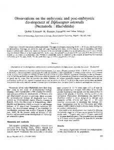
1723
Development 124, 1723-1732 (1997) Printed in Great Britain © The Company of Biologists Limited 1997 DEV1168
Sequence and embryonic expression of the amphioxus engrailed gene (AmphiEn): the metameric pattern of transcription resembles that of its segment-polarity homolog in Drosophila Linda Z. Holland1,*, Mamata Kene2, Nic A. Williams3 and Nicholas D. Holland1 1Marine Biology Research Division, Scripps Institution of Oceanography, La Jolla, CA 92093-0202, USA 2Department of Biology, University of Southern California, Los Angeles, CA 90089-0371, USA 3Department of Animal and Microbial Sciences, University of Reading, Whiteknights, Reading RG6 2AJ,
UK
*Author for correspondence (e-mail:
[email protected])
SUMMARY Vertebrate segmentation has been proposed as an evolutionary inheritance either from some metameric protostome or from a more closely related deuterostome. To address this question, we studied the developmental expression of AmphiEn, the engrailed gene of amphioxus, the closest living invertebrate relative of the vertebrates. In neurula embryos of amphioxus, AmphiEn is expressed along the anteroposterior axis as metameric stripes, each located in the posterior part of a nascent or newly formed segment. This pattern resembles the expression stripes of the segment-polarity gene engrailed, which has a key role in establishing and maintaining the metameres in embryos of Drosophila and other metameric protostomes. Later, amphioxus embryos express AmphiEn in non-metameric patterns — transiently in the embryonic ectoderm and dorsal nerve cord. Nerve cord expression occurs in a few cells approximately midway along the rostrocaudal axis and also in a conspicuous group of anterior cells in the
cerebral vesicle at a level previously identified as corresponding to the vertebrate diencephalon. Compared to vertebrate engrailed expression at the midbrain/hindbrain boundary, AmphiEn expression in the cerebral vesicle is relatively late. Thus, it is uncertain whether the cerebral vesicle expression marks the rostral end of the amphioxus hindbrain; if it does, then amphioxus may have little or no homolog of the vertebrate midbrain. The segmental expression of AmphiEn in forming somites suggests that the functions of engrailed homologs in establishing and maintaining a metameric body plan may have arisen only once during animal evolution. If so, the protostomes and deuterostomes probably shared a common segmented ancestor.
INTRODUCTION
metameres, are developmental compartments. However, arguments for such a common segmented ancestor appeared undermined by the balance of the molecular genetic evidence. For example, Hox gene homologs are expressed along the rostrocaudal axis of non-metameric animals like Caenorhabditis (Dickinson, 1995). Moreover, insect metamerism is established via pair-rule and segment-polarity genes, whereas vertebrate homologs of such genes are expressed in metameres only after these structures have become well defined morphologically (Patel, 1994; De Robertis and Sasai, 1996). Even so, the question of a common segmented ancestor for vertebrates and insects has not been laid to rest. Müller et al. (1996a) reported that her-1, a zebrafish homolog of hairy, a Drosophila pair-rule gene, is expressed in a pair-rule pattern in posterior mesoderm preceding any morphological differentiation of somites. This intriguing result led Kimmel (1996) to suggest anew that insects and vertebrates shared a common, metameric ancestor. Should this be so, the question is: why are most vertebrate homologs of Drosophila pair-rule and segment-polarity genes apparently not involved in establishing vertebrate segmentation?
The evolutionary origin of rostrocaudal segmentation in vertebrates is a central question in schemes deriving vertebrates from invertebrates. Some authors (e.g. Leydig, 1864; Dohrn, 1875; Patten, 1912) speculated that vertebrate segmentation is inherited from some protostome, often an arthropod or annelid. In contrast, others (e.g. Bateson, 1886; Garstang, 1928; Jefferies, 1986) argued that vertebrate segmentation originated in some deuterostome group, a scheme that connotes independent origins for vertebrate and protostome segmentation. Advances in molecular biology have strongly impacted ideas about the origin of the vertebrates in general and of vertebrate metamery in particular (Gee, 1996). The evolution of insects and vertebrates from a common segmented ancestor was proposed following landmark discoveries in developmental genetics: first by McGinnis et al. (1984) after the discovery of conserved homeoboxes in Drosophila homeotic genes and vertebrate Hox genes, and again by Lawrence (1990) after the demonstration that vertebrate rhombomeres, like insect
Key words: amphioxus, engrailed, segmentation, metamery, AmphiEn, Drosophila
1724 L. Z. Holland and others To address this question, we are studying amphioxus (phylum Chordata; subphylum Cephalochordata), widely believed to be the closest living invertebrate relative of the vertebrates (Wada and Satoh, 1994) and a useful stand-in for the proximate invertebrate ancestor of the vertebrates. The genomic organization of amphioxus is vertebrate-like, but simpler since amphioxus lacks the extensive gene duplications correlated with the marked increase in vertebrate structural complexity (Garcia-Fernàndez and Holland, 1994; Holland et al., 1994b). In addition, because the overall body plan of amphioxus is also vertebrate-like, the expression domains of developmental genes can help indicate homologies between body parts (Holland, 1996). For example, expression domains of amphioxus homologs of Pax-1, Brachyury, Distal-less, Otx, Hox-1, and Hox-3 support homologies of amphioxus structures with vertebrate gill slits, notochord, forebrain and hindbrain (Holland et al., 1992; Holland et al., 1994a; N. D. Holland et al., 1995; P. W. H. Holland et al., 1995; Holland and Holland, 1996; N. D. Holland et al., 1996; Williams and Holland, 1996). The present paper concerns the engrailed gene of amphioxus. Homologs of engrailed are widespread in animals generally, constitute a distinctive class of homeobox genes encoding DNA-binding transcription factors and are expressed at several developmental stages in diverse tissues in all three germ layers. engrailed genes have been most studied in their earliest expression domains — the midbrain/hindbrain junction of vertebrates (Ekker et al., 1992; Danielian and McMahon, 1996) and the forming segments of insects (Kornberg and Tabata, 1993) — as well as in one later expression domain, the forming wing of insects (Hidalgo, 1996). Biochemical and genetic studies of engrailed have concerned upstream promoters of the gene (Logan et al., 1993; Song et al., 1996) or modulators and downstream targets of the gene product (Mann, 1994; Serrano et al., 1995; Peltenburg and Murre, 1996), which is typically a repressor of activated transcription (Jaynes and O’Farrell, 1991; Han and Manley, 1993). Suggested functions for engrailed include roles in neurogenesis, axon targeting, establishment/maintenance of compartment boundaries, cell cycle control and cytodifferentiation (Logan et al., 1993; Condron et al., 1994; Friedman and O’Leary, 1996; Hidalgo, 1996; Loomis et al., 1996; Rétaux et al., 1996). During Drosophila development, the earliest expression of engrailed is in a banded pattern in the ectoderm (and more transiently in the early mesoderm), where it acts as a segmentpolarity gene establishing and maintaining the metameres; later expression is in a subset of neural cells and in a few other tissues (Lawrence, 1992). In contrast, during vertebrate development, the earliest expression of engrailed homologs is not banded, but occurs in a single broad stripe in the neuroepithelium at the midbrain/hindbrain boundary (Patel et al., 1989b; Davis et al., 1991). Later in development, vertebrate engrailed genes are transcribed in widely distributed neural cells as well as in several other tissues. Some of these later expression domains of vertebrate engrailed may appear in an iterated pattern (e.g. the muscle pioneer cells), but only in structures that have already become morphologically segmented (Hatta et al., 1991; Ekker et al., 1992). Thus, comparisons of engrailed expression between vertebrates and other animals have been consistent with an independent origin of segmentation in protostomes and deuterostomes. In contrast to the vertebrate pattern, our present results show that amphioxus
engrailed has an early developmental expression in the forming segments and a later expression in the nervous system and ectoderm. The early transcription is in reiterated stripes along the rostrocaudal axis; each stripe is in the posterior part of a nascent or newly formed segment in a pattern resembling the expression domains of engrailed homologs of metameric protostomes (e.g. annelids and arthropods). This pattern is consistent with suggestions that protostomes and deuterostomes evolved from a common segmented ancestor.
MATERIALS AND METHODS Obtaining amphioxus and purification of DNA and RNA Adults of the Florida amphioxus (Branchiostoma floridae) were collected in Tampa Bay, Florida. Ripe males and females were spawned and the embryos were cultured at 23°C as previously described (Holland and Holland, 1993). Genomic DNA for library construction and Southern blotting was extracted from 25 adults in GuSCN and purified by ultracentrifugation (L. Z. Holland et al., 1996). For cDNA library construction, RNA was extracted from embryos by the method of Chomczynski and Sacchi (1987). DNA probes; construction and screening of libraries The polymerase chain reaction (PCR) was used to amplify a 233 base pair (bp) fragment (including the homeobox) from pooled genomic DNA of Branchiostoma floridae. The forward primer B and reverse primer D were, respectively: GA(A/C)AAGCGGCCGCGCAC(A/G)GCCTTC and TGGTTGTCAG(A/C/G/T)CCCTG(A/C/G/T)GCCATGAG (Holland and Williams, 1990). The PCR product was cloned into pUC18 and subcloned into pT7T3 (Pharmacia, Piscataway, NJ, USA). Eight clones were sequenced. For genomic library construction, DNA digested with Sau3A was cloned into the XhoI site of Lambda Fix II (Stratagene, La Jolla, CA, USA). About 250,000 clones of the once-amplified library were screened with the insert of one PCR clone labeled with 32P by random priming. Hybridization was at 65°C in 6× SSC, 10× Denhardt’s, 0.1% SDS, 100 µg/ml tRNA, 1 mM EDTA with 106 cts/minute/ml of probe (specific activity of 108 cts/minute/µg). After hybridization, plaquelifts were washed 3× 20 minutes in 1× SSC, 0.1% SDS at 60°C. A cDNA library was constructed in the pSPORT vector (GIBCOBRL, Gaithersburg, MD, USA) from RNA pooled from 26-hour embryos. Approximately 60,000 unamplified clones were screened with a SalI-Eco0109 fragment (862 bp) of a genomic clone; the fragment, comprising the coding region 3′ of the intron (including the homeobox) and the most 5′ two-thirds of the 3′ untranslated region (UTR), was labeled with 32P by random priming. Colony lifts were hybridized in 6.95% SDS, 1 mM EDTA, 0.1 mg/ml tRNA, 0.5 M sodium phosphate buffer (pH 7.2) at 65°C with 106 cts/minute/ml of 32P-labeled probe and washed as already described for the genomic screening. Southern blot analysis Fifteen 10 µg aliquots of genomic DNA were each digested with a different restriction enzyme, subjected to electrophoresis and transferred to Hybond N+ (Amersham Life Sciences, Cleveland, OH, USA) according to L. Z. Holland et al. (1995). The Southern blot was hybridized at low stringency (50°C) with the same probe used for screening the CDNA library and in the same hybridization solution for genomic library screening. Washes were 2× 20 minutes at 55°C in 2× SSC, 0.1% SDS (L. Z. Holland et al., 1995). In situ hybridization and histology Expression of amphioxus engrailed was determined by in situ hybridizations on embryos of Branchiostoma floridae fixed at intervals
Amphioxus engrailed gene 1725 during the first 2 days of development (L. Z. Holland et al., 1996). Fertilization envelopes were removed from prehatching stages to facilitate penetration of reagents. Two antisense riboprobes were combined to maximize the signal. A 3′ probe was synthesized with a SalI genomic subclone as a template and a 5′ probe was synthesized from a partial cDNA clone (see results). After being photographed as whole mounts, labeled embryos were prepared as histological sections. In addition, some embryos were hybridized with riboprobes for both engrailed and for the amphioxus Distal-less homolog (N. D. Holland et al., 1996).
RESULTS Sequence analysis Amplification by PCR of the engrailed homeobox of Branchiostoma floridae yielded two types of clones with identical deduced amino acid sequences, but somewhat different base sequences. Screening a B. floridae genomic library with one of the PCR clones yielded 12 strongly hybridizing clones.
Although no two clones gave exactly the same restriction pattern when mapped with 6 different restriction enzymes, sequencing with a primer to the homeobox revealed that all were engrailed. More extensive sequencing of three clones showed that 3′ of the intron splice site, all had the same deduced amino acid sequence, although the base sequences differed somewhat (approximately one out of 100 positions in the coding region and one out of 50 in the 3′ UTR and intron). In addition, there were short (up to 3 bp) deletions and insertions in the 3′ UTRs and longer (up to 20 bp) insertions and deletions in the intron. The base sequences of the two most similar genomic clones corresponded to the two types of clones obtained by PCR. Sequencing of one genomic clone revealed that the intron exceeded 4,000 bp. Therefore, to obtain the coding region 5′ of the intron plus the 5′ UTR, we screened a cDNA library with part of one genomic clone. One cDNA clone was obtained — a partial clone including the entire cDNA 5′ of the intron splice site plus 72 bp 3′ of the splice site. Since the base sequence of the cDNA in the overlap zone
1 62 143
AACCTAGTTTCCTACAATCACTGTCAGGGAGAGGGGAAACACTGAGCTAAGTGACTCAAGC GCTCCGACCCCAGGGCAGTTGTTTATAGCCGGCGAACTCTGAGCACCGGGAGATTATAAGTAGGAGTAATAGTGACTGACA TTTTGGATTCGCGTGCGTCTTCGGTCCTTTCAATATAGGGTCGAAGTTCTTGGAGACAGAAGCGAGTAGGAGGACCGTAAG
61 142 223
224 1
ATGGCGAACAGTAGCGCGGATGAGAACGGGAACCCACCGGCGAGCCCGCCGCCTCCTCACAGCCCGGTCCAACCGGAGGAC M A N S S A D E N G N P P A S P P P P H S P V Q P E D
304 27
305 28
AGTCCGAGCCCTGACGGCCCGCGCACCACCAACTTTTCCATCGCGAACATCCTCCGGCCGGAGTTCGGCGCTCGCAAGAGA S P S P D G P R T T N F S I A N I L R P E F G A R K R
385 54
386 55
GACAGTAAAAGTTGCGACTCCTCTCCCGTGTCCAGTCCGGGGAAACCGGCACCGGGAGACCTCTCCCCCTCCCTGCCGGCC D S K S C D S S P V S S P G K P A P G D L S P S L P A
466 81
467 82
AGCCCGGGCGGAGTCCCGCAGGAGAACACGACGACAGCCGATGGACTCCGCCTCGAGGATGACCCCGCCAAGCCCGTTACG S P G G V P Q E N T T T A D G L R L E D D P A K P V T
547 108
548 109
EH2 GCAGACGATAAGGACGGTAAACCGGTGCCCCAGCCTATGATCTGGCCAGCGTGGGTCTACTGTACCCGCTACTCGGACCGC A D D K D G K P V P Q P M I W P A W V Y C T R Y S D R
628 135
629 136
EH3 CCTTCTTCTGTTCGCACCGCAGGACCCCGAACGCGGAAAGCTCGGCCCAAAGATCCGAAGAAAGCCGAGGAGAAGCGGCCG P S S V R T A G P R T R K A R P K D P K K A E E K R P
709 162
710 163
EH4 = homeobox AGAACGGCGTTCACTTCCGAACAGCTCCAGCGCCTGAAAAAGGAGTTCCAAGAGAACCGTTACCTCACAGAGCAACGGCGG R T A F T S E Q L Q R L K K E F Q E N R Y L T E Q R R
790 189
791 190
EH4 = homeobox CAGGATTTGGCAAGAGAGCTGAAGCTAAACGAGTCACAGATCAAGATTTGGTTTCAGAATAAGCGTGCGAAAATCAAGAAG Q D L A R E L K L N E S Q I K I W F Q N K R A K I K K
871 216
872 217
EH5 GCGGCTGGAGTACGGAACGGACTAGCACTCCACCTCATGGCGCAGGGGCTCTACAACCATTCCACCATGCCGACAATGGGA A A G V R N G L A L H L M A Q G L Y N H S T M P T M G
952 243
953 244
GACGAGCACGGGCTCGATATGCATGACTAGTGCGGCAAACAAACCTCGGCGGCGAACCGGCCCGTGAAATTGTGGACAGCC 1033 D E H G L D M H D 252
1034
GGACATCAGAAACATCTTACGGACAAGCATTGTCTGGACATATCGTCTAACTGACCAACTGAATCTCAGAGCTATGTTCCT 1114
1115 1196 1277 1358 1439 1520
TGTTTCAATACAAAAAAGCATCAAGTAAGCTTAATGGTTTAGAAGGTAAGGTCTCAAAATTTTTGCTAGTGTCTTAATTTA TTGTGACGATATTTTTGGCAACAGAAAAGGGTAGATTCTTTTGCGGACGAAAAAAGAATAATGAATGGACAGTACAAAAGA AGGAATCGTTTGATCAGTCTACCTTGATATATTTACATATCCATAGCGGACAGAAGAGAGATATTATTTTGTCAGTCAGTC TCTGAATTCTGTGATCTTTTGACAAGTGAAATTTGTGTTTTTCTGTTCGTGTGTCTATAGAATGAAATAGTTGTAAATACC CGTACAATTGCACGTCGTTGTCACCGTGCGAGCAAGTTAGTCAATATCTCTTTTTTCTCTTCTTTATCTCTTTTTTTGTTG TAGTCGACGGATCGATAAGCTTATATCTAAAGGGAGTCGACTCGATCCTCTATCTGTTCGAATTCCTGCAGCCCGGGGGAT
1601 1682
CCACTGAGTTCTTCTAGAGCGGCCGCCACCGGTGAGCTCAATTAACCCTCATGTATGGGGGCTCAAAAGTGACAATGGAAG 1681 1760 TCAATTCTATCATAACGCTGTTATCTGTTACTGACCGTCCAGATTTAATAAACCAAACCGTAATATATTCAATAAAAAA
EH1
Fig. 1. Base and deduced amino acid sequences of amphioxus AmphiEn (GenBank accession number U82487). Composite base sequence from genomic (3′ of the intron) and cDNA clones (5′ of intron). Location of poly(A) tail is assumed to coincide with consensus (t)5 21 bp downstream of the polyadenylation signal (underlined) in genomic clone. Inframe stop codons upstream from translational start site also underlined. Boxed regions (EH1, EH2, EH3, EH4 = homeobox, and EH5) are highly conserved among most metazoan engrailed homologs. Open arrowhead, most 5′ possible site for intron; closed arrowhead, nearest consensus sequence for 3′ intron splice site.
1195 1276 1357 1438 1519 1600
1726 L. Z. Holland and others
Fig. 2. Amino acid sequences of amphioxus AmphiEn and engrailed proteins of some other animals. Alignment with Clustal V; amino acid identities indicated by colons; other notations as in Fig. 1. Zebrafish (zf) sequences (Ekker et al.,1992); mouse (mo) sequences (Logan et al., 1992); Drosophila sequence (Poole et al., 1985); brine shrimp (Artemia) sequence (Manzanares et al., 1993); flatworm (Schistosoma) sequence (Webster and Mansour, 1992).
AmphiEn zfEn-1 zfEn-2 zfEn-3 mo En-1 mo En-2 Drosoph Artemia fluke
MANSSADENG----------------------------------------------------------------NPPASPPPPH MEDQRRGQGEEEDDSGS-------------------------------------------------------LPS::LLPA--MDENEQSARDVEQRGASDESNSAIRPL--------------------------------------------------------MEENDHSNRDVERQDSGDESNRAILPL--------------------------------------------------------MEEQQPEPKSQRDSGLGAVAAAAPSGLSLSLSPGASGSSGSDGDSVPVSPQPAPPSPPAAPCLPPLAHHPHLPPH::PP::::P MEEKDSKP--------SETAAEAQRQ------PEPSSGGGSGGGSSPSDSDTGRRRALMLP----------------------M:LEDRCSPQSAPSPITLQMQHLHHQQQQQQQQQQQMQHLHQLQQ...79 aa...DMSFHNQTHTTNEEEEAEEDDDIDVDVD MGSAIFEPGPLSLLNLACSNLTERYDGPSPLSASTPGPSPDRPGS...40 aa...TGFPTLAAIQSGHLAFRQLV:TL:FNTMFKLLDNF:EKSKSIMIEQMKQQYCLLQYDKE...78 aa...TTTTTSTTTTTSGIIPLSSTIIPSTIISPVKTSLSSLSMMT EH1
AmphiEn zfEn-1 zfEn-2 zfEn-3 mo En-1 mo En-2 Drosoph Artemia fluke
SPVQPEDSPSPDG------PRTTNFSIANILRPEFGARK--------RDSKSC---------------DSSPVSSPGKPAPGDL -------------------H:N:D:F:D:::::D::CKRERE--KVT:::GVRPTALPDSRSDGVSSSA::T::::VSSR--------LQA:GNLQLP----H:I:::F:D:::::D::RK:EANIT:YED--------------------------------NHGA -----LQA:GNV-LP----H:I:::Y:D:::::D::R::EGS-R:DEI--------------------------------NIVE P:P:HLAA:AHQPQPAAQLH:::::F:D:::::D::CK:EQPLPQLLVASAAAGGGAAAGGGSRVERDRGQTGAGRDPVHSLGT ---EVLQA:GNHQHP----H:I:::F:D::::::::R::DAG-----TCCAGAGGARGGEGGAGTTEGGGGGAGGAEQL--LGA DTSAGGRLPPPAHQQQSTAKPSLA:::S:::SDR::DVQKPGKSIENQASIFRPF...87 aa...KSALGSLCKAVSQIGQPA VKSSEGQVKEVVSTQ--SQKKPLA:::DS::::D::K-------------------------ETNEVKRRHASPHREEPKKK-IDDLQNDISKINYEEDFFSKLLKCTL:R:ENSYSNEIEQLSLSSSSSSS:SSSSSSSSSSCSTNSSSSVYMSEKKANGFFVKDI
AmphiEn zfEn-1 zfEn-2 zfEn-3 mo En-1 mo En-2 Drosoph Artemia fluke
-----------------------SPSLPASPGGVPQENTTTADGLRLEDDPAKPVTADDKDG-----------KP--------QSNKVEQGSSKSSSPS-------------------------------------------------------------------RENHNPT---------GPSTG-----QVGSTVPAEEAS::HTSSGGK:AEIES----------------EEPL::RGENVDQ-RENRCPS---------APGSG-----QV:P-VSGEGTSSPRAVNASKKT:IST----------------DESL:SRAETGDQ-RASGAASLLCAPDANCGPPDGSQPATAVGAGASKAGNPAAAAAAAAAAAAA:VAAA:AAASKPSDGGGSGGNAGSPGAQGAKFP RESR-------PNPACAPSAG-------GTLSAAAGDPAVDGE:GSKTLSLH----GGAKKPGDPGGSLDGVL:ARGLGGGDLS APTMTQPPLSSSASSLASPPPASNASTISSTSSVATSSSSSSSGCSSAASSLNSSPSSRLGASGSGVNASSPQPQPIPPPSAVS VQYIEQMKKKEEIKEEARTESRL:SSSKD:VPDNDKI:P--------------------------------------------LSFDKHKVIRRQKTDDSFEKEVLVKGRDEEKK...52 aa...VGSEEEEDNDDIN:AAKNNNTNYLRKTVSDDGMKLKSNRNH
Amphien zfEn-1 zfEn-2 zfEn-3 Mo En-1 Mo En-2 Drosoph Artemia fluke
---------------------VPQPMIWPAWVYCTRYSDRPSSVRTAG--PRTRKARPKDPKKA-EEKRPRTAFTSEQLQRLKK -------------------KDSQKQIL::::::::::::::::----:--:::::LKK:NNNTESDD::::::::A:::::::A -----CLGSESDSSQSNSNGQTG:G:L::::::::::::::::----:--::S::PKK:A--ASK:D::::::::A:::::::A -----CLSSDSDCSQRC-AAQAK:::L::::::::::::::::----:--::S::PKK:T--PTK:D::::::::A:::::::N EHNPAILLMGSANGGPVVKTDSQ::LV::::::::::::::::----:--:::::LKK:K--NEK:D::::::::A:::::::A -------VSSDSDSSQASATLGA:::L::::::::::::::::----:--::S::PKK:N--PNK:D::::::::A:::::::A RDSGMESSDDTRSETGSTTTEGGKNEM::::::::::::::::----:--::Y:RPK-QPKDKTND::::::::S::::A:::R -------------------PLP:EASK:::::F::::::::::----:RS::C:RMKK:KAITP-D:::::::::A:::S:::H NKKLGSRGRIHEITSSKLNTND:SRLHL::::F::::::::::----:--::I::P:MNRSNDELNL:::::S::VP::K::SQ
AmphiEn zfEn-1 zfEn-2 zfEn-3 mo En-1 mo En-2 Drosoph Artemia fluke
EFQENRYLTEQRRQDLARELKLNESQIKIWFQNKRAKIKKAAGVRNGLALHLMAQGLYNHSTMPTMGDEHG--------LDMHD :::TS::I::::::A:::::G:::::::::::::::::::SS:FK:A::MQ:::::::::::TTIQEE:D-------------N :::T::::::::::S::Q::G::::::::::::::::::::S::K::::I::::::::::::TSKEDKSDS------------H :::N::::::::::A::Q::G::::::::::::::::::::T:NK:T::V::::::::::A:VTKDDKSDS------------: :::A:::I::::::T::Q::S::::::::::::::::::::T:IK:::::::::::::::::TTVQDKDES------------E :::T::::::::::S::Q::S::::::::::::::::::::T:NK:T::V::::::::::::TAKE:KSDS------------E ::N:::::::R:::Q:SS::G:::A:::::::::::::::ST:SK:P:::Q:::::::::T:V:LTKE:EE-LEMRMNGQ--IP ::N:::::::R:::::::::G:H:N::::::::N:::L::SS:QK:P:::Q:::::::::::I::ED:EDD-EISSTSLQARIE ::EK::::D:L::KK::T::D:R:::V::::::::::T:::S:AQ:C:::::::E::::::VRVRSDI:EDEEDSDDMNTSEKE
EH2
EH4 = homeodomain
matched most closely the base sequence of the genomic clone used to screen the cDNA library, we chose the latter clone to make a composite cDNA sequence of the amphioxus engrailed gene, which we named AmphiEn. Fig. 1 shows the complete cDNA and deduced amino acid sequences of AmphiEn. The cDNA is 1,760 bp long with a polyadenylation signal 21 bp upstream from the poly(A) tail. There are several in-frame stop codons upstream of the indicated translational start site. The open triangle in Fig. 1 shows where the base sequences of the cDNA (CCCTTCT↓TCTGTTC) and genomic DNA clones (CCGATGC↓TCTGTTC) diverge. This position is, therefore, the most 5′ possible for the intron. However, the base sequence at this location does not correspond to the consensus sequence for the 3′ intron splice site (TXCAG↓), the nearest CAG being 14 bp downstream at a site corresponding to an intron in engrailed genes in other species (Figs 1, 2, closed arrowhead). If the 3′ intron splice site in AmphiEn is at this consensus sequence, then an unusually large number of bases (16) must be repeated at the 3′ end of the preceding exon. Although the available data do not rule out the presence of an additional, more 5′ intron, such a second intron is unlikely, because more 5′ introns are lacking in engrailed genes of other animals. The longest open reading frame codes for a protein of 252 amino acids (Fig. 1) that includes a homeodomain (EH4) of
EH3
EH5
the engrailed class as well as four additional conserved regions, EH1, EH2, EH3 and EH5 (Logan et al., 1992) (Fig. 2). In Drosophila, EH1 and EH5 are required for full repression activity, and EH2-EH3 influence targeting of engrailed (Peltenberg and Murre, 1996; Smith and Jaynes, 1996). AmphiEn contains an insert of four amino acids in EH2, VRTA (Fig. 2), which, being located in EH2, might influence targeting of the protein. AmphiEn, like mouse En-1, includes a proline-rich region upstream from EH1 that has been suggested as a potential activation domain (Logan et al., 1992). Southern blot analysis Fig. 3 shows a Southern blot hybridized at low stringency with an 862 bp stretch of AmphiEn comprising the coding region 3′ of the intron (including the homeobox and regions EH3 and EH5) plus the most 5′ two-thirds of the 3′ untranslated region. Digestion by six out of 12 restriction enzymes yielded a single major hybridization band. This result, together with the results from sequencing, suggests that AmphiEn is the sole engrailed gene in Branchiostoma floridae. The two bands from digestion by KpnI and SpeI, as well as the faint band at 6 kb from SmaI are presumably due to polymorphism. Developmental expression of AmphiEn No AmphiEn transcripts are detectable in the blastula or gastrula
Amphioxus engrailed gene 1727
Fig. 3. Genomic Southern blot analysis of pooled amphioxus DNA. Numbers at top indicate digestion in: 1, BglII; 2, BstEII; 3, BstXI; 4, Eco0109I; 5, EcoRI; 6, HindIII; 7, KpnI; 8, NcoI; 9, PstI; 10, SalI; 11, SmaI; 12, SpeI. Blot probed at low stringency with 862 bp fragment of AmphiEn including homeobox. Size markers in kilobases at left.
only in the dorsal nervous system. Expression continues in the two small groups of neural tube cells (Fig. 4J,M, arrowheads) midway along the rostrocaudal axis just caudal of the first pigment spot. Within the anterior end of the neural tube, the medullary canal has dilated slightly, thus defining a region called the cerebral vesicle. There is a conspicuous zone of AmphiEn expression in the wall of the cerebral vesicle about 50 µm caudal of the neuropore (Fig. 4J,K). Histological cross sections show that the expressing cells are concentrated ventrolaterally on either side of the cerebral vesicle (Fig. 4L). In 24-hour embryos hybridized simultaneously with riboprobes recognizing AmphiEn and amphioxus Distal-less (N. D. Holland et al., 1996), the zone of AmphiEn expression is offset slightly rostrally from the strong zone of dorsal expression of the Distal-less gene (Fig. 4N). By 30 hours of development, AmphiEn transcripts are no longer detectable anywhere in the embryo by whole-mount in situ hybridization. DISCUSSION
stages. Expression is first seen in early neurulae at 9.5 hours (Fig. 4A), as paraxial mesoderm evaginates from the archenteron to form presomitic grooves on either side of the midline. Anteriorly, the first myogenic somite (numbering system of Conklin, 1932) has been formed by a constriction of the wall of the groove, and the second somite has partly formed. AmphiEn is expressed in three stripes — the most anterior in cells constituting the posterior third of the first somite, the next in posterior cells of the second somite as it is forming and the most posterior in the wall of the presomitic groove at a rostrocaudal level where the posterior part of the third somite will be located after constricting off from the groove. Significantly, at this last site, AmphiEn expression precedes any morphological sign of a posterior boundary for the third somite. The pinching-off of definitive somites from the presomitic groove progresses in a caudal direction. Just before constriction produces a new somite, expression of AmphiEn begins in the future posterior cells of the somite. By hatching (11 hours), there are three or four definitive somites on either side (Fig. 1B,C). The posterior region of each somite expresses AmphiEn with an intensity progressively decreasing from rostral to caudal. By 12 hours, AmphiEn is expressed in the posterior portion of each of the first six somites on either side of the midline (Fig. 4D,E) and, by 13.5 hours, AmphiEn is expressed posteriorly in the first eight somites on either side (Fig. 4F). At later developmental times, the subsequently forming somites never express AmphiEn. Further, in the late (15-hour) neurula, as the ninth somite begins to form, expression in the first eight somites diminishes rapidly, although it lingers longest in the posterior part of the first somite on the right side (Fig. 4G,H, arrow). At 15 hours, expression of AmphiEn begins in two small groups of cells (one to three cells each) located in the ventrolateral wall of the neural tube about midway along the rostrocaudal axis (Fig. 4G,H, arrowhead). By 17 hours, AmphiEn expression is no longer detectable in the right first somite; however, transcripts are still found in a few neural tube cells (out of the plane of focus in Fig. 4I) midway along the rostrocaudal axis, and weak, transient expression appears in a band of epidermal cells approximately in register with the second somite (Fig. 4I, arrow). In the 24-hour embryo, AmphiEn expression is detectable
Sequence comparisons between AmphiEn and engrailed of other animals In the genome of Branchiostoma floridae, AmphiEn is evidently the only member of the engrailed family. A single engrailed gene appears to be a plesiomorphic feature of animals generally (reviewed by Wray et al., 1995); the known exceptions are higher insects (two engrailed genes), a cephalopod mollusc (two engrailed genes) and most vertebrates (two or sometimes three engrailed genes). The gene duplications almost certainly arose independently in the insects, molluscs and vertebrates, and the presence of a single engrailed gene in amphioxus, the most likely sister group of the vertebrates, is consistent with this conclusion. A comparison of the amino acid sequences of engrailed proteins among the animals in Fig. 2 shows that AmphiEn shares the most identities with vertebrate engrailed proteins, somewhat fewer with arthropod engrailed and considerably fewer with flatworm engrailed. AmphiEn is probably a fair representative of the single engrailed protein present in the earliest vertebrates before gene duplications took place. However, AmphiEn is not obviously related more closely to any one vertebrate homolog (En-1, En-2 or En-3) than to any other. In spite of structural similarities among engrailed proteins of different animals (Fig. 2), AmphiEn-containing cells in amphioxus were not labeled by antibodies against engrailed proteins of foreign species. The monoclonal antibody against Drosophila engrailed (4D9) evidently fails to recognize AmphiEn, because recognition requires a glycine at homeodomain position 40, where AmphiEn has a lysine. Polyclonal antibodies raised against mouse (αEnhb-1) and leech (αht-en) engrailed proteins did recognize proteins in some neural tube cells, but in a pattern unrelated to cells transcribing AmphiEn. Moreover, each polyclonal recognized a distinctly different subpopulation of cells. Neural expression compared between AmphiEn and engrailed homologs of other animals The most straightforward comparison of expression of engrailed homologs between amphioxus and vertebrates is the relatively late transcription observed in scattered cells in the central nervous system. In amphioxus, transcripts occur in rel-
1728 L. Z. Holland and others
atively few cells in a region that Holland et al. (1992) proposed as equivalent to part of the vertebrate hindbrain. In vertebrates, the later neural expression is in cells scattered within the hindbrain and spinal cord (Davis et al., 1991; Hatta et al., 1991; Gardner and Barald, 1992). Davis et al. (1991) proposed that engrailed expression in such cells may be involved in the differentiation of a set of spatially defined neurons. Neural expression of engrailed homologs is also observed as a conspicuous stripe in the cerebral vesicle of amphioxus (present results) and at the midbrain/hindbrain boundary of vertebrates (Patel et al., 1989b; Davis et al., 1991; Hatta et al., 1991; Ekker et al., 1992; Gardner and Barald, 1992). However,
this major neural expression domain of AmphiEn differs in three ways from that of vertebrate engrailed: AmphiEn (1) is transcribed relatively late in development, (2) is not transcribed dorsally in the neural tube and (3) is expressed only 50-100 µm from the rostral end of the nerve cord — within a region thought (on the basis of neuroanatomy and expression of amphioxus homologs of the vertebrate forebrain markers Otx and Dll) to be equivalent to diencephalon (Lacalli, 1996; N. D. Holland et al., 1996; Williams and Holland, 1996). Thus, if the conspicuous AmphiEn expression in the cerebral vesicle of amphioxus truly marks a homolog of the vertebrate midbrain/hindbrain junction, one would be forced to conclude
Amphioxus engrailed gene 1729 Fig. 4. AmphiEn expression in amphioxus embryos; all except L are whole-mount in situs; anterior toward left; unless otherwise noted, all scale lines are 50 µm (A) Frontal view of 9.5 hour neurula; on either side of midline, first somite has formed from rostral end of presomitic groove; second somite is pinching off; arrows indicate AmphiEn expression in first two somites and presumptive third somite. (B) Side view of 11 hour neurula; single arrows indicate AmphiEn expression in first four somites; tandem arrow indicates neuropore. (C) Frontal view of 11 hour neurula; single arrows indicate AmphiEn expression in first four somites. (D) Frontal view of 12 hour neurula expressing AmphiEn in first six somites. (E) Enlargement of D showing AmphiEn expression in posterior region of each somite; scale line 25 µm. (F) Frontal view of 13.5 hour neurula expressing AmphiEn in first eight somites. (G) Partial frontal view and (H) side view of 15 hour neurula; AmphiEn expression persists only in first somite (arrow) on right side and is also detectable in a few neural tube cells (arrowhead) midway along rostrocaudal axis. (I) Side view of 17 hour neurula; arrow indicates epidermal cells weakly expressing AmphiEn. (J) Side view of 24 hour embryo; AmphiEn expressed inconspicuously in a few neural tube cells (arrowheads) just posterior to first pigment spot (p); conspicuous expression near rostral end of cerebral vesicle (top left). (K) Anterior of 24 hour embryo in side view showing AmphiEn expression in cerebral vesicle a short distance caudal to neuropore (tandem arrow). (L) 24 hour embryo cross-sectioned in plane x-x′ in K; AmphiEn expressed ventrolaterally within wall of cerebral vesicle, which encloses neural canal (arrow) and is dorsal to notochord (n); dark granules in epidermal cells are pigment, not positive reaction for AmphiEn transcripts. (M) Enlarged side view of 24 hour embryo showing AmphiEn-expressing neural tube cells (arrowheads) just posterior to first pigment spot (p). (N) Side view of anterior end of 24 hour embryo double-labelled for both AmphiEn and amphioxus Distal-less; within cerebral vesicle, the latter (d) is strongly expressed dorsal and slightly caudal to the former (e); tandem arrow, neuropore.
that amphioxus has little or no homolog of the vertebrate midbrain. Such a conclusion would conflict with neuroanatomical data suggesting that amphioxus has a midbrain as well as a diencephalon (Lacalli, 1996). This discrepancy emphasizes the need for further mapping of the amphioxus brain with genetic markers specific for vertebrate midbrain, such as amphioxus homologs of Pax-7 (Jostes et al., 1991) or her-5 (Müller et al., 1996b). Alternatively, the expression domain of AmphiEn in the amphioxus cerebral vesicle might be comparable to one of the three bands of engrailed expression that appear in arthropod brains relatively late in development (Hirth et al., 1995; Scholtz, 1995). When engrailed genes are compared across the animal kingdom, their most widespread function appears to be in neurogenesis. This commonality has led many to assume that, during animal evolution, engrailed originally functioned in neurogenesis and was only later co-opted for other functions, such as establishing a segmented body plan (Patel et al., 1989b; Joyner and Hanks, 1991; Holland, 1992; Webster and Mansour, 1992; Lans et al., 1993; Condron et al., 1994; Gee, 1996). The validity of this evolutionary scenario still requires testing by studies of the structure and function of engrailed homologs in a wide spectrum of lower invertebrate phyla. Early segmental expression compared between AmphiEn and engrailed homologs of other animals The genetic basis of segmentation is best understood for
Drosophila, an advanced insect (Lawrence, 1992): sequential expression of maternal, gap, pair-rule and segment-polarity genes subdivides the anteroposterior axis into successively smaller regions, culminating in a virtually simultaneous appearance of a series of parasegments. In each nascent parasegment, the anteriormost cells begin to express the segment-polarity gene, engrailed. The morphologically obvious segments appearing later in development are out of register with the parasegments such that the engrailed-expressing cells are posteriorly located in each segment. In studies of other protostomes, it is usually presumed that parasegments are the initial lineage units of metameres, although this is rarely established rigorously and the metameres actually described are usually segments. The general rule, to which AmphiEn conforms, is that engrailed homologs playing a role in establishing segments are expressed posteriorly in each metamere. From one group of metameric protostomes to the next, different germ layers express engrailed homologs during segment formation. In centipedes (Whitington et al., 1991) and annelids (Wedeen and Weisblat, 1991; Lans et al., 1993), expression is both mesodermal and ectodermal. In molluscs (Jacobs and De Salle, 1994; Jacobs, personal communication), crustaceans (Patel et al., 1989a; Scholtz et al., 1994; Manzanares et al., 1993, 1996) and insects expression is ectodermal — if one disregards transient transcription of engrailed homologs in the mesoderm at the onset of insect segmentation (Baylies et al., 1995). In comparing these germ layer differences in engrailed expression during segment formation, Lans et al. (1993) concluded that the mesodermal site of action was ancestral and an ectodermal site was derived. This implies a transfer of the site of action from one germ layer to another, which is an instance of ‘homeogenetic induction’ as defined by De Robertis et al. (1989). In this process, cells transcribing genes specifying position somehow induce neighboring cells in a different germ layer to express the same genes — in this instance, mesoderm cells homeogenetically induce adjacent ectoderm cells. Presumably, in some animals (e.g. arthropods), once the genetic fiducial marks for segmentation were activated in the ectoderm, those in the mesoderm became inactive. The virtually simultaneous formation of all the segments at once, as in Drosophila, is atypical of animals in general. Most metameric protostomes, including lower insects, add many or all segments one at a time in an orderly rostrocaudal sequence from a growth zone near the posterior end of the body (Brown et al., 1994; Klingler, 1994; Nagy, 1994; Patel, 1994). In vertebrates, early somites usually take shape sequentially within the paraxial segmental plates (Tam and Trainor, 1994); later somites may be generated sequentially from a posterior growth zone associated with the tail bud (De Robertis et al., 1994). In amphioxus, the formation of the first eight somites on either side of the midline appears comparable to the segmental plate stage of vertebrate somite formation, and the later somites arise sequentially from a posterior growth zone. Perhaps it is significant that AmphiEn is expressed only in the genesis of the first eight somites, which are formed by enterocoely, whereas the more posterior somites are formed from solid blocks of mesoderm that later hollow out by schizocoely (Jefferies, 1986). During vertebrate development, as already mentioned, engrailed homologs can be expressed in metameric structures, but only after such structures have already become morpho-
1730 L. Z. Holland and others logically segmented (Hatta et al., 1991; Ekker et al., 1992); thus vertebrate engrailed homologs are not considered candidate genes for controlling segmentation. Unlike vertebrates, many metameric protostomes begin to express engrailed homologs in presumptive segmental regions that only later become morphologically distinguishable as segments. In this respect, amphioxus resembles not vertebrates, but metameric protostomes, because AmphiEn expression commences in the nascent somites before they become morphologically distinct. The possibility that AmphiEn has homologous functions with the Drosophila segment-polarity gene engrailed should be addressed by studies of the relations of amphioxus AmphiEn to its upstream promoters and downstream targets. Evolutionary questions raised by a common segmented ancestor for metameric protostomes and deuterostomes Substantive questions arise from the proposal that metameric protostomes and deuterostomes evolved from a common ancestor in which mesodermal expression of engrailed played a role in segment development. For example: what became of the ancestral segmentation in some lower deuterostomes, and why doesn’t engrailed play a role in the establishment of vertebrate segmentation? If the ancestral deuterostome had extensive, protostome-like segmentation, this feature must have been partially or entirely lost in echinoderms, hemichordates and tunicates. For echinoderms, one can propose that a common segmented ancestor gave rise to the extinct segmented species (homalozoans) and also, by the reduction of segmentation, to the living species, which retain only embryonic trimery. In living echinoderms, developmental expression of engrailed homologs appears limited to cells scattered in the radial nerves (Popodi et al., 1996; Wray, 1996). For hemichordates, one can propose that a segmented ancestor lost most traces of segmentation (except for trimery and gill slits) in modern species. To date there have been no studies of hemichordate engrailed expression. For tunicates, the segmentation of larval ascidians and appendicularians is inconspicuous, if present at all. If one proposes a segmented ancestor for tunicates, the vague muscular segmentation would be a vestige of an originally metameric condition. Recent support for this interpretation comes from Wada et al. (1996), who found iterated ectodermal and neural expression of tunicate Pax-3/7 and suggested descent from a more elaborate segmental plan. Clearly, there is a pressing need for studies of developmental expression of engrailed and other segment-polarity genes in tunicates. Compared to the proposed loss of segmentation in some deuterostomes, the apparent loss of engrailed participation in forming the segmented body plan of vertebrates is a much more complex problem. In vertebrates the relationship between segmentation of the nerve cord and mesoderm is far from clear at both the morphological and the molecular levels. The picture is complicated by differences between head and trunk segmentation and between lower (lampreys) and higher vertebrates. Morphologically, somite formation in lower vertebrates resembles that in amphioxus, the anterior somites (called head cavities) being enterocoelic and the trunk somites being schizocoelic; these relationships led Gilland and Baker (1993) to homologize the 8-9 somite embryo of amphioxus
with the head of lower vertebrates. In higher vertebrates, head cavities are lacking and the head mesoderm is transiently segmented into somitomeres, condensations of mesodermal cells visible only with scanning electron microscopy. In the chick, the first true somite is at the level of rhombomere 7 in the hindbrain. In spite of differences in the mode of formation, in amphioxus and in both lower and higher vertebrates, the anterior and posterior mesodermal segments are considered to be serial homologs. In addition, the head cavities of lower vertebrates and the somitomeres of higher vertebrates are thought to be homologous even though they differ both in number and in the way they are formed. To complicate the situation further, there is not a 1:1 correspondence between mesodermal segments and segments of the neuroectoderm. Nevertheless, there have been several schemes for aligning the segments in amphioxus and vertebrates. The most recent takes into consideration Hox gene expression in the nerve cord as well as anatomy (Gilland and Baker, 1993). In this scheme, the first somite of amphioxus is equated with the mandibular head cavity (second somite) of the lamprey and shark and to the first three somitomeres of the chick, the second through the fifth amphioxus somites are equated with somites 3-5 of the lamprey and shark and to somitomeres 4-7 of the chick, while the sixth somite of amphioxus is equated with the sixth somite in the lamprey and shark and to the first true somite in the chick. The genes involved in establishing metamerism in the vertebrate nerve cord and mesoderm are poorly understood: En-1 and Wnt-1 evidently have roles in establishing the midbrain/hindbrain boundary (McMahon et al., 1992; Wurst et al., 1994); Krox-20 and kreisler appear to be involved in segmentation of the hindbrain (Lumsden and Krumlauf, 1996;) and a homolog of the Drosophila pair-rule gene hairy may function in establishing metamerism of the mesoderm (Müller et al., 1996a). However, there is no evidence of a role for engrailed in establishing mesodermal segmentation. If vertebrates did arise from amphioxus-like ancestors in which mesodermal expression of engrailed played a role in establishing segmentation, then a role for engrailed in establishing vertebrate segmentation has apparently been lost. It is surprising, if the anterior somites of amphioxus are indeed homologous to the mesodermal head segments of vertebrates, that the genetic basis of vertebrate segmentation is so different from that in amphioxus. Baker (1992) has suggested that the primary metameric plan of vertebrates resided in the mesoderm early in their phylogeny, but became transferred to the neuroectoderm and our present results are compatible with this idea. In terms of homeogenetic induction, mesodermal cells using genes (including engrailed) to help specify segmentation should have induced expression of the same genes in neighboring neuroectodermal cells. One possible explanation for the loss of a role for engrailed in segmentation of the vertebrate mesoderm is that specification of segments may have been strongly influenced by the epithelium-to-mesenchyme transition that precedes the formation of most somites (Keynes and Stern, 1988). Perhaps the additional genetic controls needed for such a transition are somehow incompatible with the establishment of metamery by segmentation genes. This idea is consistent with the expression of lamprey engrailed in the posterior wall of the forming mandibular head cavity, which gives rise to the muscles of the
Amphioxus engrailed gene 1731 mandibular arch (Holland et al., 1993). This engrailed expression might be a last vestige of the involvement of this segment-polarity gene in vertebrate somite formation. In conclusion, the embryo of amphioxus, which is a deuterostome, becomes striped with zones of AmphiEn expression, each located in the posterior part of a nascent or newly formed segment. This resembles the expression pattern of engrailed homologs in embryos of metameric protostomes. Therefore, functions of engrailed in establishing and maintaining a metameric body plan may have arisen only once in evolution — in a common segmented ancestor of protostomes and deuterostomes. This insight, along with recent genetic information suggesting a reversal of the dorsoventral axes between Drosophila and vertebrates (Jones and Smith, 1995; De Robertis and Sasai, 1996), favors phylogenetic scenarios deriving the vertebrates from annelid-like or arthropod-like body plans. We thank Ray Wilson for laboratory facilities at University of South Florida (St Petersburg). Antibodies were generously supplied by Clay Davis (αEnhb-1), Nipam Patel (4D9) and Cathy Wedeen (αht-en). Our research was supported by NSF research grant IBN 96-309938 (to N. D. H. and L. Z. H.). M. K. was supported by the NSF Research Experience for Undergraduates Program.
REFERENCES Baker, R. (1992). A contemporary view of the phylogenetic history of eye muscles and motoneurons. In Vestibular and Brain Stem Control of Eye, Head and Body Movements. (ed. H. Shimazu and Y. Shinoda). pp. 3-19. Tokyo: Japan Scientific Societies Press. Bateson, W. (1886). The ancestry of the Chordata. Quart. J. Microscop. Sci. 26, 535-571. Baylies, M. K., Martinez Arias, A. and Bate, M. (1995). wingless is required for the formation of a subset of muscle founder cells during Drosophila embryogenesis. Development 121, 3829-3837. Brown, S. J., Patel, N. H. and Denell, R. E. (1994). Embryonic expression of the single Tribolium engrailed homolog. Dev. Genet. 15, 7-18. Chomczynski, P. and Sacchi, N. (1987). Single-step method of RNA isolation by acid guanidinium thiocyanate-phenol-chloroform extraction. Anal. Biochem. 162, 156-159. Condron, B. G., Patel, N. H. and Zinn, K. (1994). engrailed controls glial/neuronal fate decisions at the midline of the central nervous system. Neuron 13, 541-554. Conklin, E. G. (1932). The embryology of amphioxus. J. Morphol. 54, 69-151. Danielian, P. S. and McMahon, A. P. (1996). Engrailed-1 as a target of the Wnt-1 signalling pathway in vertebrate midbrain development. Nature 383, 332-334. Davis, C. A., Holmyard, D. P., Millen, K. J. and Joyner, A. L. (1991). Examining pattern formation in mouse, chicken and frog embryos with En-specific antiserum. Development 111, 287-298. De Robertis, E. M., Oliver, G. and Wright, C. V. E. (1989). Determination of axial polarity in the vertebrate embryo: homeodomain proteins and homeogenetic induction. Cell 57, 189-191. De Robertis, E. M., Fainsod, A., Gont, L. K. and Steinbeisser, H. (1994). The evolution of vertebrate gastrulation. Development, 1994 Supplement, 117-124. De Robertis, E. M. and Sasai, Y. (1996). A common plan for dorsoventral patterning in Bilateria. Nature 380, 37-40. Dickinson, W. J. (1995). Molecules and morphology: where’s the homology. TIG 11, 119-121. Dohrn, A. (1875). Der Ursprung der Wirbelthiere und das Princip des Functionswechsels; Geneologische Skizzen. Leipzig: Engelmann. Ekker, M., Wegner, J., Akimenko, M. A. and Westerfield, M. (1992). Coordinate embryonic expression of three zebrafish engrailed genes. Development 116, 1001-1010. Friedman, G. C. and O’Leary, D. D. M. (1996). Retroviral misexpression of engrailed genes in the chick optic tectum perturbs the topographic targeting of retinal axons. J. Neurosci. 16, 5498-5509.
Garcia-Fernàndez, J. and Holland, P. W. H. (1994). Archetypal organization of the amphioxus Hox gene cluster. Nature 370, 563-566. Gardner, C. A. and Barald, K. F. (1992). Expression patterns of Engrailed-like proteins in the chick embryo. Dev. Dynam. 193, 370-388. Garstang, W. (1928). The morphology of the Tunicata and its bearings on the phylogeny of the Chordata. Quart. J. Microsc. Sci. 72, 51-187. Gee, H. (1996). Before the Backbone, Views on the Origin of the Vertebrates. London: Chapman and Hall. Gilland, E. and Baker, R. (1993). Conservation of neuroepithelial and mesodermal segments in the embryonic vertebrate head. Acta Anat. 148, 110-123. Han, K. and Manley, J. L. (1993). Functional domains of the Drosophila engrailed protein. EMBO J. 12, 2723-2733. Hatta, K., BreMiller, R., Westerfield, M. and Kimmel, C. B. (1991). Diversity of expression of engrailed-like antigens in zebrafish. Development 112, 821-832. Hidalgo, A. (1996). The roles of engrailed. TIG 12, 1-4. Hirth, F., Therianos, S., Loop, T., Gehring, W. J., Reichert, H. and Furukubo-Tokunaga, K. (1995). Developmental defects in brain segmentation caused by mutations of the homeobox genes orthodenticle and empty spiracles in Drosophila. Neuron 15, 769-778. Holland, L. Z., Pace, D. A., Blink, M. L., Kene, M. and Holland, N. D. (1995). Sequence and expression of amphioxus alkali myosin light chain (AmphiMLC-alc) throughout development: implications for vertebrate myogenesis. Dev. Biol. 171, 665-676. Holland, L. Z. and Holland, N. D. (1996). Expression of AmphiHox-1 and AmphiPax-1 in amphioxus embryos treated with retinoic acid--insights into evolution and patterning of the chordate nerve cord and pharynx. Development 122, 1829-1838. Holland, L. Z., Holland, P. W. H. and Holland, N. D. (1996). Revealing homologies between body parts of distantly related animals by in situ hybridization to developmental genes: amphioxus versus vertebrates. In Molecular Zoology: Advances, Strategies, and Protocols. (ed. J. D. Ferraris and S. R. Palumbi), pp. 267-282; 473-483. New York: Wiley. Holland, N. D. (1996). Homology, homeobox genes, and the early evolution of the vertebrates. Mem. Calif. Acad. Sci. 20, 63-70. Holland, N. D. and Holland, L. Z. (1993). Embryos and larvae of invertebrate deuterostomes. In Essential Developmental Biology: a Practical Approach. (ed. C. D. Stern and P. W. H. Holland). pp. 21-32. Oxford: IRL Press. Holland, N. D., Holland, L. Z., Honma, Y. and Fujii, T. (1993). Engrailed expression during development of a lamprey, Lampetra japonica: a possible clue to homologies between agnathan and gnathostome muscles of the mandibular arch. Dev. Growth Differen. 35, 153-160. Holland, N. D., Holland, L. Z. and Kozmik, Z. (1995). An amphioxus Pax gene, AmphiPax-1, expressed in embryonic endoderm, but not in mesoderm: implications for the evolution of class I paired box genes. Mol. Mar. Biol. Biotech. 4, 206-214. Holland, N. D., Panganiban, G., Henyey, E. L. and Holland, L. Z. (1996). Sequence and developmental expression of AmphiDll, an amphioxus Distal-less gene transcribed in the ectoderm, epidermis and nervous system: insights into evolution of craniate forebrain and neural crest. Development 122, 2911-2920. Holland, P. W. H. (1992). Homeobox genes in vertebrate evolution. BioEssays 14, 267-273. Holland, P. W. H. and Williams, N. A. (1990). Conservation of Engrailed-like homeobox sequences during vertebrate evolution. FEBS Let. 277, 250-252. Holland, P. W. H., Holland, L. Z., Williams, N. A. and Holland, N. D. (1992). An amphioxus homeobox gene: sequence conservation, spatial expression during development and insights into vertebrate evolution. Development 116, 653-661. Holland, P. W. H., Garcia-Fernàndez, J., Holland, L. Z. and Holland, N. D. (1994a). The molecular control of spatial patterning in amphioxus. J. Mar. Biol. Assoc. U. K. 74, 49-60. Holland, P. W. H., Garcia-Fernàndez, J., Williams, N. A. and Sidow, A. (1994b). Gene duplications and the origins of vertebrate development. Development 1994 Supplement, 125-133. Holland, P. W. H., Koschorz, B., Holland, L. Z. and Herrmann, B. G. (1995). Conservation of Brachyury (T) genes in amphioxus and vertebrates — developmental and evolutionary implications. Development 121, 4283-4291. Jacobs, D. K. and DeSalle, R. (1994). Engrailed: homology of metameric units, molluscan phylogeny and relationship to other homeodomains. Dev. Biol. 163, 536. Jaynes, J. B. and O’Farrell, P. H. (1991). Active repression of transcription by the engrailed homeodomain protein. EMBO J. 10, 1427-1433.
1732 L. Z. Holland and others Jefferies, R. P. S. (1986). The Ancestry of the Vertebrates. London: British Museum (Natural History). Jones, C. M. and Smith, J. C. (1995). Revolving vertebrates. Curr. Biol. 5, 574-576. Jostes, B., Walther, C. and Gruss, P. (1991). The mouse paired box gene, Pax-7, is expressed specifically during the development of the nervous and muscular system. Mech. Dev. 33, 27-38. Joyner, A. L. and Hanks, M. (1991). The engrailed genes: evolution of function. Sem. Dev. Biol. 2, 435-445. Keynes, R. J. and Stern, C. D. (1988). Mechanisms of vertebrate segmentation. Development 103, 413-429. Kimmel, C. B. (1996). Was Urbilateria segmented? TIG 12, 329-331. Klingler, M. (1994). Segmentation in insects: how singular is Drosophila? BioEssays 16, 391-392. Kornberg, T. B. and Tabata, T. (1993). Segmentation of the Drosophila embryo. Curr. Opin. Genet. Dev. 3, 585-593. Lacalli, T. C. (1996). Frontal eye circuitry, rostral sensory pathways and brain organization in amphioxus larvae: evidence from 3D reconstructions. Phil. Trans. Roy. Soc. Lond. B 351, 243-263. Lans, D., Wedeen, C. J. and Weisblat, D. A. (1993). Cell lineage analysis of the expression of an engrailed homolog in leech embryos. Development 117, 857-871. Lawrence, P. A. (1990). Compartments in vertebrates? Nature 344, 382-383. Lawrence, P. A. (1992). The Making of a Fly. Oxford, Blackwell. Leydig, F. (1864). Vom Bau des thierischen Körpers. Handbuch der vergleichenden Anatomie V1. Tübingen: Laupp and Siebek. Logan, C., Hanks, M. C., Noble-Topham, S., Nallainathan, D., Provart, N. J. and Joyner, A. L. (1992). Cloning and sequence comparison of the mouse, human, and chicken engrailed genes reveal potential functional domains and regulatory regions. Dev. Genet. 13, 345-358. Logan, C., Khoo, W. K., Cado, D. and Joyner, A. L. (1993). Two enhancer regions in the mouse En-2 locus direct expression to the mid/hindbrain region and mandibular myoblasts. Development 117, 905-916. Loomis, C. A., Harris, E., Michaud, J, Wurst, W., Hanks, M. and Joyner, A. L. (1996). The mouse Engrailed-1 gene and ventral limb patterning. Nature 382, 360-363. Lumsden, A. and Krumlauf, R. (1996). Patterning the vertebrate neuraxis. Science 274, 1109-1115. Mann, R. S. (1994). engrailed-mediated repression of Ultrabithorax is necessary for the parasegment 6 identity in Drosophila. Development 120, 3205-3212. Manzanares, M., Marco, R. and Garesse, R. (1993). Genomic and developmental pattern of expression of the engrailed gene from the brine shrimp Artemia. Development 118, 1209-1219. Manzanares, M., Williams, T. A., Marco, R. and Garesse, R. (1996). Segmentation in the crustacean Artemia: engrailed staining studied with an antibody raised against the Artemia protein. Roux’s Arch. Dev. Biol. 205, 424-431. McMahon, A. P., Joyner, A. L., Bradley, A. and McMahon, J. A. (1992). The midbrain-hindbrain phenotype of Wnt-1-/Wnt-1- mice results from stepwise deletion of engrailed-expressing cells by 9.5 days postcoitum. Cell 69, 581595. McGinnis, W., Hart, C. P., Gehring, W. J. and Ruddle, F. H. (1984). Molecular cloning and chromosome mapping of a mouse DNA sequence homologous to homeotic genes of Drosophila. Cell 38, 675-680. Müller, M., Weizsäcker, E. V. and Campos-Ortega, J. A. (1996a). Expression domains of a zebrafish homologue of the Drosophila pair-rule gene hairy correspond to primordia of alternating somites. Development 122, 2071-2078. Müller, M., Weizsäcker, E. V. and Campos-Ortega, J. A. (1996b). Transcription of a zebrafish gene of the hairy-Enhancer of split family delineates the midbrain anlage in the neural plate. Dev. Genes Evol. 206, 153-160. Nagy, L. M. (1994). A glance posterior. Curr. Biol. 4, 811-814. Patel, N. H. (1994). Developmental evolution: insights from studies of insect segmentation. Science 266, 581-590.
Patel, N. H., Kornberg, T. B. and Goodman, C. S. (1989a). Expression of engrailed during segmentation in grasshopper and crayfish. Development 107, 201-212. Patel, N. H., Martin-Blanco, E., Coleman, K. G., Poole, S. J., Ellis, M. E., Kornberg, T. B. and Goodman, C. S. (1989b). Expression of engrailed proteins in arthropods, annelids, and chordates. Cell 58, 955-968. Patten, W. (1912). The Evolution of the Vertebrates and Their Kin. Philadelphia: P. Blakiston’s Son. Peltenburg, L. T. C. and Murre, C. (1996). Engrailed and Hox homeodomain proteins contain a related Pbx interaction motif that recognizes a common structure in Pbx. EMBO J. 15, 3385-3393. Popodi, E., Ferkowicz, M. J., Andrews, M. E. and Raff, R. A. (1996). Expression of engrailed and Wnt5 in the developing sea urchin nervous system. Abstr. 9th Internat. Echinoderms Conf., San Francisco 1996, 103. Poole, S. J., Kauver, L. M., Drees, B. and Kornberg, T. (1985). The engrailed locus of Drosophila: Structural analysis of an embryonc transcript. Cell 40, 37-43, Rétaux, S., McNeill, L. and Harris, W. A. (1996). Engrailed, retinotectal targeting, and axonal patterning in the midbrain during Xenopus development: an antisense study. Neuron 16, 63-75. Scholtz, G. (1995). Head segmentation in Crustacea — an immunocytochemical study. Zoology 98, 104-114. Scholtz, G., Patel., N. H. and Dohle, W. (1994). Serially homologous engrailed stripes are generated via different cell lineages in the germ band of amphipod crustaceans (Malacostraca, Peracarida). Int. J. Dev. Biol. 38, 471-478. Serrano, N., Brock, H. W., Demeret, C., Dura, J. M., Randsholt, N. B., Kornberg, T. B. and Maschat, F. (1995). polyhomeotic appears to be a target of Engrailed regulation in Drosophila. Development 121, 1691-1703. Smith, S. T. and Jaynes, J. B. (1996). A conserved region of engrailed, shared among all en-, Nk1-, Nk-2 and msh-class homeoproteins, mediates transcriptional repression in vivo. Development 122, 3141-3150. Song, D. L., Chalepakis, G., Gruss, P. and Joyner, A. L. (1996). Two Pax-binding sites are required for early embryonic brain expression of an Engrailed-2 transgene. Development 122, 627-635. Tam, P. P. L. and Trainor, P. A. (1994). Specification and segmentation of the paraxial mesoderm. Acta Embryol. 189, 275-305. Wada, H. and Satoh, N. (1994). Details of the evolutionary history from invertebrates to vertebrates, as deduced from the sequence of 18S rDNA. Proc. Natl. Acad. Sci. USA 91, 1801-1804. Wada, H., Holland, P. W. H. and Satoh, N. (1996). Origin of patterning in neural tubes. Nature 384, 123. Webster, P. J. and Mansour, T. E. (1992). Conserved classes of homeodomains in Schistosoma mansoni, an early bilateral metazoan. Mech. Dev. 38, 25-32. Wedeen, C. J. and Weisblat, D. A. (1991). Segmental expression of an engrailed-class gene during early development and neurogenesis in an annelid. Development 113, 805-814. Whitington, P. M., Meier, T. and King, P. (1991). Segmentation, neurogenesis and formation of early axonal pathways in the centipede, Ethnostigmus rubripes (Brandt). Roux’s Arch. Dev. Biol. 199, 349-363. Williams, N. A. and Holland, P. W. H. (1996). Old head on young shoulders. Nature 383, 490. Wray, C. G., Jacobs, D. K., Kostriken, R., Volger, A. P., Baker, R. and DeSalle, R. (1995). Homologs of the engrailed gene from five molluscan classes. FEBS Lett. 365, 71-74. Wray, G. A. (1996). Origin and diversification of echinoderm body architecture: insights from the expression of body-patterning genes. Abstr. 9th Internat. Echinoderms Conf., San Francisco 1996, 125. Wurst, W, Auerbach, A. B. and Joyner, A. L. (1994). Multiple developmental defects in Engrailed-1 mutant mice: an early mid-hindbrain deletion and patterning defects in forelimbs and sternum. Development 120, 2065-2075.
(Accepted 25 February 1997)