mated by the automated Kodak Ektachem system and meas- ured by a manual method. ... tical software package (Crunch Software Corp., Oakland, CA. 94618) which .... ogy (1986) Health Sciences Marketing Division Publication. MP2-40.
39
Technical note Eur. J. Clin. Chem. Clin. Biochem. Vol. 30, 1992, pp. 39—41 © 1992 Walter de Gruyter & Co. Berlin · New York
TECHNICAL NOTE
Serum Delta Bilirubin Estimation by an Automated Method By P. Rosenthal and Mary Theresa Jennings Departments of Pediatrics and Pathology, Cedars-Sinai Medical Center and the University of California at Los Angeles School of Medicine, Los Angeles, CA, U.S.A. (Received May 6/October 21, 1991)
Summary: This study compares delta bilirubin values in 40 serum samples from patients with various diagnoses, as estimated by the automated Kodak Ektachem system and measured by a manual method. Regression analysis of the results yielded a slope = 0.832, intercept = 9.9 and r = 0.724. Withinrun standard deviation was 1.7μιηο1/1 for the automated method. In samples with predominantly unconjugated bilirubin, the Ektachem system over-estimated delta bilirubin values. In specimens with conjugated hyperbilirubinaemia, the Ektachem system and manual method were in close agreement for delta bilirubin values. We conclude that samples with predominantly unconjugated bilirubin should have the presence of delta bilirubin confirmed by the manual assay. Since the Ektachem system currently provides the only automated means of estimating delta bilirubin values, its use appears warranted.
Introduction
Fractionation of serum bilirubins is invaluable in the diagnosis and treatment of hepatic disorders, haemolysis and neonatal jaundice. Most routine automated methods for bilirubin analysis utilize diazo method modifications to fractionate bilirubin into total and direct-reacting fractions. These routine diazo methods are incapable of differentiating delta bilirubin from other direct-reacting bilirubin fractions. Delta bilirubin is the serum bilirubin fraction composed of bilirubin conjugates that are tightly and covalently bound to serum proteins. Delta bilirubin has a half-life in serum of 12 —14 days, approximating to that of albumin, the predominant serum protein (1). The Kodak Ektachem (Eastman Kodak Co., Rochester, NY 14650) dry-slide method directly measures unconjugated (Bu) and conjugated bilirubins (Be) by a rapid automated method (2). We have previously demonstrated that total and directreacting bilirubin measurements utilizing this method correlate very well with HPLC determinations (3). Additionally, the Ektachem method allows an estimate of the delta bilirubin concentration in serum by subtracting unconjugated and conjugated bilirubin values from the total bilirubin value. Here we compare the results for delta bilirubin in serum as estimated by the Ektachem system with results for delta bilirubin measured by a manual method. Eur. J. Clin. Chem. Clin. Biochem. / Vol. 30,1992 / No. 1
Materials and Methods
Our study protocol is described in detail in another publication (3). We analysed 10 serum samples from healthy individuals, 10 samples from patients with unconjugated hyperbilirubinaemia including five patients with Gilbert's syndrome, and one patient with Crigler-Najjar syndrome type I, and 20 samples from patients with conjugated hyperbilirubinaemia including one patient with Dubin-Johnson syndrome. We used a Kodak Ektachem 700 system (Eastman Kodak Co., Rochester, NY 14650) with total bilirubin (TBil) and BuBc slides to measure total bilirubin and a calculated delta bilirubin fraction [TBil-(Bu-hBc) = delta] (2, 4). Samples were run in duplicate and diluted as necessary. Quality control was performed and documented in accord with guidelines of the College of American Pathologists, according to the recommendations of Westgard & Barry (5). Instrument maintenance, calibration, and operation were performed according to the manufacturer's recommendations (2, 4). The personnel assaying the samples were experienced in the methodology used and were unaware of the bilirubin content in each sample. Delta bilirubin (bilirubin-protein conjugates) in serum was assayed by use of a manual procedure based on extraction with organic solvents for the selective removal of the bilirubins that are reversibly bound to serum proteins; the delta bilirubin in the remaining denatured protein pellet was then measured (6).
Data a n a l y s i s For statistical analysis of the data, we used the Crunch statistical software package (Crunch Software Corp., Oakland, CA 94618) which includes simple data descriptions (mean, standard deviation, standard error), Student's t-test, linear regression analysis, contingency analysis, tests for proportions, tests for correlated proportions and predictive value calculations. Comparison of the analyser estimates with the manual delta values were subjected to linear regression analysis. Precision was evaluated according to a published protocol (7).
40
Technical note
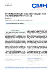
Results Accuracy Figure 1 depicts the results for the delta bilirubin estimates by the Ektachem system grouped by diagnoses in comparison with delta bilirubin measurements by the manual method. Regression analysis of the results for all 40 samples yielded a slope of 0.832, intercept of 9.9, and r of 0.724 (p < 0.001). Duplicate values were in close agreement for all determinations. The percentage of the delta bilirubin fraction contributing to the total bilirubin ranged from 0 to 65% by the Ektachem method and from 0 to 56% by the manual method.
120 |100
80
We investigated 20 samples with conjugated hyperbilirubinaemia. Delta bilirubin might be expected in these samples, depending on the time samples were collected during the course of the illness; the manual method detected delta bilirubin in 16 of these samples (range 1.7 — 78.7 μηιοΐ/ΐ); in the same 16 samples, the Ektachem system also returned positive values for delta bilirubin (range 1.7-90.6 μηιοΐ/ΐ). Precision Unfortunately, there are no standards for delta bilirubin available so we were unable to perform precision studies for different delta bilirubin concentrations. The within-run precision, based on estimates of within-run standard deviations from 40 paired observations, showed a standard deviation of 1.7 μηιοΐ/ΐ by the slide method.
Discussion
60
Currently, the Kodak Ektachem system provides the only automated means of estimating delta bilirubin in serum. Our data suggest that the Ektachem values are reasonable estimates of the delta bilirubin present when compared with the manual delta bilirubin method.
20:: 20
40
60
80
100
Delta bilirubin (manual method) [μΓτιοΙ/1]
Fig. 1. Comparison by diagnosis of delta bilirubin determinations by Ektachem (y) and manual delta method (x). Diagnoses: (·) healthy individuals, (O) unconjugated hyperbilirubinaemia, ( + ) Gilbert, (D) Crigler-Najjar, ( χ ) conjugated hyperbilirubinaemia, (*) Dubin-Johnson.
In the 20 samples with predominantly unconjugated bilirubin, in which delta bilirubin would not be expected, the manual method did not detect delta bilirubin (tab. 1). The Ektachem system estimated delta bilirubin in 16 of these 20 samples (80%) with estimates ranging from 0.86 to 35.1 μηιοΙ/1. Tab. 1. Ektachem delta bilirubin estimates in patients with no detectable delta bilirubin by the manual method Diagnosis
Ektachem delta estimate Oimol/1)
Manual method delta value (|imol/l)
1. 2. 3. 4. 5. 6. 7. 8. 9. 10. 11. 12. 13. 14. 15. 16. 17. 18. 19. 20.
2.57 9.41 0.86 11.12 10.26 0 9.41 7.7 1.71 0 20.52 3.42 35.06 1.71 9.41 0 0 6.84 7.7 5.99
0 0 0 0 0 0 0 0 0 0 0 0 0 0 0 0 0 0 0 0
Healthy Healthy Healthy Healthy Healthy Healthy Healthy Healthy Healthy Healthy Gilbert Gilbert Gilbert Gilbert Gilbert Crigler-Najjar Unconjugated Unconjugated Unconjugated Unconjugated
Previous work comparing Ektachem delta bilirubin values with HPLC delta bilirubin measurements demonstrated good correlation (8). Unfortunately, the methodology for HPLC delta bilirubin determination requires incubation with 10 per cent albumin for 20 minutes to stabilize monoconjugated and diconjugated bilirubins (1, 8, 9). Addition of albumin to the sample could result in the interaction of bilirubin conjugates with the additional albumin, causing actual formation of delta bilirubin in vitro. The advantage of the manual delta bilirubin method utilized in the present study is that it does not require albumin addition and only selects the bilirubin fraction that is tightly bound to protein (6). We purposely included serum samples from patients with unusual diagnoses (e. g., Crigler-Najjar, Dubin- Johnson syndrome) to see how the automated method would respond. Certainly, such samples would not be expected on a routine basis. As we previously reported, the Ektachem system again had difficulty with a Crigler-Najjar syndrome sample, in which unconjugated bilirubin was increased but no or little conjugated bilirubin was present (3, 10). We have observed this problem several times with several different lots of TBil and BuBc slides, and we are aware that other clinical chemists have also encountered this problem. The Kodak test methodology acknowledges that TBil may be overestimated in specimens that contain predominantly unconjugated bilirubin, as seen in patients with Crigler-Najjar and Gilbert syndromes (4). This may be the result of the mathematical transformations employed in the Ektachem methodology to linearize the reflection densities with respect to the analyte concentrations (11). Unfortunately, when patient specimens arrive in the clinical laboratory the diagnosis of the patient is not known prior to the analysis of the sample. Since Gilbert syndrome is estimated by some authorities to comprise 10% of the general population, a significant number of samples evaluated by the Ektachem system may be expected to be in error. Delta bilirubin was expected only in the serum of those patients with conjugated hyperbilirubinaemia, since delta bilirubin is composed of covalently linked bilirubin conjugates with serum proteins (1, 9, 12 — 14). Of the 20 samples with predominantly unconjugated bilirubin, in which delta bilirubin should not be present, the manual method revealed no delta bilirubin. The Ektachem system responded poorly to these specimens, returning positive values for delta bilirubin for the majority of the samples (tab. 1). For all specimens in which delta bilirubin was detected by the manual method, delta bilirubin was also detected by the Ektachem system. Eur. J. Clin. Chem. Clin. Biochem. / Vol. 30,1992 / No. 1
41
Technical note Not only was delta bilirubin detected in the serum of patients with conjugated hyperbilirubinaemia, this fraction also accounted for a considerable amount of the total bilirubin. The concentration of delta bilirubin in serum did not correlate with the total bilirubin concentration. This may reflect differences in formation and clearance of delta bilirubin compared with bilirubin monoconjugates and diconjugates. The percentage of the total bilirubin accounted for by the delta bilirubin fraction might differ considerably, depending upon the particular point in the course of the illness that serum samples were obtained (1). No discriminating value in diagnosis could be attributed to delta bilirubin measurement. Delta bilirubin estimation, while not absolutely essential for patient management, can provide useful information regarding the time-course of an illness. The presence of delta bilirubin in
serum unaccompanied by the presence of bilirubin monoconjugates and diconjugates in a patient with a prior history of monoconjugates or diconjugates in serum suggests improvement. Further, since delta bilirubin can contribute to a substantial percentage of the total bilirubin, differentiation of the contribution of delta bilirubin from that of other bilirubin serum conjugates may aid in the determination of the chronicity of an illness upon patient presentation. Caution must be exercised in the use of Ektachem delta bilirubin estimates in patients with unconjugated hyperbilirubinaemia. Since it is likely that the estimates are positively biased, samples with predominantly unconjugated bilirubin should have the presence of delta bilirubin confirmed by the manual assay. However, since the Ektachem system currently provides the only automated means of estimating delta bilirubin, its use seems warranted.
References 1. Weiss, J. S., Gautam, A., LaufT, J., Sundberg, M. W., Jatlow, P., Boyer, J. L. & Seligson, D. (1983) The clinical importance of a protein-bound fraction of serum bilirubin in patients with hyperbilirubinemia. N. Engl. J. Med. 309, 147-150. 2. Kodak Ektachem test methodology: BuBc test methodology (1986) Health Sciences Marketing Division Publication MP2-40. Eastman Kodak Company, Rochester, New York. 3. Rosenthal, P., Keefe, M. T., Henton, D., Cheng, M., Lee, C. R., Hall, R. L. & Newton, M. K. (1990) Total and direct-reacting bilirubin values by automated methods compared with liquid chromatography and with manual methods for determining delta bilirubin. Clin. Chem. 36, 788 — 791. 4. Kodak Ektachem test methodology: total bilirubin test methodology (1986) Health Sciences Marketing Division Publication MP2-39. Eastman Kodak Company, Rochester, New York. 5. Westgard, J. O. & Barry, P. L. (1986) Cost-effective quality control: managing the quality and productivity of analytical processes. American Association for Clinical Chemistry, Washington, D. C. 6. Blanckaert, N., Servaes, R. & Leroy, P. (1986) Measurement of bilirubin-protein conjugates in serum and application to human and rat sera. J. Lab. Clin. Med. 108, 77-87. 7. Bauer, S. & Kennedy, J. W. (1981) Applied statistics for the clinical laboratory: II. within-run imprecision. J. Clin. Lab. Autom. 1, 197-201.
Eur. J. Clin. Chem. Clin. Biochem. / Vol. 30,1992 / No. 1
8. Wu, T-W., Dappen, G. M., Spayd, R. W., Sundberg, M. W. & Powers, D. M. (1984) The Ektachem clinical chemistry slide for simultaneous determination of unconjugated and sugar-conjugated bilirubin. Clin. Chem. 30, 1304— 1309. 9. Lauff, J. J., Kasper, M. E., Wu, T. W. & Ambrose, R. T. (1982) Isolation and preliminary characterization of a fraction of bilirubin in serum that is firmly bound to protein. Clin. Chem. 28, 629-637. 10. Rosenthal, P., Keefe, M. T, Henton, D. & Cheng, M. (1989) Ektachem and unconjugated bilirubin measurements [Letter]. J. Clin. Chem. Clin. Biochem. 27, 829. 11. Wu, T-W, Dappen, G. M., Powers, D. M., Lo, D. M., Rand, R. N. & Spayd, R. W. (1982) The Kodak Ektachem clinical chemistry slide for measurement of bilirubin in newborns: principles and performance. Clin. Chem. 28, 2366-2372. 12. Wu, T-W. & Sullivan, S. S. (1982) Biliprotein in adult icteric serum demonstrated by extension of the alkaline methanolysis procedure. Clin. Chem. 28, 2398-2404. 13. Brett, E. M., Hicks, J. M. & Rand, R. N. (1984) Delta bilirubin in serum of pediatric patients: correlations with age and disease. Clin. Chem. 30, 1561-1564. 14. Rosenthal, P., Henton, D., Felber, S. & Sinatra, F. R. (1987) Distribution of serum bilirubin conjugates in pediatric hepatobiliary diseases. J. Pediatr. 110, 201-205. Philip Rosenthal, M. D. Director, Pediatric GI Cedars-Sinai Medical Center Suite 4310 8700 Beverly Boulevard Los Angeles, CA 90048-1869 U.S.A.
43
Technical note Eur. J. Clin. Chem. Clin. Biochem. Vol. 30, 1992, pp. 43^*5 © 1992 Walter de Gruyter & Co. Berlin · New York
TECHNICAL NOTE
Amniotic Fluid Components and Changes Due to Storage Conditions By J. L. Zaidman, M. Waron, Sonica Meyer and S. Micle Division of Laboratories, Assaf Harofeh Medical Center, Affiliated to the Sackler Faculty of Medicine, Tel Aviv University, Zerifin, Israel (Received May 31/November 13, 1991)
Summary: The composition of amniotic fluid was found to vary in dependence on storage conditions. Catalytic concentrations of creatine kinase and lactate dehydrogenase were considerably decreased after storage at — 20 °C, and the decreases were less marked after storage at — 70 °C. Other analytes were more stable. Consequently, the optimal storage conditions for amniotic fluid samples may differ for various analytes. The low inter-individual variability of some analytes (Na, K, Ca, Cl) suggests that their concentrations are homeostatically controlled during the gestational period studied. In contrast, the high variability of enzyme activities may be due to more rapid quantitative changes during normal gestation; in addition, enzyme activities may be affected by certain pathological and non-pathological conditions of the fetus. Introduction
Amniotic fluid contains many biochemical components, originating from the fetus' tissues and excretions, the placental tissues, and the maternal organism. Many of the amniotic fluid substances are used in the prenatal diagnosis of diverse pathological conditions, such as chromosomal aberrations, neural tube defects and some genetically conditioned metabolic disorders. The fact that the biochemical composition of the amniotic fluid varies at different stages of pregnancy is usually taken into consideration when evaluating the significance of the presence and concentration of the different substances. However, another important factor which may affect results from the analysis of the amniotic fluid components, namely the storage conditions of the samples, is often neglected, although its influence is well known in relation to other body fluids. Determination of the optimal storage conditions of the amniotic fluid samples is essential when frozen samples are used for establishing the real composition of this fluid. Although numerous data have been published on the amniotic fluid composition at different gestational stages (see, e.g. (1, 2)), the accumulation of additional data, particularly for the gestational stages significant in prenatal diagnosis, is important. This could contribute to the recognition of the possible significance of some constituents for the gestational prognosis, so that such data could lead to an improvement in the accuracy and scope of prenatal diagnosis. Eur. J. Clin. Chem. Clin. Biochem. / Vol. 30,1992 / No. 1
In the present study, a series of amniotic fluid components were analysed in fresh fluid and after storage at different temperatures. Material and Methods
Amniotic fluid samples were originally collected for ot-fetoprotein determinations from 34 women at 16 — 18 weeks of gestation, the stage at which amniocentesis for prenatal diagnosis is usually performed. The fluid samples were centrifuged at 3600 min"1 (900 g; 15 min) and were analysed fresh, and after 7 days storage at -20°C and -70°C. Twenty amniotic fluid components were determined under these different conditions. All pregnancies had a normal outcome with the birth of a single, apparently normal infant. Chemical and enzyme assays were performed on a random access analyser (Monarch 2000-Instrumentation Laboratory, USA) by using commercially available kits: alkaline phosphatase1), creatine kinase1), creatinine, lactate dehydrogenase1), and α-amylase1), from Boehringer Mannheim, Diagnostica, Germany; alanine aminotransferase1), aspartate aminotransferase1), cholesterol, glucose, triacylglycerol, urea-N and uric acid from Bio Merieux, France; sodium, potassium and chloride from Instrumentation Laboratory Sud, Italy; calcium from Ciba-Corning, Gilford Systems, USA; phosphate from Sigma Diagnostics USA. Radioimmunoassays for carcinoembryonic antigen (CEA), cyanocobalamin and folic acid were performed on a gamma counter (LKB, Sweden) by using commercially available kits from DPC-Diagnostic Products Corporation, USA. Lactate dehydrogenase1) isoenzymes were
!
) Enzymes Alkaline phosphatase: Orthophosphoric-monoester phosphohydrolase (EC 3.1.3.1); Creatine kinase: ATP: creatine N-phosphotransferase (EC 2.7.3.2); Lactate dehydrogenase: L-Lactate:NAD + oxidoreductase (EC 1.1.1.27); ot-Amylase: 1,4-oc-D-Glucan glucanohydrolase (EC 3.2.1.1); Alanine aminotransferase: L-Alanine: 2-oxoglutarate aminotransferase (EC 2.6.1.2); Aspartate aminotransferase: L-Aspartate: 2-oxoglutarate aminotransferase (EC 2.6.1.1).
44
Technical note
Tab. 1. Amniotic fluid components determined following different storage conditions. (N = 34) Fresh sample Analyte
Unit
Mean S.D.
Alkaline phosphatase Aspartate aminotransferase Alanine aminotransferase Lactate dehydrogenase Creatine kinase Glucose Urea Uric acid Creatinine Na K Cl Ca P
U/l
6-67 29.8 15.7 8-14 10.2 1.5 3-8 5.3 1.2 71-242 129.2 40.9 0-4 1.8 1.1 2.5 0.50 1.5-3.8 3.63 0.68 2.5-5.1 0.19 0.04 0.10-0.28 55.4 7.40 41-67 137.5 2.95 135-146 3.74 0.16 3.3-4.1 110.7 2.73 108-118 1.85 0.15 1.54-2.43 1.08 0.32 0.63-1.99
u/i
U/l U/l U/l mmol/1 mmol/1 mmol/1 mmol/1 mmol/1 mmol/1 mmol/1 mmol/1 mmol/1
Range
Storage at -70°C
Storage at -20°C
Mean S.D. Range
Mean S. D. Range
4-55 29.6 16.0 10.7 2.8 5-16 2-11 5.7 2.0 42-202 106.4 36.1 0-4 1.6 1.3 2.6 0.60 1.6-3.8 3.80 0.70 2.8-5.5 0.20 0.05 0.12-0.32 58.4 9.90 32-77 141.4 5.08 129-153 3.85 0.19 3.4-4.1 114.0 5.00 110-127 1.88 0.18 1.58-2.12 1.07 0.32 0.65-2.54
5-65 28 .8 15.5 6-16 10 .7 2.4 2-10 5.1 2.2 37-114 61 .3 17.0 0-2 0 .7 0.7 2 .6 0.60 1.6-3.8 3.72 0.71 2.6-5.6 0 .20 0.05 0.08-0.33 19-73 53 .9 12.40 140 .8 4.81 128-150 3.84 0.19 3.3-4.1 113 .1 4.93 102-125 1 .63 0.25 1.06-2.03 0.96 0.30 0.63-2.25
separated electrophoretically on Titan gel plates and scanned in a densitometer from Helena Laboratories USA. The enzyme assays were carried out at a uniform reaction temperature of 30 °C. Quality assurance: for internal quality control, we used Precipath E (Boehringer Mannheim, Diagnostica, Germany) and Liquid comprehensive chemistry control (three levels) (Beckman, USA). For external quality assessment, we used the Clinical chemistry assessment programme (Wellcome, UK) and QAP (Baxter Healthcare Corporation, Dade Division, USA). Results and Discussion Results for the concentrations of 14 amniotic fluid analytes are presented in table 1. Amylase, cholesterol, triacylglycerol, folic acid and cyanocobalamin were not detectable, while carcinoembryonic antigen was detected in very small amounts. The data presented show that the majority of the analytes considered are not substantially modified after 7 days storage at —20 °C or — 70 °C. Only the activity of two enzymes, creatine kinase and lactate dehydrogenase, undergo important changes. Of the initial activity of creatine kinase, 88.9% is preserved after 7 days storage at —70 °C, and only 38.9% after storage at — 20 °C. Of the initial activity of lactate dehydrogenase, 82.3% is preserved by storing the samples at —70 °C, and only 47.4% after storage at -20°C. The electrophoretic separation of the lactate dehydrogenase (LD) fractions show that these changes are mainly due to a decrease in the activity of the LD4 and LD5 fractions, and at a lesser extent, to the decrease of the LD3 fraction, while the LD1 and LD2 fractions are more stable. Accordingly, the percentage contribution of each fraction is modified (fig. 1). The sensitivity of these two enzymes, lactate dehydrogenase and creatine kinase, to storage temperature is known from numerous publications (e. g. (3, 4)), but there are conflicting recommendations for optimal storage conditions. It should be emphasized, however, that the published data refer to enzymes derived from different sources, such as peripheral blood, cord blood, or tissue homogenates of various origins. The activities of enzymes in amniotic fluid were also reported relatively recently by other authors (2). Comparison of their data with the results of the present work showed similar activities for alkaline phosphatase and lower activities for aspartate aminotransferase and alanine aminotransferase in our fresh fluid samples. The activities of creatine kinase and lactate dehydrogenase found by these authors, who stored part of their samples at — 20 °C, are lower than the activities we found in fresh samples, but identical with those found in the samples
ο CO ω
c o
δ _C
1
2
3
4
5
Lactate dehydrogenase isoenzymes Fig. 1. Representative scan of lactate dehydrogenase isoenzyme patterns in amniotic fluid. a) fresh Isoenzyme Fraction (%)
1 14.8
2 16.9
3 12.7
4 17.4
5 38.2
b) after storage at -20 °C Isoenzyme 1 2 Fraction (%) 29.4 30.1
3 17.1
4 4.8
5 18.5
Eur. J. Clin. Chem. Clin. Biochem. / Vol. 30, 1992 / No. 1
45
Technical note stored for 7 days at —20 °C. It may be concluded that for a correct appreciation of the amniotic fluid composition, fresh samples are the most adequate. When storage is needed, suitable conditions should be verified for each analyte. From the data presented in table 1 it is evident that some analytes such as Na, K, Ca and Cl, have very low coefficients of variation (CV). This fact suggests the existence of an essential homeostasis, controlling the concentrations of these analytes during the gestational period studied. In connection with this proposal, it should be noted that the observed concentrations of Na, K and Cl in the amniotic fluid and their concentrations in the normal human blood are similar, and that at this stage of development — as stressed by certain authors (e. g. (5)) — the fetal skin is highly permeable to water.
Urea, uric acid and creatinine are present in low concentrations, similar to the lowest limits of their normal concentrations in blood plasma; they show moderate CVs. These data show that at this stage of fetal development, 16 — 18 weeks of gestation, the contribution of fetal urine to the amniotic fluid may consist of only small quantities of primitive urine, which have a composition similar to a protein-poor filtrate of fetal serum (6). The activity of the enzymes determined is significantly lower than in blood, and highly variable, as may be deduced from table 1. The high CV of the enzyme activities may be due to differences in gestational stage of the studied subjects and suggests, probably, that normal activity limits of the enzymes in amniotic fluid should be tested at shorter time intervals (e. g. one week).
References 1. Fairweather, D. V. I. & Eskes, T. K. A. (eds.) (1973) Amniotic fluid. Excerpta Medica, Amsterdam. 2. Salgo, L. & Pall, A. (1989) Variation in some enzymes in amniotic fluid and maternal serum during pregnancy. Enzyme 41, 101-107. 3. Jacobs, E., Hissin, P. J., Propper, W., Mayer, L. & Sarkozi, L. (1986) Stability of lactate dehydrogenase at different storage temperatures. Clin. Biochem. 19, 183-188. 4. Shain, S. A., Boesel, R. W., Klipper, R. W. & Lancaster, C. M. (1983) Creatine kinase and lactate dehydrogenase: stability of isoenzymes and their activity in stored human plasma and prostatic tissue extracts and effects of sample dilution. Clin. Chem. 29, 832-835.
Eur. J. Clin. Chem. Clin. Biochem. / Vol. 30,1992 / No. 1
5. Abramovich, D. R. (1973) The volume of amniotic fluid and factors affecting or regulating this. In: Amniotic fluid (Fairweather, D. V. I. & Eskes, Τ. Κ. Α., eds.) Excerpta Medica, pp. 29-51. 6. Stanier, M. W. (1960) The function of mammalian mesonefros. J. Physiol. (London) 757, 472-478. Dr. J. L. Zaidman Department of Biochemical Pathology Assaf Harofeh Medical Center Zerifm 70300, Israel