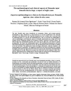
Am. J. Trop. Med. Hyg., 74(2), 2006, pp. 308–317 Copyright © 2006 by The American Society of Tropical Medicine and Hygiene
SPATIAL CLUSTERING AND EPIDEMIOLOGICAL ASPECTS OF VISCERAL LEISHMANIASIS IN TWO ENDEMIC VILLAGES, BARINGO DISTRICT, KENYA JEFFREY R. RYAN, JANE MBUI, JUMA R. RASHID, MONIQUE K. WASUNNA, GEORGE KIRIGI, CHARLES MAGIRI, DEDAN KINOTI, PHILLIP M. NGUMBI, SAMUEL K. MARTIN, SHADRAK O. ODERA, LISA P. HOCHBERG, CHRISTIAN T. BAUTISTA, AND ADELINE S. T. CHAN* Department of Entomology, Division of Communicable Diseases and Immunology, Walter Reed Army Institute of Research, Silver Spring, Maryland; Centre for Clinical Research, Kenya Medical Research Institute, Nairobi, Kenya; U.S. Army Medical Research Unit 64109, Nairobi, Kenya; U.S. Military HIV Research Program and Henry M. Jackson Foundation for the Advancement of Military Medicine, Inc., Rockville, Maryland
Abstract. Visceral leishmaniasis (VL) seroprevalence in Kenya is unknown because of the lack of a practical and accurate diagnostic test or surveillance system. A novel serological assay was used to estimate the seroprevalence of Leishmania-specific antibodies, and Global Information System and spatial clustering techniques were applied to study the presence of spatial clusters in Parkarin and Loboi villages in Baringo District in 2001. VL seroprevalences were 52.5% in Parkarin and 16.9% in Loboi. Significant associations among seropositivity and house construction, age, and proximity to domestic animal enclosures were found. A significant spatial cluster of VL was found in Loboi. The spatial distribution of cases in the two villages was different with respect to risk factors, such as presence of domestic animals. This study suggests that disease control efforts could be focused on elimination of sand fly habitat, placement of domestic animal enclosures, and targeted use of insecticides. into the vertebrate host and sand fly vector. The assay was improved and developed into an ELISA that can detect Leishmania-specific IgM and IgG antibodies in patients with visceral and cutaneous leishmaniasis.11 In preliminary studies, the sera from 129 visceral leishmaniasis patients (Brazil, Italy, North Africa, Nepal) with sera from matched controls were tested. The test reported a sensitivity of 98.2%. Testing this ELISA with small subsets of naïve North American normals, Kenyan endemic normals, malaria, African trypanosomiasis, echinococcosis, filariasis, and schistosomiasis case patients yielded a specificity of better than 97%.10–12 Visceral leishmaniasis, caused by Leishmania donovani, is endemic in Baringo District, Kenya. The first case of VL was recorded in 1948 in the District Hospital at Kabarnet.13 Some scientists believe that nomadic Turkanas may have introduced the disease into the area from the north. Others speculate that Kenyan soldiers returning from North Africa after Word War II were responsible for introduction of the parasite. Phlebotomus martini is the vector of the parasite, and man is the only known reservoir.14 Currently, the disease is widespread throughout Baringo District. A number of studies have used diagnostics, epidemiologic surveys, and Global Information System (GIS) technology to identify potential risk factors with several tropical diseases.15–17 The current study applied a multidisciplinary approach with integration of new technologies. The objectives in this study were to determine in two villages in a VLendemic area of the Baringo District of Kenya 1) the current seroprevalence of Leishmania-specific antibodies, 2) the presence of spatial clustering (hot spots) of seropositive cases, and 3) the identification of associations with certain risk factors in those same villages.
INTRODUCTION The leishmaniases are a group of mainly zoonotic infections caused by protozoan parasites of the genus Leishmania. These infections produce a variety of clinical diseases depending on the virulence or tropism of the parasite and differential host immune responses.1 The World Health Organization estimates that with 350 million people at risk worldwide, 12 million people are infected with Leishmania parasites and that as many as 2 million new cases occur each year. Recent reports detail a visceral leishmaniasis (VL) pandemic in the Horn of Africa and parts of India, Nepal, Bangladesh, and Central and South America. A major epidemic reported in the Sudan from 1989 to 1993 was responsible for the deaths of approximately 10% of that country’s population.2 Whereas the definitive diagnosis of VL leishmaniasis requires demonstration of parasites from smears or in vitro cultivation, these methods can be time consuming and involve at least one invasive, and at times risky, procedure such as lymph-node biopsy, splenic or bone marrow aspiration. A variety of serological methods, including indirect immunofluorescence (IFA),3 enzyme-linked immunosorbent assay (ELISA),4,5 and polymerase chain reaction (PCR)6 are associated with a number of problems including cross-reactivity with other pathogens, high cost and/or need for sophisticated equipment. The direct agglutination test (DAT)7 is a sensitive, specific, and simple test, but the main disadvantage is that it can have high background seropositivity in endemic areas. New diagnostic tests for VL have been a major focus of numerous research groups.8,9 Martin and co-workers reported the use of a soluble antigen prepared from Leishmania donovani as the foundation for an ELISA that could detect specific IgG antibodies in kala azar patients.10 Soluble antigens produced by Leishmania (exo-antigens) are released
MATERIALS AND METHODS Study area. Baringo District is the only region in Kenya where both VL and cutaneous leishmaniasis (CL) have been found together.18 It covers an area of approximately 10,000 km2 and is located north of the Equator in Kenya’s Rift Valley Province. Marigat Township is central to this region and a
* Address correspondence to Adeline S. T. Chan, M.P.H., Ph.D., Department of Entomology, Division of Communicable Diseases and Immunology, Walter Reed Army Institute of Research, 503 Robert Grant Ave., Silver Spring, MD 20910-7500. E-mail: adeline.chan@ na.amedd.army.mil
308
SPATIAL EPIDEMIOLOGY OF VISCERAL LEISHMANIASIS IN KENYA
site where most VL studies have been conducted in the past. Generally, the district includes a range of landscapes varying between fertile highlands as high as 2,700 m elevation and semiarid lowland at about 900 m elevation. The Tugen hills and the adjacent highlands in the southwest receive 1,200– 1,500 mm average yearly rainfall and have daily average temperatures ranging from 10°C to 32°C. In contrast, lowland areas to the west, east, and north of the Tugen Hills receive annual rainfall from 300 to 700 mm and the temperatures vary between 16°C and 42°C. The rainy season is from March to September, with maximum rainfall in May and August. Three main ethnic groups, all classified as agropastoralists, inhabit Baringo District: the Tugen, Pokot, and Njemps. The district is sparsely populated due to harsh physical and climatic conditions. There have been no reports of cutaneous or mucocutaneous leishmaniasis in the two study areas. Study sites. This epidemiologic study was conducted in the villages of Parkarin and Loboi (0°35⬘N latitude, 36°06⬘E longitude) in the Rift Valley Province, Baringo District of central Kenya, and approximately 207 kilometres northwest of Nairobi. The population of Parkarin (N ⳱ 286 inhabitants) was spread over an area of 3.3 km2. The population of Loboi (N ⳱ 643 inhabitants) was spread over an area of 2.3 km2. Both villages, being only 7 km apart, have the same subtropical climate. Outwardly, the two villages appear to be very similar and both have had a long history of kala azar. The villagers live in family units (homesteads) each consisting of several huts. Within the homestead, householders may live together in one house or in 2–3 houses. Each homestead has domestic animals including cows, goats, sheep, chickens, and dogs. Animal pens and corrals for animals are built adjacent to or near the buildings in each homestead, and in some cases the buildings were within the corral. The economic status of the population of Parkarin is uniformly low. Most villagers live in huts made of sticks and mud, tend to domestic animals, and have no income. Terrain throughout the village is very dry with scattered Acacia trees, thorn bushes, and little ground vegetation. The population of Loboi is a mix of very poor families, day laborers, shopkeepers, and professional staff working at the Lake Borgoria National Park. Loboi has a health post, a number of shops, schools, and public utilities. Parkarin has no health post, therefore, residents must travel 7 km to Marigat when in need of medical assistance. These health posts are staffed by nurses and community health workers who are responsible for patient screening for kala azar, as well as taking blood smears and providing malaria treatment to patients. Although the workers are familiar with kala azar clinically, there are no tools for diagnosing or treating this disease in the endemic area. Another dramatic difference between the two villages is the amount of vegetation. Loboi has more overhead cover from Acacia trees, whereas Parkarin has more open areas with denser scrub and a tree line of Acacia concentrated along the irrigation canal. Loboi also has more termite mounds (N ⳱ 188; 87.1 mounds/km2) than Parkarin (N ⳱ 113; 33.7 mounds/km2). Study population. This study included 489 (53%) research volunteers from the population of Loboi and Parkarin. It was composed of individuals of both sexes, 5 years of age or older, that volunteered for the study. All participants had been living in the community for at least 3 months and called the community their primary home. This study was approved by the Walter Reed Army Institute of Research (WRAIR) Hu-
309
man Use Review Committee (HURC) and the Kenya Medical Research Institute (KEMRI) Ethical Review Committee. Informed written consent was obtained from all volunteers prior to the employment of any examining, sampling, and questioning measures. Written parental consent was obtained for children under 18 years old. Children under the age of 5 years and temporary visitors to the communities were excluded from the study. Survey components. A community-wide survey consisting of a census, GIS mapping, blood sampling by venipuncture, medical histories, and epidemiologic questionnaires was conducted in the two villages during the period from May to July 2001. The epidemiologic questionnaire captured information concerning house construction; the presence and location of domestic animals; the presence of domestic dogs; the proximity of the resident from typical sand fly breeding sites (termite mounds and animal enclosures); how long the family had lived at the present site; and, the daytime occupation of each resident. Each participant was assigned a unique personal identifier and household address. All participant information was recorded in a database and linked to diagnostics results. Medical examination. A team composed of two clinicians experienced in VL diagnosis, a guide, and several clinical assistants and translators worked systematically through each village. Whereas the clinician conducted the medical examination, an assistant administered the medical history questionnaire. The study clinician recorded the participant’s condition and any clinical abnormalities and/or VL-related symptoms. Degree of splenomegaly was determined by palpitation and graded by use of the Hackett model.19 Serology. Seroprevalence was determined by an ELISA based on an antigen derived from Leishmania donovani promastigotes cultivated in a protein-free, serum-free medium.11,12 Leishmania donovani (Ld, Strain WR0130E) washed promastigotes were inoculated into 200 mL of a defined, conditioned protein-free medium (XOM) to give a final density of 1 × 10⁸ cells/mL. The parasites were incubated at 25°C for 72 hours in roller bottles. Thereafter, the spent medium was harvested by centrifugation at 11,000 × g for 20 minutes and the relative protein concentration of the soluble antigens was estimated by measuring the optical density at 280 nm.20 Plate sensitization was effected by coating polystyrene, 96well microtiter plates (Immulon 4, Dynatech Laboratories, Chantilly, VA) with 100 L of the Ld antigen solution (5 g protein per well). Plates were then blocked with 0.5% casein (Sigma Chemical Co., St. Louis, MO) in PBS for 1 hour at room temperature. The blocking buffer was removed by aspiration and the serum samples (100 L of 1:1000 dilution) and appropriate controls were added to the microtiter plate and the plate incubated at 26°C for 40 minutes. After washing with 0.05% PBS-Tween-20 (PBS-Tween) buffer four times, goat antihuman IgG (whole molecule) conjugated with horseradish peroxidase (Kirkegaard & Perry Laboratories Inc., Gaithersburg, MD) was added at a 1:5,000 dilution in blocking buffer and the plate incubated at 26°C for 30 minutes. The plate was then washed four times with PBS-Tween buffer and 100 L of TMB substrate (Kirkegaard & Perry Laboratories Inc.) was added to each well. The plate was incubated in the dark, and the optical density (OD) was periodically read at 650-nm wavelength in an ELISA plate reader (Molecular Devices,
310
RYAN AND OTHERS
Menlo Park, CA) until the OD value of a reference positive control reached 0.75. At this point, 100 L of a stop solution (1.0 M phosphoric acid) was applied to the plate and the final OD reading taken at 450 nm. A positive reference serum was used in all plates, and only interassay variation of less than 10% was accepted. The lower limit of positivity (cutoff) was determined by the mean of the negative controls subset + 3 standard deviations.21 Global Information System integration. Global Information System (GIS) and spatial clustering statistical techniques were applied to evaluate the presence of high-risk areas and spatial clustering Leishmania seroprevalence. All residences, streets, public buildings, latrines, termite mounds and other features of interest in the villages were mapped using Trimble ProXR GPS receivers to permit the calculations of latitude, longitude, and altitude. During the mapping, each house was assigned a unique household identification number and information about construction type was also recorded. Pathfinder Office software, version 1.1 (Trimble Navigation, Sunnyvale, CA) was used to perform differential correction of all feature locations and to create a locational database for use in GIS analyses. Seroprevalences were calculated by household. For the GIS analysis, ArcView software, version 3.0, (Environmental Systems Research Institute, Inc., Redlands, CA) was used to merge the locational database with the study group data. A surface analysis of cumulative Leishmania risk across the entire village at the household level was performed by using the Inverse Distance Weighted (IDW) interpolator model22 using ArcView Spatial Analyst v.1.0 (ESRI, Inc. Redlands, CA). IDW weights the contribution of each input (control) point by a normalized inverse of the distance from the control point to the interpolated point. IDW assumes that each input point (household) has a local influence that diminishes with distance. It weights the points closer to the processing points more than those farther away. A specified number of points (or all points) within a specified radius are used to determine the output value for each location. Statistical analysis. Chi-square and Fisher’s exact tests were used to compare differences in proportions. The Student t test or Mann-Whitney U, a nonparametric test, was used to compare differences in continuous variables. To evaluate ELISA results and splenomegaly size on a qualitative scale, the Bartholomew test was applied.23 Because the study population included sets of familial members, the data were presumed to violate the standard logistic regression assumption of independent response probabilities across observations. Potential risk factors and VL seropositivity were evaluated by odds ratios (OR) using random effects logistic regression for each village, where the homesteads were defined as the group variable.24 To describe the mount of aggregation existing in VL seropositivity within homesteads and/or households units, the percentage of explained variance attributed (rho) was estimated after adjusting for age and gender. All reported confidence intervals (CI) are 95%, and all reported P values are two-sided. These statistical analyses were carried out using Stata v. 6.0 (Stata Corporation, College Station, TX). Spatial analysis. To evaluate the presence of spatial disease clusters and to identify focalized areas of “high risk” for VL, we applied a spatial scan statistic.25 The spatial scan statistic is well suited for geographic disease surveillance. This method takes into account the uneven spatial geographic distribution of cases and population densities; it does not require a priori
assumptions about the number, place, or size of locations that may be identified as clusters; it adjusts for multiple testing inherent in the search for multiple clusters; and it searches for either high or low incidence or prevalence areas. The spatial scan statistic works by aggregating together the unique combinations of small-area geographies that have a high probability of being clusters. This statistical test can detect spatial clusters of any size located anywhere in the study area. The spatial scan statistic imposes a geographic circular window to perform purely spatial analysis of varying size on the map surface and allows its center to move so that at any position and size across the study area, the circular window includes different sets of adjacent households. As the circular window is placed at each household, this spatial method creates a large number of distinct geographical circles, with different sets of households areas within them, and each is a possible candidate for containing a spatial cluster of prevalent leishmaniasis cases. For any given geographic position of the household, the radius of the circular window varied continuously from zero to a previously user-defined maximum. Although the choice of maximum cluster window size is somewhat arbitrary, it is important to make the choice of maximum cluster window a priori to avoid the problems of multiple hypothesis testing. In this analysis, we chose 50% of the total study population as the maximum to consider. By choosing 50% as the maximum, we evaluated all sizes from zero percent up to 50% and adjusting for the multiple testing related to each of them. We assumed that the geographic spatial distribution of leishmaniasis cases follow a Bernoulli distribution. The most likely spatial cluster was determined by computing maximum likelihood ratios. The spatial scan statistic uses the Monte Carlo simulation to evaluate the statistical significance of the most likely spatial cluster. In this study, the simulated P value of the statistic was obtained through 9,999 Monte Carlo simulations, where the null hypothesis of no high spatial leishmaniasis clusters was rejected at an ␣ level of 0.05. Spatial risk patterns for VL was expressed as relative risk and was estimated by household as the observed VL seroprevalence/expected VL seroprevalence by using the spatial scan statistic. These spatial analyses were carried out by using the SaTScan v.2.1 software.26 RESULTS Study area and population. The census conducted at the onset of this study showed that there were a total of 643 villagers in Loboi and 286 villagers in Parkarin living in 235 homesteads: 176 in Loboi and 59 in Parkarin. Population density in Loboi was approximately four times that in Parkarin, with 279.6 and 85.4 persons per km2, respectively; 52.5% of the villagers were males, and 62.2% were less than 20 years old with a median age of 15 years. The mean number of persons per homestead in Loboi was 3.7 (standard deviation [SD] ⳱ 2.7) and in Parkarin it was 4.8 (SD ⳱ 3.8). There was no significant difference in mean age and gender between both villages. In wall construction, there was a clear and significant difference in both villages. Parkarin had more houses made of mud and sticks than Loboi (78.0% versus 31.4%, P < 0.05) (Table 1). Residents of Parkarin had lived an average of 11.5 years (SD ⳱ 8.9) in their community, whereas residents of Loboi had lived in theirs an average of 9.8 years (SD ⳱ 10.7). People generally resided longer in their houses in
311
SPATIAL EPIDEMIOLOGY OF VISCERAL LEISHMANIASIS IN KENYA
TABLE 1 Demographic characteristics of all population and participants tested by study sites in Kenya, 2001 Population Feature
Population/homestead No. of inhabitants No. of homesteads Wall construction (stick and mud) Mean (SD) number of people per homestead Gender Female Male Age group (years) < 10 10–20 21–30 31–40 > 40 Mean (SD) age (years)
Tested
All people No.
Loboi No. (%)
Parkarin No. (%)
All tested No. (%)
Lobi No. (%)
Parkarin No. (%)
929 235 101 4.0 (3.0)
643 (69.2) 176 (74.9) 55 (31.4) 3.7 (2.7)
286 (30.8) 59 (25.1) 46 (78.0)* 4.8 (3.8)
490 (52.7) 180 (76.6) 88 (87.1) 2.7 (2.1)
290 (59.2) 127 (70.6) 45 (35.7) 2.3 (1.7)
200 (40.8) 53 (29.4) 43 (81.1)* 3.8 (2.7)*
439 486
305 (47.7) 334 (52.3)
134 (46.9) 152 (53.1)
245 (55.8) 245 (50.4)
154 (53.1) 136 (46.9)
91 (45.5) 109 (54.5)
287 252 184 87 57 18.0 (14.1)
201 (33.2) 167 (27.6) 144 (23.8) 63 (10.4) 30 (5.0) 17.9 (13.6)
86 (32.8)* 85 (32.4) 40 (15.3) 24 (9.2) 27 (10.3) 18.4 (15.2)
81 (28.2) 176 (69.8) 106 (57.6) 58 (66.7) 38 (66.7) 21.3 (12.7)
47 (17.2) 99 (36.1) 74 (27.0) 38 (13.9) 16 (5.8) 21.4 (12.3)
34 (18.4)* 77 (41.6) 32 (17.3) 20 (10.8) 22 (11.9) 21.3 (13.4)
Note: Denominators total varied slightly due to missing data; SD, standard deviation. * P < 0.05 by chi-square or Fisher’s exact test compared between villages.
Parkarin (mean 4.5 years; SD ⳱ 4.5) than in Loboi (mean 2.8 years; SD ⳱ 3.3). Sample population. From the potential study population, 289 villagers in Loboi and 200 in Parkarin volunteered to participate in this study, representing 44.9% and 69.9%, repectively. Of these, 245 (50%) were males (Table 1). The participants lived in 180 homesteads (76.6% of the total 235 homesteads). The population studied in Loboi was not significantly older than that of Parkarin (P ⳱ 0.924). The average number of people per homestead sampled in Loboi was significantly lower than in Parkarin (P < 0.001). There were also significant differences with respect to wall construction, domestic animals, presence of goats, dogs and where animals were kept at night between villages (P < 0.05) (Table 2). Clinical and serological findings. Of the 481 (98.4%) individuals examined, a total of 155 (32%) presented with some degree of splenomegaly: 90 (45% of the 200 participants) in Parkarin and 65 (23%) in Loboi. Of these 155 individuals, only 11 (7.1%) presented with other classic signs and symptoms of kala azar such as pallor, weight loss, and fever. However, we were unable to rule out other potential causes of chronic fever such as typhoid fever or malaria. The overall antibody prevalence by ELISA was 31.5% (154 of 489). The antibody prevalences were significantly different between the two villages, 52.5% and 16.9% in Parkarin and Loboi, respectively (P < 0.001). The difference in seroprevalence between females (29.4%, 72 of 245) and males (33.6%, 82 of 244) was not statistically different (P ⳱ 0.364), even when the analysis was performed for each village separately. The mean age of positive subjects (25.6 years) was greater than that of subjects without antileishmanial antibodies (19.6 years). The difference was statistically significant whether the population was analyzed as a whole (P < 0.001) or separately for each village. There was a strong linear association of increasing seroprevalence of VL with age groups (P < 0.001 by 2 for trend) in both villages (Figure 1). An association (P ⳱ 0.022 by Bartholomew test) between ELISA results and splenomegaly size on a qualitative scale was also found (Table 3). In Loboi, a significant level of aggregation was found only within homesteads units (rho ⳱ 27%, 95% CI ⳱ 13 to 48, P < 0.001) and not within household units. Households or
household aggregation did not explain a significant proportion of the variance of VL seropositivity in Parkarin. Epidemiologic factors. From the serological and epidemiologic surveys, we found a number of factors strongly associated with altered risk of leishmaniasis. For each 10-year increase in age, the likelihood of being seropositive was higher
TABLE 2 Village comparison using certain epidemiological factors of homestead by study sites in Kenya, 2001 Feature
Visited No.
No. of homesteads in the village 180 Presence of domestic animals No 71 Yes 105 Presence of cattle No 114 Yes 62 Mean (SD) number of cattle 2.5 (4.9) Presence of sheep No 156 Yes 20 Mean (SD) number of sheep 1.4 (8.3) Presence of goats No 99 Yes 77 Mean (SD) number of goats 7.6 (12.4) Presence of chicken No 91 Yes 85 Mean (SD) number of chicken 3.5 (5.2) Presence of dogs No 127 Yes 48 Animal shelters Inside house 23 Adjacent corral 76 Away from homestead 2
Loboi No. (%)
Parkarin No. (%)
127 (70.6)
53 (29.4)
56 (45.5) 67 (54.5)
15 (28.3)* 38 (71.7)
81 (65.9) 42 (34.1)
33 (62.3) 20 (37.7)
2.8 (5.6)
1.9 (3.1)
112 (91.1) 11 (8.9)
44 (83.0) 9 (17.0)
1.7 (9.8)
1.2 (2.9)
79 (64.2) 44 (35.8)
20 (37.7)* 33 (62.3)
5.7 (17.3)
12.2 (12.4)*
66 (53.7) 57 (46.3)
25 (47.2) 28 (52.8)
3.6 (5.5)
3.3 (4.5)
96 (78.7) 26 (21.3)
31 (58.5)* 22 (41.5)
21 (17.4) 44 (36.4) 1 (0.8)
2 (4.0)* 32 (64.0) 1 (2.0)
Note: Denominators total varied slightly due to missing data; SD, standard deviation. * P < 0.05 by chi-square or Fisher’s exact test compared between villages.
312
RYAN AND OTHERS
spatial scan statistic revealed the presence of a spatial cluster of Leishmania-specific seropositives in the village of Loboi (Figure 4). This spatial cluster contained 11% (16 of 152) of the total houses sampled in the village and 29% (14 of 49) of the total ELISA cases. Villagers living within this spatial cluster were 3.3 times more likely to have Leishmania-specific antibodies than people living in other areas in the village (Table 4; P ⳱ 0.0003). In addition, this spatial cluster occurred in the northwestern corner of the village in a wooded area in very close proximity to a seasonal river, a kind of setting commonly associated with VL. The rest of the village consisted of more open areas with scattered Acacia trees, shrubs, and little ground cover. There were two other spatial clusters that appear graphically (but not statistically) in Loboi, and their settings were nearly identical to that of the primary cluster. There was not a specific high risk pattern and no significant spatial cluster was found (P ⳱ 0.296). The spatial distribution of seropositives among households appeared to be homogeneous (Figure 3), and this may be due to the high seroprevalence of VL found in this village. FIGURE 1. Seroprevalence of Leishmania-specific antibody by age group and study sites in Kenya, 2001.
in Loboi (OR ⳱ 1.88, 95% CI ⳱ 1.32 to 2.69, P < 0.0001) than Parkarin (OR ⳱ 1.52, 95% CI ⳱ 1.16 to 1.99, P ⳱ 0.002). In univariate logistic regression analysis for Loboi, residents living in households made of sticks and mud, as well as presence of domestic animals, especially cattle, sheep, and goats, were more likely to be seropositive to VL. In the multiple logistic regression analysis adjusted for age and gender, these potential risk factors remained statistically significant associated to VL with the exception of cattle. In Parkarin, presence of domestic animals in households (especially sheep and goats) was negatively associated with a positive ELISA and appears to be protective in the univariate analysis (Table 5). However, only presence of goats remained negatively associated to VL in the multiple logistic regression analysis. Therefore, these data suggest an inverse odd ratio risk relationship in the two villages studied. In both villages gender, proximity to active or inactive termite mounds, and presence of dogs were not significantly associated with VL. Spatial analysis. Based on the serological findings, we generated risk pattern maps by household for Loboi (Figure 2) and Parkarin (Figure 3) that defined areas of high risk. The
TABLE 3 Splenomegaly size of visceral leishmaniasis by ELISA ELISA Spleen size category
None Slight Palpable Considerable Severe Total
Positive no.
Negative no.
Total no.
89* 30 28 3 2 152
237 38 48 5 1 329
326 68 76 8 3 481
ELISA, enzyme-linked immunosorbent assay. * P ⳱ 0.022 by Bartholomew test (association between ELISA results and splenomegaly size on a qualitative scale).
DISCUSSION To our knowledge, this study represents the first geographic epidemiologic analysis in two villages to study the microepidemiology of VL in Africa and probably in other continents. We identified microgeographic areas where there is more likely to be high Leishmania exposure and spatial cluster of seropositives through GIS mapping and spatial statistic techniques. Baringo District has been the focus of many epidemiologic studies on leishmaniasis. As early as 1983, Jahn and Diesfeld27 used a crude ELISA for detecting Leishmania specific antibodies in VL patients seen in the Baringo District Hospital at Kabarnet. With limited success, they applied this ELISA in clinical routine and sero-epidemiologic surveys. Low titers were recorded in healthy individuals from VL foci, but their values were easily distinguished from those of active patients. After that small study, Jahn and co-workers, using the same ELISA, showed that children between 2 and 15 years old were the most affected age group for VL and that male patients predominated slightly at 57%.28 All VL cases came from the semiarid and arid parts of the district below 1500 m, where pastoralism predominated. A positive correlation was reported for active cases and the proximity of homes to seasonal rivers. However, no satisfying explanation was found for the clustering of cases. ELISA values above the cutoff, taken as the borderline nonspecific reaction, were found in about half of the population, suggesting that asymptomatic infections were common.29 Schaefer and colleagues tested Baringo District inhabitants from 26 clusters, averaging 97 people, with the leishmanin skin test (LST).30 There was an obvious aggregation of LST positivity centered on recent VL cases with approximately 11% of the 2,411 individuals tested having a marked DTH reaction. The level of LST positivity was twice as high in males as females. LST positivity increased with age to a stable level after 15 years of age, reflective of an endemic situation. Schaefer and co-workers later measured Leishmania-specific antibodies using the direct agglutination test (DAT) in a defined, endemic rural area of Baringo District composed of 30
SPATIAL EPIDEMIOLOGY OF VISCERAL LEISHMANIASIS IN KENYA
FIGURE 2.
313
Spatial risk patterns for visceral leishmaniasis in Loboi, Kenya, 2001. This figure appears in color at www.ajtmh.org.
clusters, each averaging 98 people.31 From 2,934 individuals samples, 78 were DAT seropositive; 54 of those had a history of VL. The seroconversion rate was 9 of 1,000 person-years of observation among 2,332 seronegative individuals retested the following year. During the entire study period, VL was diagnosed in only 10 patients, with an incidence rate of 2.2 of 1,000 person-years of observation. Household contacts of individuals with previously confirmed VL had a higher frequency of DAT positivity than the rest of the population. These results suggested domiciliary transmission. Using another approach, Schaefer and colleagues used PCR with capillary blood samples dried on filter paper.32 Assaying 20 parasitologically confirmed VL case-patients, 21 subclinical cases, and 11 healthy controls from a longitudinal study of anthroponotic VL in Baringo District, they were able to detect Leishmania DNA 10 months before diagnosis and up to 3 years after treatment that was classified as successful. Alarmingly, these findings showed that subclinical and treated cases may remain potential reservoirs for long periods. The study results suggest that males and females have similar risks to VL, and seropositive cases are clustered within households. The results of this study are interesting and, at the same time, confounding. We see dramatic differences in seroprevalences between the two villages (16.9% versus
52.5%). GIS mapping and spatial scan statistic showed the presence of a spatial cluster only in the village with the lowest seroprevalence. This spatial cluster found may be related to the wooded environment of the northwestern corner of Loboi that provides a good habitat for sand flies.33 When merging ELISA data with the results of the epidemiologic survey, we find strong positive associations between seropositivity, age, and poor living conditions. House building materials (mud and sticks) related to lower socioeconomic status were associated with higher risk. However, we find that the presence of domestic animals is only associated with increased risk in Loboi. The figures from Parkarin indicate that there was an inverse relationship between the presence of animals and exposure to Leishmania. For both villages, there was a significant association between ELISA seroprevalence and distance to the nearest corral (P ⳱ 0.015 for Loboi and P ⳱ 0.004 for Parkarin) (data not shown). We surmise that this result may be due to the fundamental differences between the two villages with respect to proximity of rivers, vegetation cover, population density, and economic make-up of the population. However, there may be limitations in the statistical analysis due to the extremely high seroprevalence found in Parkarin and the small size of this village. Our data suggests strategies for targeted control and for prioritization of scarce resources. A community-based VL
314
RYAN AND OTHERS
FIGURE 3.
Spatial risk patterns for visceral leishmaniasis in Parkarin, Kenya, 2001. This figure appears in color at www.ajtmh.org.
FIGURE 4. Map showing location of the most likely spatial cluster of visceral leishmanisis in Loboi, Kenya, 2001. This figure appears in color at www.ajtmh.org.
315
SPATIAL EPIDEMIOLOGY OF VISCERAL LEISHMANIASIS IN KENYA
for goats.35 Zooprophylaxis explains the situation in Parkarin where the higher ratio of goats appears to have a protective effect on the human population. VL is a grave public health problem in this area that imposes an additional strain on the local health authorities and is unlikely to be resolved by the current strategies. Understanding the transmission dynamics of VL could lead to sustainable prevention and control measures. Alternative methods of vector control, other than the conventional indoor spraying of houses with residual insecticide (which can be prohibitively expensive) should be considered. Repeated pesticide applications to the whole community can reduce sand fly numbers, but as the reduction is short lived, this method is used only in epidemics.43 The termite hills associated with leishmaniasis transmission in Kenya38 are common throughout the two villages. Although no association was shown in this study, the termite hill habitat is the favored breeding and resting sites of P. martini.44,45 This points to targeted control strategies such as insecticidal applications to resting habitats such as termite mounds46 and insecticide barrier spraying.47 The feeding propensity of P. martini on livestock and the efficacy of zooprophylaxis could be enhanced by using the livestock not only to divert the vector from humans to animals but to attract them to contact with insecticide-treated livestock.48,49 Because P. martini is active during the night when people sleep, the use of treated bed nets to block transmission could provide considerable protection.39 A prospective intervention study would be needed to evaluate the effectiveness of targeting control interventions at high-risk areas identified by this study.
TABLE 4 Most likely spatial cluster of visceral leishmaniasis by study sites in Kenya, 2001 Feature
Study area No. of houses No. of inhabitants No. of positive inhabitants by ELISA Seroprevalence Most likely spatial cluster No. of houses No. of inhabitants No. of positive inhabitants by ELISA Seroprevalence Overall relative risk* P value
Loboi
152 290 49 16.9 16 25 14 56.0 3.28 0.0003
Parkarin
75 200 105 52.5 5 11 10 90.9 1.73 0.2961
ELISA, enzyme-linked immunosorbent assay. Seroprevalence was estimated as the number of positive inhabitants by ELISA/total inhabitants tested. * Relative risk ⳱ observed VL seroprevalence/expected VL seroprevalence.
control or suppression program could be formulated on educating residents of this endemic area about the risk associated with house construction and the proximity of domestic animals to one’s living quarters. These points are likely related to sand fly habitat, whereby the sand flies seek out a favorable habitat in this arid setting that would be supportive of egg laying and survival of immature forms.33 Most residents kept their animals at night in corrals no more than 30 m from their homes. A higher and statistically significant difference (P ⳱ 0.001) of the ratio of animals to people was found in Parkarin (median ⳱ 17.5) than in Loboi (median ⳱ 5). The animals serve as a blood source,34–36 and the accumulation of animal dung may be attractive to the sand fly, drawing the vectors into closer association with the humans and increasing the risk of being bitten. In addition, the edges of the corral constructed from cut thorn bushes provide good areas for rodent burrows and nesting sites. Positive correlation of disease and the presence of domestic animals have been shown in some studies,37 whereas others have shown no significant effect38 or have noted an inverse relationship between domestic animals and disease presence.35 However, the nature of the relationship between disease transmission and domestic animals is complex.39–41 Among the factors that can affect feeding behavior, host odor, heat loss, and CO2 production are important stimuli for orientation of blood sucking insects in the open (i.e., nonforest) situations,42 diverting blood feeding from people to animals. Blood meal studies of P. martini, the vector of kala azar in the study area, has shown it to have a broad host range among domestic animals, with a preference
Received October 8, 2004. Accepted for publication March 31, 2005. Acknowledgments: The authors thank the kind people of Baringo and the tribal elders of Parkarin and Loboi for their perseverance and kindness throughout the course of this study. Additionally, we express our gratitude to Dr. H. A. Lodenyo and Samuel Chirchir of the Kenya Medical Research Institute for their kind support and consultation and Sebastian A. for his technical assistance. Partial results of this study were presented at the ASTMH 51st Annual Meeting, Denver, Colorado, November 10–14, 2002 (Abstract No 1239). Financial support: The work was supported in part by funding received from the U.S. Department of Defense Gulf War Illnesses Research Program (PE 0601105D8Z). Disclaimer: The opinions or assertions contained herein are the private views of the authors and are not to be construed as official or as reflecting the views of the U.S. Department of Defense, of the U.S. Department of the Army, of the Kenyan Ministry of Health, of the Henry M. Jackson Foundation for the Advancement of Military Medicine, Inc., or any other organization listed.
TABLE 5 Potential risk factors associated with visceral leishmaniasis seropositivity by study sites in Kenya, 2001 Loboi Feature
Wall construction type Stone, wood, sheet metal (sticks only, mud) Presence of domestic animals (no.) Presence of cattle (no.) Presence of sheep (no.) Presence of goats (no.)
Parkarin
OR (95% CI)
P value
AOR (95% CI)
P value
OR (95% CI)
P value
AOR (95% CI)
P value
4.3 (2.1–8.9) 3.8 (1.8–8.2) 2.0 (1.1–3.7) 2.6 (1.2–5.7) 3.5 (1.8–7.0)
< 0.001 0.001 0.036 0.014 < 0.001
5.5 (2.4–12.5) 4.1 (1.6–10.4) 1.9 (0.9–4.6) 3.3 (1.2–8.9) 4.5 (2.1–9.9)
< 0.001 0.003 0.103 0.018 < 0.001
2.0 (0.9–4.2) 0.2 (0.1–0.5) 0.6 (0.3–1.1) 0.4 (0.2–0.9) 0.3 (0.2–0.7)
0.083 0.001 0.091 0.026 0.003
1.6 (0.8–3.2) 0.4 (0.2–0.8) 0.7 (0.4–1.4) 0.6 (0.3–1.3) 0.4 (0.2–0.8)
0.165 0.016 0.387 0.178 0.016
ELISA, enzyme-linked immunosorbent assay; OR, odds ratio; AOR, adjusted odds ratio by gender and age; 95% CI, 95 percent confidence interval. Categories in parentheses describe the reference group for odds calculations.
316
RYAN AND OTHERS
Authors’ addresses: Jeffrey R. Ryan, Lisa P. Hochberg, and Adeline S. T. Chan, Department of Entomology, Division of Communicable Diseases and Immunology, Walter Reed Army Institute of Research (WRAIR), 503 Robert Grant Ave., Silver Spring, MD 20910-7500, Telephone: (301) 319-9784, Fax: (301) 319-9290, E-mail: adeline
[email protected]. Jane Mbui, Juma R. Rashid, Monique K. Wasunna, George Kirigi, Charles Magiri, Dedan Kinoti, and Phillip M. Ngumbi, Kenya Medical Research Institute Center for Clinical Research, P.O. Box 20778, Nairobi, Kenya, Telephone: (254) 202726460, E-mail:
[email protected]. Samuel K. Martin and Shadrak O. Odera, United States Army Medical Research UnitKenya, Unit 64109, APO AE 09831-64109, Telephone: (254) 202713689. Christian T. Bautista, Department of Epidemiology and Threat Assessment, The U.S. Military HIV Research Program at the WRAIR and the Henry M. Jackson Foundation for the Advancement of Military Medicine, Inc., 1 Taft Court, Suite 250, Rockville, MD 20850, Telephone: (301) 251-5033, Fax: (301) 294-1898, E-mail:
[email protected]. Reprint requests: Adeline S. T. Chan, M.P.H., Ph.D., Department of Entomology, Division of Communicable Diseases and Immunology, Walter Reed Army Institute of Research, 503 Robert Grant Ave., Silver Spring, MD 20910-7500, Telephone: (301) 319-9784; Fax: (301) 319-9290, E-mail:
[email protected].
REFERENCES 1. Magill AJ, 1995. Epidemiology of the leishmaniases. Dermatol Clin 13: 505–523. 2. Berman JD, 1997. Human leishmaniasis: clinical, diagnostic, and chemotherapeutic developments in the last 10 years. Clin Infect Dis 24: 684–703. 3. Badaro R, Reed SG, Carvalho EM, 1983. Immunofluorescent antibody test in American visceral leishmaniasis: sensitivity and specificity of different morphological forms of two Leishmania species. Am J Trop Med Hyg 32: 480–484. 4. Hommel M, Peters W, Ranque J, Quilici M, Lanotte G, 1978. The micro-ELISA technique in the serodiagnosis of visceral leishmaniasis. Ann Trop Med Parasitol 72: 213–218. 5. el Safi SH, Evans DA, 1989. A comparison of the direct agglutination test and enzyme-linked immunosorbent assay in the sero-diagnosis of leishmaniasis in the Sudan. Trans R Soc Trop Med Hyg 83: 334–337. 6. Piarroux R, Gambarelli F, Dumon H, Fontes M, Dunan S, Mary C, Toga B, Quilici M, 1994. Comparison of PCR with direct examination of bone marrow aspiration, myeloculture, and serology for diagnosis of visceral leishmaniasis in immunocompromised patients. J Clin Microbiol 32: 746–749. 7. el Harith A, Kolk AH, Leeuwenburg J, Muigai R, Huigen E, Jelsma T, Kager PA, 1988. Improvement of a direct agglutination test for field studies of visceral leishmaniasis. J Clin Microbiol 26: 1321–1325. 8. Sundar S, Reed SG, Singh VP, Kumar PC, Murray HW, 1998. Rapid accurate field diagnosis of Indian visceral leishmaniasis. Lancet 21: 563–565. 9. Zjilstra EE, Daifalla NS, Kager PA, Khalil EAG, El-Hassan AM, Reed SG, Ghalib HW, 1998. rK39 enzyme-linked immunosorbent assay for diagnosis of Leishmania donovani infection. Clin Diagn Lab Immunol 5: 717–720. 10. Martin SK, Thuita-Harun L, Adoyo-Adoyo M, Wasunna KM, 1998. A diagnostic ELISA for visceral leishmaniasis, based on antigen from media conditioned by Leishmania donovani promastigotes. Ann Trop Med Parasitol 92: 571–577. 11. Rajasekariah GH, Ryan JR, Hillier SR, Yi LP, Stiteler JM, Cui L, Smithyman AM, Martin SK, 2001. Optimization of an ELISA for the serodiagnosis of visceral leishmaniasis using in vitro derived promastigotes antigens. J Immunological Methods 252: 105–119. 12. Ryan JR, Smithyman AM, Rajasekariah GH, Hochberg L, Stiteler JM, Martin SK, 2002. Enzyme-linked immunosorbent assay based on soluble promastigote antigen detects IgM and IgG antibodies in visceral and cutaneous leishmaniasis. J Clin Microbiol 40: 1037–1043. 13. McKinnon JA, Fendall NR, 1956. Kala azar in the Baringo District of Kenya. Progress Report. J Trop Med Hyg 59: 208–212.
14. Wijers DJ, Kiilu G, 1984. Studies on the vector of kala azar in Kenya, VIII. The outbreak in Machakos District; epidemiological features and a possible way of control. Ann Trop Med Parasitol 78: 597–604. 15. Werneck GL, Costa CH, Walker AM, David JR, Wand M, Macguire JH, 2002. The urban spread of visceral leishmaniasis: Clues from spatial analysis. Epidem 13: 364–367. 16. Boelaert M, Criel B, Leeuwenburg J, Van Damme W, Le Ray D, Van der Stuyft P, 2000. Visceral leishmaniasis control: a public health perspective. Trans Roy Soc Trop Med Hyg 94: 465–471. 17. Khalil EA, Zijlstra EE, Kager PA, El Hassan AM, 2002. Epidemiology and clinical manifestations of Leishmania donovani infection in two villages in an endemic area in eastern Sudan. Trop Med Internat Hlth 7: 35–44. 18. Schaefer KU, Kurtzhals JA, Sherwood JA, Githure JI, Kager PA, Muller AS, 1994. Epidemiology and clinical manifestations of visceral and cutaneous leishmaniasis in Baringo District, Rift Valley, Kenya. A literature review. Trop Geogr Med 46: 129–133. 19. Manson-Bahr PE, Bell DR, 1988. Manson’s Tropical Diseases. 19th edition. London: WB Saunders Company. 20. Peterson GL, 1983. Determination of total protein. Methods Enzymol 91: 95–119. 21. Kurstak E, 1985. Progress in enzyme immunoassays: production of reagents, experimental design and interpretation. Bull Wld Hlth Org 63: 793–811. 22. Isaaks EH, Srivastava RM, 1989. An Introduction to Applied Geostatistics. Oxford University Press, New York, NY, 249– 277. 23. Bartholomew D, 1959. A test of homogeneity for ordered alternatives. Biometrika 41: 36–48. 24. Conway MR, 1990. A random effects model for binary data. Biometrics 46: 317–328. 25. Kulldorff M, Nagarwalla N, 1995. Spatial disease clusters: detection and inference. Stat Med 14: 799–810. 26. Kulldorff M, Rand K, Gherman G, Williams G, DeFrancesco D, 1998. SaTScan v2.1: Software for the spatial and space-time scan statistics. Bethesda, MD: National Cancer Institute. 27. Jahn A, Diesfeld HJ, 1983. Evaluation of a visually read ELISA for serodiagnosis and sero-epidemiological studies of kala-azar in the Baringo District, Kenya. Trans R Soc Trop Med Hyg 77: 451–454. 28. Jahn A, Lelmett JM, Diesfeld HJ, 1986. Seroepidemiological study on kala-azar in Baringo District, Kenya. J Trop Med Hyg 89: 91–104. 29. Schaefer KU, Khan B, Gachihi GS, Kager PA, Muller AS, Verhave JP, McNeill KM, 1995. Splenomegaly in Baringo District, Kenya, an area endemic for visceral leishmaniasis and malaria. Trop Geogr Med 47: 111–114. 30. Schaefer KU, Kurtzhals JA, Kager PA, Gachihi GS, Gramiccia M, Kagai JM, Sherwood JA, Muller AS, 1994. Studies on the prevalence of Leishmania skin test positivity in the Baringo District, Rift Valley, Kenya. Am J Trop Med Hyg 50: 78–84. 31. Schaefer KU, Kurtzhals JA, Gachihi GS, Muller AS, Kager PA, 1995. A prospective sero-epidemiological study of visceral leishmaniasis in Baringo District, Rift Valley Province, Kenya. Trans R Soc Trop Med Hyg 89: 471–475. 32. Schaefer KU, Schoone GJ, Gachihi GS, Muller AS, Kager PA, Meredith SE, 1995. Visceral leishmaniasis: use of the polymerase chain reaction in an epidemiological study in Baringo District, Kenya. Trans R Soc Trop Med Hyg 89: 492–495. 33. Basimike M, Mutinga MJ, Kumar R, 1991. Distribution of sandflies (Diptera: Psychodidae) in three vegetation habits in the Marigat area, Baringo District, Kenya. J Med Entomol 28: 330– 333. 34. Basimike M, Mutinga MJ, 1997. Studies on the vectors of leishmaniasis in Kenya: Phlebotomine sand flies of Sandai location, Baringo district. East Afr Med J 74: 582–585. 35. Ngumbi PM, Lawyer PG, Johnson RN, Kilu G, Asiago C, 1992. Identification of phlebotomine sandfly bloodmeals from Baringo District, Kenya, by direct enzyme-linked immunosorbent assay (ELISA). Med Vet Entomol 6: 385–388. 36. Mutinga MJ, Basimike M, Kamau CC, Mutero CM, 1990. Epidemiology of leishmaniasis in Kenya. Natural host preference of wild caught phlebotomine sand flies in Baringo District, Kenya. East Afr Med J 67: 319–327.
SPATIAL EPIDEMIOLOGY OF VISCERAL LEISHMANIASIS IN KENYA
37. Southgate BA, Oriedo BV, 1962. Studies in the epidemiology of East African leishmaniasis. 1. The circumstantial epidemiology of kala-azar in the Kitui District of Kenya. Trans R Soc Trop Med Hyg 56: 30–47. 38. Wijers DJB, Mwangi S, 1966. Studies on the vector of kala-azar in Kenya VI. Environmental epidemiology in Meru District. Ann Trop Med Parasitol 60: 373–391. 39. Bern C, Joshi AB, Jha SN, Das ML, Hightower A, Thakur GD, Bista MB, 2000. Factors associated with visceral leishmaniasis in Nepal: bed-net use is strongly protective. Am J Trop Med Hyg 63: 184–188. 40. Alexander B, de Carvalho RL, McCallum H, Pereira MH, 2002. Role of the domestic chicken (Gallus gallus) in the epidemiology of urban visceral leishmaniasis in Brazil. Emerg Infect Dis 8: 1480–1485. 41. Saul A, 2003. Zooprophylaxis or zoopotentiation: the outcome of introducing animals on vector transmission is highly dependent on the mosquito mortality while searching. Malar J 2: 32. 42. Lehane MJ, 1991. Biology of Blood-Sucking Insects. First edition. London: HarperCollins Academic. 43. WHO, 1990. Control of Leishmaniasis. Report of a WHO Expert Committee. Geneva; World Health Organization. (WHO/ LEISH/98.9). 44. Robert LL, Schaefer KU, Johnson RN, 1994. Phlebotomine sand
45.
46. 47.
48.
49.
317
flies associated with households of human visceral leishmaniasis cases in Baringo District, Kenya. Ann Trop Med Parasitol 88: 649–657. Ngumbi PM, Irungu LW, Robert LL, Gordon DM, Githure JI, 1998. Abundances and nocturnal activities of phlebotomine sandflies (Diptera: Psychodidae) in termite hills and animal burrows in Baringo District, Kenya. Afr J Health Sci 5: 28–34. Robert LL, Perich MJ, 1995. Phelotomine sand fly (Diptera: Psychodidae) control using a residual pyrethroid insecticide. J Am Mosq Control Assoc 11: 195–199. Perich MJ, Hoch AL, Rizzo N, Rowton ED, 1995. Insecticide barrier spray for the control of sand fly vectors of cutaneous leishmaniasis in rural Guatemala. Am J Trop Med Hyg 52: 485–488. Rowland M, Durrani N, Kenward M, Mohammed N, Urahman H, Hewitt S, 2001. Control of malaria in Pakistan by applying deltamethrin insecticide to cattle: a community randomized trial. Lancet 357: 1837–1841. Nasci RS, McLaughlin RE, Focks D, Billodeaux JS, 1990. Effect of topically treating cattle with permethrin on blood feeding of Psorophora columbiae (Diptera: Culicidae) in a southwestern Louisiana rice-pasture ecosystem. J Med Entomol 27: 1031– 1034.