Introduction. Baclofen [y-amino-fl-(p-chlorophenyl)- butyric acid] is a derivative of the inhibitory neuro- transmitter y-aminobutyric acid (GABA) but, unlike. GABA ...
2065
Acta Cryst. (1982). B38, 2065-2067
Structure and Absolute Configuration of (R)-Baclofen Monohydrochloride BY CHONG-HWAN CHANG,* DANIEL S. C. YANG,* CHUNG Soo Yoo, BI-CHENG WANG, JAMES PLETCHER, MARTIN SAX AND CHRISTOPHER F. TERRENCE
Crystallography Department, University of Pittsburgh, Pittsburgh, PA 15260 and Biocrystallography Laboratory, VA Medical Center, Pittsburgh, PA 15240, USA (Received 9 September 1981; accepted 9 February 1982)
Abstract. C~oHI3C1NO+.C1 -, M, = 250.13, orthorhombic, P2~2t2 ~, a = 6.373 (1), b = 7.318 (2), c = 25.699 (5) A, 2(Cu) = 1.54180 A, V = 1198.5 A 3, Z = 4, D c - 1.386 g cm -3, ~ = 47.35 c m - k The phase problem was solved by the direct method ( M U L T A N 78); R(F) -- 0.029 for 1169 reflections. The molecules are linked into infinite chains along the b axis by hydrogen bonding. There is no significant ring stacking.
Introduction. Baclofen [y-amino-fl-(p-chlorophenyl)butyric acid] is a derivative of the inhibitory neurotransmitter y-aminobutyric acid (GABA) but, unlike GABA, it can cross the blood/brain barrier (Birkmayer, 1972). It has been shown that baclofen reduces excitatory transmitter effects, especially substance P (Pier & Zimmerman, 1973; Polc & Haefely, 1976; Potashner, 1979; Saito, Konishi & Otsuka, 1975). Baclofen has become the drug of choice in the treatment of spasticity of spinal origin due to its antispastic efficacy at doses which do not produce sedation, its low frequency of serious side effects and its lack of organ toxicity (Sachais & Logue, 1977). Recent studies have shown baclofen to be a promising new drug in the treatment of the paroxysmal pain of trigeminal neuralgia (Fromm, Terrence, Chattha & Glass, 1980). White, tabular crystals were grown from water by slow evaporation. The space group was uniquely determined from Weissenberg photographs as P212~21. The unit-cell parameters were obtained from a leastsquares fitting of the setting angles for 12 reflections measured on a Picker FACS-1 diffractometer. The intensity data were collected using graphite-monochromated Cu Ka radiation with the 0:20 scan technique. Within the 20 range of 5.0 ° to 125.0 °, 68 out of 1 169 reflections were considered unobserved by the criterion I < 3o(1). The E map generated by M U L T A N 78 (Main, 1978) revealed positions of two CI atoms. All C and N atoms were located from a * In partial fulfillment of the requirements for a PhD degree, University of Pittsburgh. 0567-7408/82/072065-03501.00
subsequent Fourier synthesis. The structure was refined by the full-matrix least-squares refinement procedures and all H atoms (with the exception of those of the NH 3 group) were located in a difference Fourier map. A close look at the difference Fourier map around N showed vague positions of three H atoms. The coordinates of these H atoms were fixed from consideration of the hydrogen bonding to CI-. In the final stages of the refinement all atoms except H were refined with anisotropic temperature factors. The final R factor is 0.029 while the weighted R factor, Table 1. Final atomic positions (and e.s.d.'s) of nonhydrogen atoms (x 104) and hydrogen atoms (x 103)
and isotropic temperature factors For non-hydrogen atoms Beq = ] Zi Y-jfie at" aj.
B~,~/B Cl Cl0(1) o(2) N c(1) c(2) c(3) c(4) c(5) C(6) C(7) c(8) c(9) c(10) H(O) H 1(2) H2(2) H(3) Hl(4) H2(4) H(6) H(7) H(9) H(10) HI(N) H2(N) H3(N)
x
y
9043 (2) 684 (1) 1496 (4) 4706 (4) 5744 (5) 3358 (6) 3516 (5) 5755 (5) 5960 (6) 6468 (5) 5401 (6) 6168 (6) 8031 (5) 9100 (6) 8311 (5) 158 (8) 296 (6) 264 (5) 671 (5) 739 (7) 512 (6) 433 (7) 560 (8) 27 (8) 906 (5) 685 597 457
4977 (1) 938 (1) 3394 (4) 2595 (3) 9032 (4) 3731 (4) 5679 (5) 6306 (4) 8376 (4) 5817 (4) 6448 (5) 6186 (5) 5275 (4) 4575 (5) 4861 (5) 235 (7) 636 (5) 586 (5) 579 (4) 873 (6) 900 (5) 712 (5) 657 (5) 396 (6) 436 (5) 833 1019 860
z -1729 (0.4) -4693 (0.3) -4411 (1) -4140 (1) -4501 (1) -4195 (1) -4019 (1) -3898 (1) -3951 (1) -3350 (1) -2915 (1) -2415 (1) -2354 (1) -2775 (2) -3272(1) -445 (2) -424 (1) -369 (1) -414 (1) -384 (1) -374 (1) -294 (1) -211 (2) -275 (2) -354 (1) -459 -461 -465
© 1982 International Union of Crystallography
(A2) 5.3 3. I 4.4 4.3 2.9 3.0 2-7 2.6 3.0 2-6 3.2 3.7 3.4 4.0 3.5 5.5 3.1 2.9 1.9 4-5 3-9 3.2 4-6 5.4 3.6 11.0 11.8 11.2
2066
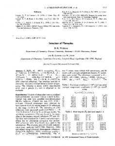
(R)-BACLOFEN M O N O H Y D R O C H L O R I D E H3(N) H2(NlCX~/p N~HI(N)
Rw __ [~ w(iFol _ iFci)2/wF2o]1/2, is 0.034. The atomic scattering factors were taken from International Tables for X-ray Crystallography (1974). The corrections for anomalous scattering (International Tables for X-ray Crystallography, 1974) were included for CI and O atoms. The highest peak in the final difference Fourier maps was less than 0.32e,h,-3. The final atomic parameters with their e.s.d.'s are listed in Table 1. The absorption-correction program used was originally written by L. Templeton (based on the analytical method of Alcock, 1970). The absolute configuration was determined by the Bijvoet method (Bijvoet, Peerdeman & van Bommel, 1951). The structure factors of both configurations had been calculated.* The R factor was 0.029 for the correct configuration and 0.047 for its enantiomorph. According to Hamilton's (1965) statistical test, the configuration with lower R value has a probability of being correct at a significance level higher than 99,5%.
HI(g)
H(hl
C16~ ('(?)
H(7)%
HI(2)
O( I )~-~,-~ H(O~
ICt5) ~ H(IO) • ('( I0} "~('('4~
cJ J
Fig. 1. Perspective view of (R)-baclofen with the numbering scheme used in this paper showing thermal ellipsoids at the 50% probability level for non-hydrogen atoms. H atoms are represented by spheres of arbitrary radii.
o
Discussion. An ORTEP II (Johnson, 1970) drawing of the molecule illustrating the atom numbering is shown in Fig. 1. The benzene ring is planar and the deviation of C1 from the ring is - 0 . 0 5 3 ,A while that of C(3) is - 0 . 1 7 8 7~. The N - C ( 4 ) bond length (Table 2) is longer than the standard value (1.47A), confirming the presence of a charged -NH~- group, while the two C - O bond lengths (1.332, 1.203 A) are consistent with an uncharged carboxyl group as found in cyanoacetic acid (1.327 and 1. 199 A; Kanters, Roelofsen &Straver, 1978). The molecules are linked into infinite chains along the b axis by O(1)-H(O). • • CI- and O(2)...H3(N) hydrogen bonds. Each molecule also forms salt bridges with the CI- through its charged -NH~- group. The hydrogen-bonding distances are listed in Table 3. There is no significant ring stacking in this molecule as found in p-(p-chlorophenyl)propionic acid (Glusker, Zacharias & Carrell, 1975) and pchlorobenzoic acid (Miller, Paul & Curtin, 1974). A conformational comparison of the aminobutyric acid backbones in baclofen and in protonated and free aminobutyric acid as obtained from crystallographic analysis is shown in Fig. 2. Although Fig. 2(a) and (d) are different from (b), (e), (e) and (f), one baclofen enantiomorph [(R) form] can attain the conformation of GABA shown in Fig. 2(c) and ( f ) by freely rotating about the single bonds. On the other hand, the (S) form cannot assume the same conformation of 2(c) and ( f ) because of steric hindrance due to the configuration around C(3). Inspection of CPK models shows that all GABA conformations are assumable by racemic * Lists of structure factors and anisotropic thermal parameters have been deposited with the British Library Lending Division as Supplementary Publication No. SUP 36736 (8 pp.). Copies may be obtained through The Executive Secretary, International Union of Crystallography, 5 Abbey Square, Chester CH 1 2HU, England.
oN oO (a)
(b)
(d)
(e)
(f)
Fig. 2. A conformational comparison of the aminobutyric acid backbone in baclofen (a,d) and in protonated (b,e) and free aminobutyric acid (c,f). Projections are viewed along the bonds C(4)-C(3) (a,b,e) and C(3)-C(2) (d,e,f). The torsion angles at bonds C(1)-C(2), C(2)-C(3) and C(3)-C(4) are 165.5 (4), 155.5 (3), 67.2 (5)° for baclofen, and 167.3, 73.6, -175.2 ° for GABA at room temperature (Tomita, Higashi & Fujiwara, 1973) and 170.3, 73.6, -175.2 ° for GABA at low temperature (Steward, Player & Warner, 1973a) and 167.9, 169.3, 178.0° for GABA. HCI (Steward, Player & Warner, 1973b). baclofen but in certain cases by one of the enantiomorphs only. Consequently, if the aliphatic-chain conformation were the critical element determining the activity of GABA, racemic baclofen would be expected to react at the same receptor as GABA with at least one half the activity. However, the pharmacological evidence indicates that they act on entirely different receptors with the singular exception of the zona retieulata of the rat substantia nigra (Waddington & Cross, 1979). Clearly, the failure of racemic baclofen to interact at the GABA receptor is not due to limitations on the conformation of the common moiety imposed by the ]~-(p-chlorophenyl) substituent. The most probable explanation lies in the concept that GABA receptors can accommodate only molecules approximating GABA in size (Galli, Zilletti, Scotton, Adembri & Giotti, 1979, 1980; Honore, Hjeds, Krogsgaard-Larsen & Christiansen, 1978; Nicholson, Suckling & Iversen, 1979). Ironically, the addition of the chlorophenyl group, which was made to enable the drug to penetrate the blood/
(R)-BACLOFEN M O N O H Y D R O C H L O R I D E Table 2. Bond distances (A) and angles (o) with e.s.d.'s in parentheses Ring C(5)-C(6) C(6)-C(7) C(7)-C(8) C(8)-C(9) C(9)-C(10) C(10)-C(5) C(8)-CI C(5)-C(3)
1.387 (5) 1.388 (5) 1.371 (5) 1.377 (5) 1.390(5) 1.382(5) 1.743 (3) 1.523 (4)
Ring C(5)-C(6)-C(7) C(6)-C(7)-C(8) C(7)-C(8)-C(9) C(8)-C(9)-C(10) C(9)-C(10)-C(5) C(10)-C(5)-C(6) C(7)-C(8)-C! CI-C(8)-C(9) C(3)-C(5)-C(6) C(10)-C(5)-C(3)
121.8(3) 118.6(3) 121.2(3) 119.2 (3) 121.1 (3) l l8.0 (3) 119.2 (3) 119.6 (3) 121.4(3) 120.4 (3)
Side chain C(1)-C(2) C(2)-C(3) C(3)-C(4) C(1)-O(I) C(1)-O(2) C(4)-N
1.500 (5) 1.530 (5) 1.527 (4) 1.332 (4) 1.203 (4) 1.499 (4)
Side chain O(1)-C(1)-O(2) O(1)-C(1)-C(2) O(2)-C(1)-C(2) C(1)-C(2)-C(3) C(2)-C(3)-C(4) C(2)-C(3)-C(5) C(4)-C(3)-C(5) C(3)-C(4)-N
123.8 (3) 111.2 (3) 124.9 (3) 114-1 (3) 111-0 (3)
113.3 (3) 106.8 (3) 113.2 (3)
Table 3. Hydrogen bonds (e.s.d.'s ~ 0 . 0 0 3 / k ) A-H...B N-HI(N).-.CI-I0001* N-H2(N).-. CI-[01115" N-H3(N)... CI-[ 1001" O(l)-H(O)-.-Cl-[0i0]* N-H3(N)...O(2)[010]*
A...B 3.272 A 3.246 3-196 3-069 2.845
H...B 2.55 A 2.80 2.58 2.33 2.28
* Lattice translation. t (½+ x, ½- y, ,~) + lattice translation. brain barrier by increasing its lipid solubility, also incapacitated its interaction with the G A B A receptors because it simultaneously increased the size of the molecule too much. In the single exception mentioned above, it v'as observed that both (R)- and (S)-baclofen interacted equally effectively at low-affinity G A B A receptors in the substantia nigra of the rat but neither was active at the high-affinity receptors. Presumably, a large steric tolerance distinguishes the low- from the high-affinity receptors in the unusual case. We thank Dr W. Bencze for providing these compounds and helpful discussions concerning the results. We gratefully acknowledge the use of computing facilities at the University of Pittsburgh.
2067 References
ALCOCK, N. W. (1970). Crystallographic Computing, edited by F. R. AHMED,pp. 271-288. Copenhagen: Munksgaard. BIJVOET, J. M., PEERDEMAN, A. F. • VAN BOMMEL, A. J. ( 1951). Nature (London), 168, 271-272. BIRKMAYER, W. (1972). Spasticity - A Topical Survey. Bern: Huber. FROMM, G. H., TERRENCE, C. F., CHATTHA, A. S. & GLASS, J. D. (1980). Arch. Neurol. (Chicago), 37, 768-771. GALLI, A., ZILLETTI, M., SCOTI'ON, M., ADEMBRI, G. St, Glorrk A. (1979). J. Neurochem. 32, 1123. GALLI, A., ZILLETTI, M., SCOTTON, M., ADEMBRI, G. & GIOTTI, A. (1980). Pharmacol. Res. Commun. 12, 267-272. GLUSKER, J. P., ZACHARIAS, D. E. & CARRELL, H. L. (1975). J. Chem. Soc. Perkin Trans. 2, pp. 68-74. HAMILTON, W. C. (1965). Acta Cryst. 18, 502-510. HONORE, T., HJEDS, H., KROGSGAARD-LARSEN, P. & CHRISTIANSEN, T. R. (1978). Eur. J. Med. Chem. Chim. Ther. 13, 429-433. International Tables for X-ray Crystallography (1974). Voi. IV. Birmingham: Kynoch Press. JOHNSON, C. K. (1970). ORTEP II. Report ORNL-3794, 2nd revision. Oak Ridge National Laboratory, Tennessee. KANTERS, J. A., ROELOFSEN, G. & STRAVER, L. H. (1978). Acta Cryst. B34, 1393-1395. MAIN, P. (1978). MULTAN 78. A System of Computer Programs for the Automatic Solution of Crystal Structures from X-ray Diffraction Data. Univ. of York, England. MILLER, R. S., PAUL, I. C. 8/. CURTIN, D. Y. (1974). J. Am. Chem. Soc. 96, 6334-6339. NICHOLSON, S. H., SUCKLING, C. J. St, IVERSEN, L. L. (1979). J. Neurochem. 32, 249-252. PIER, F. K. & ZIMMERMAN, P. (1973). Brain Res. 54, 376-380. POLC, P. & HAEFELY, W. (1976). Naunyn Schmiedeberg's Arch. Phamacol. 294, 121-131. POTASHNER, S. J. (1979). J. Neurochem. 32, 103-109. SACHAIS, B. A. & LOGUE, J. N. (1977). Arch. Neurol. (Chicago), 34, 422-428. SAITO, K., KONISHI, S. & OTSUKA, M. (1975). Brain Res. 97, 177-180. STEWARD, E. G., PLAYER, R. B. & WARNER, D. (1973a). Acta Cryst. B29, 2825-2826. STEWARD, E. G., PLAYER, R. B. t~ WARNER, D. (1973b). Acta Cryst. B29, 2038-2040. TOMITA, K., HIGASHI, H. & FUJIWARA, T. (1973), Bull. Chem. Soc. Jpn, 46, 2199-2204. WADDINGTON, J. L. & CROSS, A. L. (1979). Neurosci. Left. 14, 123-127.