Apr 18, 1983 - Mouse teratocarcinoma cells express neither H-2 heavy chains nor ,32-microglobulin (32-m). We have constructed two genomic libraries, one ...
The EMBO Joumal Vol.2 No.7 pp. 1061 - 1065, 1983
Structure and expression of the mouse f2-microglobulin gene isolated from somatic and non-expressing teratocarcinoma cells
Francoise Daniel*, Dominique Morello, Odile Le Bail, Philippe Chambon, Yvon Cayre and Philippe Kourilsky Unite de Biologie Moleculaire du Gene, E.R. C.N.R.S. 201 and S.C. I.N.S.E.R.M. 20, Institut Pasteur, 25, rue du Dr. Roux, 75724 Paris CUdex 15, France Communicated by P.Kourilsky Received on 16 March 1983; revised on 18 April 1983
Mouse teratocarcinoma cells express neither H-2 heavy chains nor ,32-microglobulin (32-m). We have constructed two genomic libraries, one from PCC4-aza-RI embryonal carcinoma cells and the other from their adult syngenic counterpart 129/Sv liver cells (H-2bc). The libraries were screened with a full length mouse $2-m cDNA probe which we isolated and sequenced. Two cosmid clones carrying the entire ,82-m gene were isolated, one from each library. There was no detectable difference in structure between the two genes. Furthennore, both were shown to be active and to restore (B2-m synthesis upon transfer into mutant cells deflcient in (2-m. Irreversible DNA alterations in or around the (32-m gene are thus unlikely to account for the lack of $2-m gene expression in embryonal teratocarcinoma cells. Key words: ,B2-microglobulin/gene transfer/molecular cloning/teratocarcinoma cells
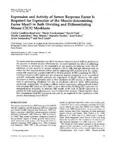
et al., 1981). Four out of -2000 clones were found to hybridize under low stringency conditions (see Materials and methods). The one which carried the longest insert, p,B2-m2, was further studied and sequenced. The restriction map and the strategy used to determine the nucleotide sequence are shown in Figure 1. The sequence (711 bp) is indicated in Figure 2. p32-m2 contains the entire $2-m coding sequence including the signal sequence, 52 bp upstream of the ATG and 238 bp of the 3' non-coding region followed by a poly(A) stretch of 64 residues. There is identity with the sequence of exons in the $2-m gene described by Parnes and Seidman (1982), except at position - 3 of the amino acid sequence where we found a TTG instead of CTG, both triplets coding for leucine. Therefore, our data confirm the existence of a signal peptide of 20 amino acids with Met in position - 20 and Ala in position - 19 rather than Ser, as reported by Lingappa et al. (1979). Mapping of the promoter of the (32-m gene has not yet been reported. Unless a very long 5' -untranslated region exists, p32-m2 is likely to be a full length or nearly full length cDNA clone, but this cannot be ascertained. The 3 '-untranslated region contains two polyadenylation signals (AATAAA, underlined in Figure 2). The (32-m gene is known to be transcribed into (at least) two major RNA species (Croce et al., 1981), 800 and 1000 nucleotides long (Morello et al., 1982) but it is not yet possible to correlate precisely p,B2-m2 with either of these transcripts. The 85th amino acid underlined in the sequence (Figure 2) is Asp, coded by a GAC triplet. p(32-m2, therefore, encodes the more acidic form of $2-m (02-ma), while the more basic form (32-mb) is specified by an Ala residue (Gates et al., 1981). This allelism correlates with the existence (b form) or the absence (a form) of a BglI restriction site in the gene (our unpublished results). The PstI-PstI and PstI-EcoRI fragments which served as 3'- (probe A) and 5'- (probe B) specific probes, respectively, are shown in Figure 1. Probe C was a mixture of the two 350-bp PstI-PstI fragments encompassing the entire insert. Isolation of the 32-m gene from PCC4-aza-RI and 129/Sv liver libraries A cosmid vector was engineered from plasmid pSV2 (Berg, -
Introduction Mouse (2-microglobulin (032-m) is a polypeptide 99 amino acids long found associated with a variety of membrane glycoproteins which include the class I transplantation antigens. (32-m, as well as H-2 antigens, is absent from the fertilized egg, but clearly detectable at the surface of day 7 embryos (Buc-Caron et al., 1978). However, it has been reported to be present in stage two cell embryos (Sawicki et al., 1981). (32-m is serologically undetectable in embryonal carcinoma cells (EC cells) (Dubois et al., 1976; Morello, 1978). There is no accumulation of H-2 and/or (32-m poly(A) + RNA either in the cytoplasm (Croce et al., 1981; Morello et al., 1982) or in the nucleus of EC cells (Morello et al., 1983). When differentiation of EC cells is induced in vitro, it appears (like H-2) in increasing amounts on an increasing number of cells (reviewed in Gachelin, 1978). To analyze whether the :2-m gene isolated from EC cells can be expressed upon introduction into adult cells, we have cloned it from a cosmid library prepared with DNA of PCC4-aza-RI EC cells. As a control, we also isolated the (32-m gene from a library of 129/Sv liver DNA. We show that genes from both sources are structurally indistinguishable and both can be expressed upon transfer into ,B2-m - mutant cells.
i
ffi
IC
8
=
Z
a
4 $$
w
.' +
4.0 cr 0
L
c
Results Isolation of a mouse (2-m cDNA probe A cDNA library, constructed in plasmid pBR327 from poly(A) + RNA extracted from DBA/2 mouse liver (Lalanne et al., 1983) was screened by in situ hybridization with a human (32-m cDNA probe kindly provided by S.Suggs (Suggs *To whom reprint requests should be sent.
© IRL Press Limited, Oxford, England.
100 p
Fig. 1. Restriction endonuclease analysis of p,B2-m2. pBR327 vector DNA is represented as a thick line. The strategy used to determine the nucleotide sequence of p,B2-m2 is described above the restriction map. Arrows indicate the sequenced fragments, tails representing the 5' or 3' labelled ends. Fragments A, B and C below the restriction map were used as probe in in situ hybridizations and Southern blotting experiments.
1061
F.Daniel et al. M
A
S
R
T
V
L
V
F
L
V
L
V
S
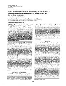
ATTTCAC,TGGCTGCTACTC(GGCGCTTCAGTCGCGGTCGCTTCAGTCGTCAGC ATG GCT CGC TCG GTG ACC CTA GTC TTT CTG GTG CTT GTC TCA -20 P M P C P M G K I L M Q I Q V Y S R H P E L Y A I K T T G Q L CTG ACC GGC TTG TAT GCT ATC CAG AAA ACC CCT CM ATT CAA GTA TAC TCA CGC CAC CCA CCG GAG MT GGG MG CCG MC ATA CTG MC TGC 20 1 M M D F P K I K V E S S H I E I L K M G K H P M V T F P Q y Q TAC GTA ACA CAG TTC CAC CCG CCT CAC ATT GM ATC CM ATG CTG MG MGC GGG AM AAA ATT CCT AAA GTA GAG ATG TCA GAT ATG TCC TTC
40 A M C T E T D T R V K H D H T F T Y I L A P F Y E S S K D W S AGC MG GAC TGG TCT TTC TAT ATC CTG GCT CAC ACT GM TTC ACC CCC ACT GAG ACT GAT ACA TAC GCC TGC AGA GTT MG CAT GAC AGT ATG 80 60 W D R D M K T V E Y P A GCC GAG CCC AAG ACC GTC TAC TGG GAT CGA GAC ATG TGATCAAGCATCATGATGCTCTGMGATTCATTTGMCCTGCTTMTTACMATCCAGTTTCTAATATGCTATAC 99
TAAGTATTTTGATCAGAATAATAMTATMTTTTMMGAACAAAAAAAAAAAAAA
...................
Fig. 2. Nucleotide sequence of pf2-m2. The coding sequence is represented in triplets according to the genetic code. Letters represent the corresponding amino acids. Negative numbers are used for the signal peptide. The 85th amino acid characteristic of the ,32-ma allele and the two polyadenylation signals are underlined. iLk
a
_F-W
a
t3
c
a
b
-
d
s1
L-1; b c. d zD
e f.
-
as_
.l
0...
4.
:
a' *4.
4*
Fig. 3. Southern blot hybridization of c32-ml DNA with 3' (A) and
a' 5'
(B)
p032-m2 probes. Hybridizations were performed as described in Materials
Fig. 4. Southern blot hybridization of c,B2-m2 DNA with 3' (A) and 5 (B)
p,32-m2 probes. Experiments and size determinations were performed as
and methods. c,32-ml DNA was digested by: lane a: BamHI; lane b: BamHI and EcoRI; lane c: EcoRI; lane d: EcoRI and HindIII; lane e: HindlII; lane f: HindIII and BamHI. Sizes in kb (within 10% error) shown in the margins were determined from stained HindIII digests of phage XS7 DNA.
described in Figure 3 except that the c,B2-m2 DNA was digested from the left to the right, by: lane a: EcoRI and BamHI; lane b: BamHl; lane c: Hindlll and BamHI; lane d: HindIII; lane e: EcoRI and HindIII; lane f: EcoRI.
1981) as outlined in Materials and methods, and kindly provided by J.Papamatheakis. It is 7 kb long and carries the Ecogpt gene. Following the method of Grosveld et al. (1981), we built two genomic libraries containing large inserts (30-40 kb) of PCC4-aza-RI and 129/Sv liver DNA. Each library contained -200 000 independent clones and was screened with probe C (Figure 1). One positive recombinant cosmid (ci32-ml) was isolated from the PCC4-aza-RI library, and another one (c32-m2) from the liver library. KpnI was convenient to establish the overall restriction maps of purified c32-ml and c,B2-m2 DNAs. Three restriction enzymes (BamHI, EcoRI, HindIII) were also used alone or in combinations. Southern blotting experiments were performed (Figures 3 and 4), using the 3' probe (A) and the 5'
probe (B) to elaborate the restriction maps of the two genes and of their surrounding regions (Figure 5). The inserts in cf32-ml and c,B2-m2 are 35 kb and 39 kb long, respectively, within < 1O0o error. Both contain an entire ,B2-m gene. Their restriction maps with BamHI, EcoRI, HindlII and KpnI in and around the gene, are identical and fit perfectly the map of the ,B2-m gene established by Parnes and Seidman (1982), except that the HindIII site, at the 3' end of the gene, is, in our case, located 200 bp closer to exon 4. Thus, the ,32-m gene in non-expressing PCC4-aza-RI cells has a structure similar, or identical, to that in liver cells. This conclusion is further strengthened by the visualization of heteroduplexes between restriction fragments of cfl2-ml and c,32-m2 under the electron microscope which revealed no difference in several kilo-
1062
Structure and expression of the K
K
K
I
I
I
K
K
K
K
i
I
1
K
K
-1kb
i
K
K
I
Cell line
Mean fluorescence
070
RI
336 422 577 161 201 602 272 429 747 309 471 762
100 77 42 100 70 5 100 58 25 100 61 30
,~~~T-
:~~~~~~~~~~~~~ H
K
K
i
H KH
I
E
IU
E
RI (j32)
H
K v
H
E44
t E
4
E
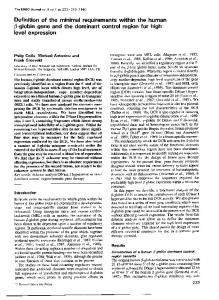
Fig. 5. Restriction endonuclease analysis of c,B2-m2 (A) and cf2-ml (B) using Kpnl restriction enzyme. Thick lines represent vector DNA. Restriction endonuclease analysis of ,B2-m gene and surrounding region (C): K = Kpnl; B = BamHI; H = Hindlll; E = EcoRI. Black boxes 1-2-34 define the regions of ,B2-m genes represented in mature mRNA (as defined by Parnes and Seidman, 1982).
A
C
D
B
Fig
6
Fluorescence
micrographs of parental and transformed RI(_32-m
cells tested with rabbit anti (32 anti rabbit
A
transformed with
m
antiserum and fluorescein
parental
RI cells
c02n
D
B
=RI(32-m
bases at the 5' end of the gene
Transient expression mouse
R1Q32-m
=
) cells, C
conjugated goat RI(32-m =
) transformed with c_32-m2.
(data
of the cloned
not
(32-rn
shown). gene in
RI((32-m
cells
We undertook to transfer the two genes into
recipient cells had
no
appropriate they are both functional. Since we detect specifically a (32-Ma polypeptide in a
to examine if
reagent
to
(32-Mb background, we could not introduce our (32-Ma genes into (32-mb cells. We, therefore, turned to mutant cells defi(32-in. One such cell
line, named by immunoselection as a TL -, derivative of a C58 thymoma (Hyman and Stallings, 1976). Parnes and Seidman (1982) demonstrated that these cells
cient in the R1Q32-m -)
synthesis of
was
isolated
show alterations in the (32-in gene of both chromosomes.
c(32-mlI
c(32-mn2
DNA into these cells, we protoplast fusion technique described by Rassoulzadegan et al. (1982), as explained in Materials and methods. The expression of (32-in at the cell surface was monitored by an indirect immunofluorescence assay, using a rabbit anti-mouse (32-in serum adsorbed on To introduce
devised
a
and
modification of the
gene
Table I. Expression of ,32-m in parental RI and RI(32-m ) cells transfected either with c(32-ml or c,B2-m2.
.---'
a
32-niicroglobulin
K
1Kb
-
B
K
I
A
mouse
R1 (J32) c,2-ml
RI (32
c,B2-m2
For each selected window (see Materials and methods), the mean fluorescence and percentage of corresponding fluorescence cells were calculated. Fusion of RI(j32-m -) cells with a cosmid carrying an H-2 gene (to be published) give results which are identical to the RI(J2-m -) cells results (data not shown).
Daudi cells. Observation under the fluorescence microscope, 72 h after protoplast fusion, showed that both cosmids restore 32-m expression in the R132-m -) cells (Figure 6). These results were quantitated by use of a fluorescence activated cell sorter. About 50%o of the cells expressed ,B2-m at the surface with a fluorescence intensity identical to that of the RI(32-m +) parental cell line and several fold higher than that of the RI(2-m -) parental cell line. About 200%o of the transformed cells showed a fluorescence intensity higher than that of the RI(32-m +) cell line (Table I). Discussion Here we describe the isolation and sequence of a 32-m cDNA clone from DBA/2 liver mRNA which is likely to be full length or nearly so, and encodes the more acidic form of $2-m. We used it as a probe to clone the ,B2-m gene from two sources: PCC4-aza-RI cells, which do not express ,82-m and H-2, and their adult syngenic counterpart, 129/Sv liver cells. The genomic libraries contained 200 000 clones each, i.e., more than one genome equivalent. We found only one 32-m gene in each library and no $2-m related gene or ,B2-m pseudogene. Thus, our results are compatible with the existence of a single ,82-m gene per haploid genome, a conclusion reached by Parnes and Seidman (1982) from the analysis of the genetic defects of the R1Q2-m -) cell line. Our two cosmid clones, c(2-ml and c,B2-m2 show identical restriction maps in and around the (32-m gene, as judged from restriction analysis and Southern hybridization with 5' and 3' specific probes, but discrete structural differences between the (2-m gene isolated from EC cells and adult cells would not have been detected. We, therefore, proceeded to show that both genes are active. The experiment required gene transfer into a lymphoid cell line, which had not been achieved before, and which we performed through a modification of the protoplast fusion technique. (32-m expression was detected with both genes, more reliably 72 h rather than 48 h after fusion. (32-m was less easily detected and in a smaller population of transformed cells than SV40 T antigen in control experiments (Y.Cayre and B.Cami, unpublished data). Many factors could interfere with ,B2-m expression and/or detection. Protoplast fusion for the transfer of cosmid DNA from 1063 -
F.Daniel et al.
a recA - Escherichia coli host may be less efficient. The synthesis and/or transport of f2-m to the membrane may depend on the cellular cycle or the physical state of the membrane altered by the fusion, or be limited by the synthesis of the presently unidentified heavy chain presumably associated with it. Together, the finding that the 32-m gene extracted from EC cells has a structure similar to that extracted from adult cells, and the demonstration that it can be expressed in f2m - mutant cells, argue very strongly that there is no DNA rearrangement associated with its activation in vivo. Other structural modifications may, however, take place, such as partial demethylation, which we know occurs in the case of H-2 genes (Morello et al., 1983). Further analyses on the structure and expression of the ,B2-m gene transferred into EC cells should shed some light on the developmentally controlled expression of the $2-m gene.
Materials and methods Materials Bacterial strains E. coli BHB 2688 and BHB 2690 (Hohn, 1980) were used for preparation of the lambda packaging mix. Packaged cosmid DNA was transfected and propagated in E. coli 1046 (803 rk -mk - su3 + recA -) (Murray et al., 1977). PCC4-aza-RI and its growth conditions are described in Jakob et al. (1973). RI and RI (132-mr-) (Hyman and Stallings, 1976) cells were grown in RPMI 1640 medium (Gibco) supplemented with glutamine (2 mM) and 10% fetal calf serum. Restriction endonucleases were from Boehringer Mannheim, nitrocellulose filters BA 85 from Schleicher and Schull. [ae-32P]nucleotides (2000- 3000 Ci/mmol), [-y_32P]nucleotides (2000 Ci/mmol) and cordycepin (5000 Ci/mmol) were purchased from Amersham. T4 nucleotide kinase was from P.L. Biochemicals and deoxynucleotidyl terminal transferase from B.R.L. Rabbit anti-12-m antiserum (Natori et al., 1974), a kind gift from Dr.Tanigaki, was absorbed on Daudi cells. Isolation and sequence of pf32-m2 Poly(A)+ RNA was extracted from DBA/2 mouse liver and complementary cDNAs were cloned as described by Lalanne et al. (1983). In situ hybridizations were performed for 16 h at 60°C in 1 ml per filter of 2 x SSC, 2 X 10⁻³ M EDTA 1 x Denhardt solution, potassium phosphate buffer pH 7.2, 0.025 M, 0.5% SDS with 2 x 10⁶ c.p.m. of radiolabelled PstI fragment of human $2-m cDNA (Suggs et al., 1981). Filters were washed at 60°C in 1 x SSC and 0.50%o SDS (4 times 45 min). p,B2-m2 was analyzed by restriction mapping and its DNA sequence was determined according to Maxam and Gilbert (1980) after either 5' labelling by -y exchange or 3' labelling by addition of cordycepin with terminal transferase (Maxam and Gilbert, 1980). Construction of cosmid libraries High mol. wt. DNA was extracted from PCC4-aza-RI cells and mouse 129 liver as described by Gross-Bellard et al. (1973). These two DNAs were partially digested by Sau3A and fractionated in a sucrose gradient according to Grosveld et al. (1981). The size of the DNA in each fraction was measured in a 0.2% agarose gel. Fractions of - 35 kb were used for ligation. The cosmid vector, pSV2cos, was constructed by J.Papamatheakis (unpublished work) as follows: pHC79 DNA (Hohn and Collins, 1980) was digested by BglII. The 1.8-kb fragment containing the cos site was recovered from a 1% agarose gel. pSV2 DNA (Berg, 1981) was digested by EcoRI and treated with bovine alkaline phosphatase (Worthington). Both DNAs were treated with the Klenow enzyme (Boehringer Mannheim) and ligated. pSV2cos was recovered after in situ hybridization using the 'cos' fragment of
pHC79 as a probe. Vector and genomic DNAs were ligated and packaged in vitro as described by Grosveld et al. (1981). In situ hybridization - Southern blotting DNA fragments used as probes were recovered from low gelling agarose gels (Wieslander, 1979). They were labelled with [a-32P]dTTP and [a-32P]dCTP by nick translation to a specific activity in the range of 1-2 x 10⁸ c.p.m./4g (Rigby et al., 1977). Nitrocellulose filters carrying lysed bacterial colonies were prepared according to Hanahan and Meselson (1980). Hybridizations were performed for 16 h at 68°C in 1 ml per filter of: 2 x SSC, 2 x 10⁻³ M EDTA, 1 x Denhardt
1064
solution, potassium phosphate buffer 0.025 M, pH 7.2, 0.5% SDS with 2 x 10⁶ c.p.m. of radiolabelled probe per ml. Filters were washed at 68°C in 2 x SSC, 1 x Denhardt solution, 0.5% SDS (4 times, 45 min) and then once in 0.1 x SSC for 30 min. Filters were dried and exposed at - 70°C using a XAR film (Kodak) with intensifying screens (Dupont de Nemours). After complete digestion by various enzymes, recombinant cosmid DNAs were run on 0.8% agarose gels and transferred to nitrocellulose filters according to Southern (1975). Hybridizations were carried out, as above, using 2 x 10⁶ c.p.m. of radiolabelled probe in 5 ml of buffer per filter. Protoplast fusion Protoplasts were prepared according to the procedure of Rassoulzadegan et al. (1982) with some modifications. E. coli bacteria (strain 1046) carrying the cosmid recombinants were grown up in L broth to a density of 2 x 10⁸ cells/ml. (In our hands, chloramphenicol amplification was not required.) 50 ml of culture were centrifuged, resuspended in 500 1u of 50 mM Tris-HCl,
pH 8.0, containing 20% sucrose and allowed to stay on ice for 10 min. 100 /d of a freshly made solution of lysozyme (5 mg/ml) (Sigma) were added and the suspension was incubated for 10 min at 0°C. 250 Id of 0.25 M EDTA, pH 8.0, were added and the conversion of bacteria into protoplasts was followed under the microscope. The suspension was then diluted by addition of 750 tl of 50 mM Tris-HCl, pH 8.0, containing 7% sucrose. Protoplasts were centrifuged at 2000 r.p.m. during 10 min. At the same time, S x 106 RI(32-m -) cells were washed with RPMI 1640 medium without fetal calf serum and collected by centrifugation. Protoplasts were resuspended in 1 ml of a 41.7% PEG 1500 solution (20 g of PEG 1500 dissolved in 28 ml of Dulbecco's phosphate buffered saline 1 x containing 15% dimethylsulfate) and this suspension was added on the RI(32-m -) cells pellet, mixed gently during 1 min at room temperature. 10 ml of RPMI 1640 without fetal calf serum were added slowly (during - 5 min), the cells were then washed and resuspended in normal medium containing 250 ug/ml of kanamycin. Indirect immunofluorescence tests were performed 48 h and 72 h after fusion. Cells were allowed to react with the rabbit anti-mouse 32-m antiserum and with fluorescein immunoconjugated goat anti-rabbit immunoglobulins, using the conventional indirect immunofluorescence procedure (Pope and Rowe, 1964). ,B2-m expression in transformed and parental cells was determined either by fluorescence microscopy or by flow cytofluorimetry as follows: stained cells were analyzed for the percentage of bright fluorescent cells in a cytofluorograph 50 HH (Ortho Diagnostic Systems, Westwood, MA) using the 488 nm line with 400 mW power from an Arganion laser (Lexel model 95.4). In this analysis, cells with a given size class were selected in such a way as to discriminate for living cells (dead cells have a different shape and are, therefore, not taken into account). The low angle light scattering was used for gating, thus eliminating the contribution of the debris from the fluorescence histograms. The exciting light was excluded by the dichroic filter and narrow band pass (514- 540 nm) filter for fluorescein. For each sample, data obtained from -25 000 cells were analyzed on a microprocessor MP 2000 (2150 Ortho Diagnostic Systems). For each cell line, three 'windows' were selected to define three subpopulations with different mean fluorescence (see Table I).
Acknowledgements We are grateful to Dr.J.L.Lalanne who provided the cloned cDNA library, to Dr.Suggs for his generous gift of the human 12-m cDNA clone and to J.Papamatheakis, B.Cami and other colleagues in the laboratory for helpful discussions. We also thank Dr.P.Metezeau for use of the Cell Sorter (S.C. I.N.S.E.R.M.-Pasteur 'Trieur de Cellules'). F.Daniel is C.R. of I.N.R.A. This work was supported by grants of C.N.R.S. (E.R. 201 and A.T.P. 95.5039), I.N.S.E.R.M. (S.C. 20 and P.R.C. 124031) and the Fondation pour la Recherche Nationale Fran,aise.
References Berg,P. (1981) Science (Wash.), 213, 296-302. Buc-Caron,M.H., Condamine,H. and Jacob,F. (1978) J. Embryol. Exp. Morphol., 47, 149-160. Croce,C.M., Linnenbach,A., Hebner,K., Parnes,J.R., Margulies,D.H. Appella,E. and Seidman,J.G. (1981) Proc. Natl. Acad. Sci. USA, 78, 5754-5758.
Dubois,P., Fellous,M., Gachelin,G., Jacob,F., Kemler,R., Pressman,D. and Tanigaki,M. (1976) Transplantation, 22, 467473. Gachelin,G. (1978) Biochim. Biophys. Acta, 516, 27-60. Gates,F.T., Coligan,J.E. and Kindt,T.J. (1981) Proc. Natt. Acad. Sci. USA,
77, 7395-7399. Gross-Bellard,M., Oudet,P. and Chambon,P. (1973) Eur. J. Biochem., 36, 32-38. Grosveld,F.G., Hans-Henrik,M.D., Boer,E. and Flavell,R.A. (1981) Gene,
Structure and expression of the mouse j32-microglobulin gene 13, 227-237. Hanahan,D. and Meselson,M. (1980) Gene, 10, 63-67. Hohn,B. (1980) Methods Enzymol., 68, 299-308. Hohn,B. and Collins,J. (1980) Gene, 11, 291-298. Hyman,R. and Stallings,V. (1976) Immunogenetics, 3, 75-84. Jakob,H., Boon,T., Gaillard,J., Nicolas,J.F. and Jacob,F. (1973) Ann. Microbiol. Inst. Pasteur, 124B, 269-282. Lalanne,J.L., Delarbre,C., Gachelin,G. and Kourilsky,P. (1983) Nucleic Acids, Res., 11, 1567-1577. Lingappa,V.R., Cunningham,B.A., Jazwinski,S.M., Hopp,T.P., Blobel,G. and Edelman,G.M. (1979) Proc. Natl. Acad. Sci. USA, 76, 3651-3655. Maxam,A. and Gilbert,W. (1980) Methods Enzymol., 65, 499-560. Morello,D. (1978) Transplantation, 26, 119-125. Morello,D., Daniel,F., Baldacci,P., Cayre,Y., Gachelin,G. and Kourilsky,P. (1982) Nature, 2%, 260-262. Morello,D., Gachelin,G., Daniel,F. and Kourilsky,P. (1983) Cold Spring Harbor Symp. Quant. Biol., 10, in press. Murray,N.E., Brammar,W.J., Murray,K. (1977) Mol. Gen. Genet., 150, 53-61. Natori,T., Katagiri,M., Tanigaki,N. and Pressman,D. (1974), Transplantation, 18, 550-557. Parnes,J.R. and Seidman,J.G. (1982) Cell, 29, 661-669. Pope,J.M. and Rowe,W.P. (1964) J. Exp. Med., 120, 121-125. Rassoulzadegan,M., Binetray,B. and Cuzin,F. (1982) Nature, 295, 257-259. Rigby,P.W.J., Dieckmann,M., Rhodes,C. and Berg,P. (1977) J. Mol. Biol., 113, 237-251. Sawicki,J.A., Magnuson,T. and Epstein,C.J. (1981) Nature, 294, 450451. Southern,E.M. (1975) J. Mol. Biol., 98, 503-517. Suggs,S.V., Wallace,R.B., Hirose,T., Kawashima,E.H. and Itakura,K. (1981) Proc. Natl. Acad. Sci. USA, 78, 6613-6617. Wieslander,L. (1979) Anal. Biochem., 98, 305-309.
1065