Sub-cellular Feature Detection and Automated Extraction of Collocalized Actin and Myosin Regions Justin Martineau
Ronil Mokashi
David Chapman
University of Maryland Baltimore County 1000 Hilltop Circle Baltimore, MD 21250 410-455-6338
University of Maryland Baltimore County 1000 Hilltop Circle Baltimore, MD 21250 410-455-2860
[email protected] Mary Brady
University of Maryland Baltimore County 1000 Hilltop Circle Baltimore, MD 21250 410-455-6338
[email protected] Yelena Yesha
University of Maryland School of Medicine 100 S. Paca Street, 6th Floor, #200 Baltimore, MD 21201
National Institute of Standards and Technology 100 Bureau Drive Gaithersburg, MD 301-975-4094
[email protected] Antonio Cardone
University of Maryland Baltimore County 1000 Hilltop Circle Baltimore, MD 21250 410-455-3542
[email protected] Alden Dima
University of Maryland Baltimore County 1000 Hilltop Circle Baltimore, MD 21250 410-455-2669
University of Maryland Institute for Advanced Computer Studies AVW Building College Park, MD 20742 301-975-4463
National Institute of Standards and Technology 100 Bureau Drive Gaithersburg, MD 301-975-2450
[email protected] Michael Grasso
[email protected] Yaacov Yesha
[email protected]
[email protected]
[email protected]
ABSTRACT
1.
We describe a new distance-based metric to measure the strength of collocalization in multi-color microscopy images for user-selected regions. This metric helps to standardize, objectify, quantify, and even automate light microscopy observations. Our new algorithm uses this metric to automatically identify and annotate a donut shaped actomyosin stress fiber bundle evident in vascular smooth muscle cells on certain types of surfaces. Both the metric and the algorithm have been implemented as an open source plugin for the popular ImageJ toolkit. They are available for download at http://code.google.com/p/actin-myosin-plugin/. Using cells stained for the cytoskeletal proteins actin and myosin, we show how characteristics of the identified stress fiber bundle are indicative of the kind of surface the cell is placed upon, and prove that weak spots in this structure are correlated with local membrane extensions. Given the relationship between membrane extension, cell migration, vascular disease, embryonic development, and cancer metastasis we provide that these tools to enable biological research that could improve our quality of life.
This study introduces a metric to standardize, quantify, and objectify light microscopy colocalization observations. It is important to standardize light microscopy colocalization observations observations so biologists can quickly, and robustly compare cells and subcellular regions. With standard metrics for colocalization we can define algorithms to automatically identify structural features in cells. For example, in Figure 1 there is an interesting stretched donut shaped actomyosin stress fiber bundle as annotated in blue in Figure 2. Algorithms that can identify these structural sub-cellular features coupled with metrics to determine their strength provide useful insights into cell migration, which is central to many vital biological processes, including vascular disease, embryonic development, and cancer metastasis. To understand cell migration we examine the correlation between cytoskeletal organization and cell morphological features such as membrane protrusions. Membrane protrusions are structures that extend from the cell surface and are good indicators for cell migration. The areas roughly indicated in blue from Figure 4 are examples of membrane protrusions in our dataset. The cytoskeleton is a three-dimensional network of structural fibers found within the cytoplasm of a cell. It is responsible for cell movement and shape stability. Actin microfilaments (shown in red) are one of the three major types of fibers that form the cell cytoskeleton. Through their association with the motor protein myosin (shown in green) these microfilaments carry out cellular movements including gliding, contraction, and cytokinesis. Cells can sense and respond to the mechanical stiffness and the chemical identity of the surfaces that they attach to.
Permission to make digital or hard copies of all or part of this work for personal or classroom use is granted without fee provided that copies are not made or distributed for profit or commercial advantage and that copies bear this notice and the full citation on the first page. To copy otherwise, to republish, to post on servers or to redistribute to lists, requires prior specific permission and/or a fee. IHI’12, January 28–30, 2012, Miami, Florida, USA. Copyright 2012 ACM 978-1-4503-0781-9/12/01 ...$10.00.
INTRODUCTION AND RELATED WORK
Figure 1: Smooth muscle cell with a stretched donut shaped actomyosin stress fiber bundle. Actin is stained in red. Myosin is stained in green. Yellow areas are overlapping actin and myosin.
Figure 2: The structure we want to identify has been labeled and superimposed in blue upon the actomyosin stress fiber bundle.
Figure 4: Membrane protrusions are circled in blue.
Light microscopy is a powerful approach to study cytoskeletal responses to the extracellular matrix because the cytoskeleton encodes for underlying signals from the extracellular matrix it rests on. Cell microscopy methods often
Figure 3: The proposed structure produced by our algorithm is annotated in blue. Thicker and brighter shades of blue represent higher metric scores indicating greater structure strength.
involve the visual inspection of nuclei, organelles, and morphology. Using this approach, observations are made based on variations in cells from their expected appearance. However, this approach requires human judgments which are subjective due to observer variability, a lack of standardization, and a limited feature set. Consequently, we define a set of quantitative measures that correlate well with the visual appearance of cells and that allow for both intracellular and intercellular comparisons to address this subjectivity. While there are many different statistical measures of dependence and correlation, this is the first measure of correlation or dependence for items adhering to a geometric structure. This kind of measure provides a new kind of information that can improve computational image classification accuracy. Computational image classification techniques have been successfully applied to a number of clinical problems [5] [4] [2]. Computational image classification is used to categorize a raster image into a finite set of classes based on computationally extracted features. When considering meaningful features for describing an image, the three fundamental features include spectral, textural, and contextual features. Spectral features describe the tonal variations that can be measured as a distribution and represented as a histogram [9]. Textural features contain information about the spatial distribution of tonal variations. These tonal variations can be represented as a co-occurrence matrix [8]. The first two types of features, spectral and textural, are essentially non-geometric image features based on tonal variations and tonal distributions. Contextual features are more complex. They are used to extract structural information from the image context. This paper defines new structural features for image retrieval, classification, and other kinds of analysis building on our previous work in this area [7].
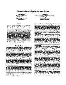
011/2%% !"#
%$3,)4"/+5'%
6)"(#7,)4%7),4% &)"2#8"/'%*,%9':%
!"#$%&'($$)*%
!"#$%% &'(')"*+,(%
-)%.+/*')%
!');'%