Electronic Supplementary Material (ESI) for RSC Advances This journal is © The Royal Society of Chemistry 2013
Supplementary Information for:
Surface Properties of Nanostructured Bio-Active Interfaces: Impacts of Surface Stiffness and Topography on Cell-Surface Interactions Ilia Platzman, ‡,*,a,b Christine Anna Muth,,‡,a,b Cornelia Lee-Thedieck,a,b,c Diego Pallarola,a,b Ralitsa Atanasova,a Ilia Louban,a,b Eva Altrock a and Joachim P. Spatz a,b a
Department of New Materials and Biosystems, Max Planck Institute for Intelligent Systems
Heisenbergstr. 3, Stuttgart 70569, Germany; b Department of Biophysical Chemistry, University of Heidelberg, Heidelberg 69120, Germany; c Karlsruhe Institute of Technology, Institute of Functional Interfaces, Hermann-von Helmholtz-Platz 1, 76344 Eggenstein-Leopoldshafen, Germany. Keywords: Biofunctionalization; Block copolymer nanolithography; Cell adhesion; Mechanical properties; PEG hydrogel; Surface roughness.
*
Corresponding author E-mail:
[email protected] (IP)
1
Electronic Supplementary Material (ESI) for RSC Advances This journal is © The Royal Society of Chemistry 2013
20 n nm
5 nm m
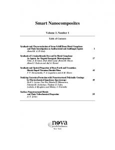
Figure S1. TEM crross-sectionn micrographhs with diffferent resoluutions of Auu/Glass confformation. mage showss a gold naanoparticle partially (~ ~25 %) em mbedded in the glass High-resolution im surface.
micrographs of the caantilever ussed for indeentation meeasurementss with the Figure S2. SEM m o about 8 µ µm in diameter. sphericaal indenter of
2
Electronic Supplementary Material (ESI) for RSC Advances This journal is © The Royal Society of Chemistry 2013
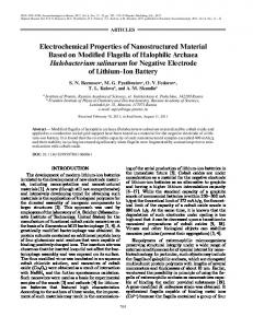
RGDfK) peeptide used w within this study. The cyclically Figure S3. Molecuular structurre of the c(R arrangedd amino aciids are argiinine (R), glycine (G) and aspartaate (D). A) PEG spacerr (6 units) was useed to breachh between thhe peptide aand the cystteine. B) Aminohexanooic (Ahx) sppacer was used to breach betw ween the pepptide and the thiol.
D micrograpphs showingg the rough morphologgy of PEG7000-DA hydrrogel (Rrms Figure S4. AFM 2D m) (A) and smooth moorphology of PEG-passsivated glasss substrate (Rrms = 1.6 nm) with = 17 nm interparrticle spacings of 60 ± 115 nm (B).
3
Force (N)
Electronic Supplementary Material (ESI) for RSC Advances This journal is © The Royal Society of Chemistry 2013
32.0n 30.0n 28.0n 26.0n 24.0n 22.0n 20.0n 18.0n 16.0n 14.0n 12.0n 10.0n 8.0n 6.0n 4.0n 2.0n 0.0 -60.0n
PEG(700)-DA (2m/sec) PEG(700)-DA (1m/sec) PEG(700)-DA (0.5m/sec) PEG(700)-DA (0.1m/sec) PEG(700)-DA (0.01m/sec)
-40.0n
-20.0n
0.0
20.0n
Distance (m)
40.0n
60.0n
Figure S5. Representative force distance curves derived during indentation measurements on 45 nm spacing gold nanostructured PEG700-DA hydrogel substrate. The substrate was immersed in PBS solution at ambient conditions and measured with different rates of tip approach.
4
32.0n 30.0n 28.0n 26.0n 24.0n 22.0n 20.0n 18.0n 16.0n 14.0n 12.0n 10.0n 8.0n 6.0n 4.0n 2.0n 0.0 -60.0n
Bare PEG(700)-DA (1m/sec) Gold Nanostructured PEG(700)-DA (1m/sec) 1300 1200 1100 1000 900 800 700 600 500 400 300 200 100 0
E Y (KPa) PEG(700)-DA
Force (N)
Electronic Supplementary Material (ESI) for RSC Advances This journal is © The Royal Society of Chemistry 2013
Gold Nanostructured PEG700-DA Bare PEG700-DA
10 15 20 25 30 35 40 45 50 55 60
Indentation (nm)
-40.0n
-20.0n
0.0
20.0n
Distance (m)
40.0n
60.0n
Figure S6. Representative force distance curves derived during indentation measurements on bare (red, left) and 45 nm spacing gold nanostructured (black, right) PEG700-DA hydrogel substrates. Two substrates were immersed in PBS solution at ambient conditions and measured with the same rate of tip approach (1 µm/sec). The insert shows Young’s modulus dependence on indentation depth measured with different rates of tip approach on gold nanostructured PEG700-DA and bare hydrogels.
5
Electronic Supplementary Material (ESI) for RSC Advances This journal is © The Royal Society of Chemistry 2013
Figure S7 Phase-ccontrast imaages of KG11a cell obseerved at 1 h after platinng on nanoostructured GD functionnalized PEG G-passivatedd glass (left ft column) aand PEG7000-DA hydroogel (right and cRG column)) surfaces. T The inserts show the suurfaces conssisted of a ggold nanostrructured areea beneath the dippping line annd the area with no goold nanoparrticles abovve the dippiing line (miicroscopic images)), the cells can be seen aas bright spoots on a grey backgrounnd.
6
Electronic Supplementary Material (ESI) for RSC Advances This journal is © The Royal Society of Chemistry 2013
( obbserved at 4 h after Figure S8. Immunnofluoresceence microggraphs of ffibroblasts (REF52) m cRGDplating on nanostrructured annd cRGD ffunctionalizzed PEG-paassivated gllass (62 nm nanopattternes, uppeer row), PEG700-DA hhydrogel (622 nm cRGD D-nanopatterrnes, middlee row) and PEG7000-DA (110 nm cRGD--nanopatternnes, lower rrow) surfaces. REF52 cells were fixed and immunoostained witth primary aantibodies against a zyxiin and paxilllin, followeed by Alexaa 594- and 647-connjugated seccondary anttibodies, resspectively. Filamentouus actin andd nuclei werre labeled with Aleexa 488-connjugated phaalloidin andd DAPI, resppectively. Scale bars: 30 µm.
7