Akerman (1982), Dufresne-Dube, Metivier, Dube & Guerrier (1983) have demon- strated that Con A elicites Ca2+ fluxes. On the other hand, in the light of experi-.
/. Embryol. exp. Morph. 86, 39-51 (1985) Printed in Great Britain. © Company of Biologists Limited 1985
39
Target cell surface glycoconjugates and neural induction in an amphibian LYDIE GUALANDRIS, PIERRE ROUGE AND ANNE-MARIE DUPRAT* Laboratoire de Biologie ginerale and Laboratoire de Biologie Cellulaire, Faculty Sciences Pharmaceutiqu.es, Universite Paul Sabatier, 118 Route de Narbonne, 31062 Toulouse Cedex, France SUMMARY
The possible involvement of target membrane specific receptor(s) in the transmission of the neural signal leading to activation of the intracellular machinery involved in the process of neural determination, has been examined using lectin probes (Con A, succinylated-ConA, LcA, PsA and SB A). Not only Con A binding sites but many different glycoconjugated molecules (a-Dgalactose, N-acetyl-D-galactosamine, a-D-fucose, N-acetyl-D-glucosamine, etc.) would have to be involved, if neural receptor(s) are invoked to explain initiation of neural induction. We show here that the close involvement of such receptor molecules in neural induction is so far hypothetical and remains to be demonstrated. Moreover we are inclined to the view of Barth and others who suggested that ionicfluxesand physicochemical and electrophysiological properties of the target membrane could play a crucial role in neural induction. INTRODUCTION
The molecular mechanism of the neuralization of ectodermal cells during gastrulation is an important and yet unsolved problem of neuroembryology. It is now recognized that a neuralizing factor (or stimulus) exerts its first effect only at the cell surface of target cells (Tiedemann & Born, 1978; Yamamoto & Ozawa, 1981). Several authors have shown recently the important role played by the target cell membrane in the onset of the neural induction process (Grunz & Staubach, 1979; Takata etal. 1981; Takato, Yamamoto, Ishii & Takahashi, 1984; Duprat, Gualandris & Rouge, 1982; Gualandris, Rouge & Duprat, 1983). Recently, using in vitro association of blastoporal lip with presumptive ectoderm covered (inner side) or not (outer side) by extracellular material, we have shown that the extracellular matrix (covering the neural target tissue) is not necessary for the transmission of the neuralizing signal and seems therefore not directly involved in the process of neural induction (Duprat & Gualandris, 1984). The molecular components of the competent presumptive ectoderm surface (neural target tissue) have a specific pattern especially in their glycoconjugated components as visualized by binding to labelled lectin probes (Nosek, 1978; *For reprints. Keywords: Neural induction, cell surface glycoconjugates, lectins, amphibian embryo, cell determination.
40
L. GUALANDRIS, P. ROUGE AND A.-M. DUPRAT
Barbieri, Sanchez & Delpino, 1980; Gualandris etal. 1983). The process of neural induction is impaired by molecular reorganization after Soybean lectin treatment of the target plasmalemma prior its in vitro association with the blastoporal lip (Duprat et al. 1982). This impairment is reversed after reconstitution of the normal molecular organization of the membrane, due to normal turnover of glycoconjugates (Gualandris etal. 1983). Therefore, glycoconjugates and /or the structural organization of the plasma membrane of target cells play a role in the onset of the molecular events in the neural inductive machinery which ultimately lead to neural determination. The aim of the present work was to discuss, in the light of new findings using lectin probes (PsA, LcA, Con A and succinylated - Con A) which bind to the same oligosaccharide residues (a-D-mannose, a-D-glucose residues), the hypothetical existence of a specific neural receptor on the competent target plasma membrane and its relationship to the molecules which bind lectins (Takata et al. 1984).
MATERIAL AND METHODS
Experimental procedure Presumptive ectoderm was isolated from early gastrulae (stage 8) of Pleurodeles waltl staged according to Gallien & Durocher (1957). Experiments were carried out in Holtfreter solution, pH8-0, Tris 5mM, containing penicillin (lOOi.u.mF1) and streptomycin (lOOjugml"1). Gastrulae were manually dejellied and the vitelline membrane removed. The microsurgically excised ectoderm was treated with different lectin solutions (50 jug ml" 1 or 300 [ig ml"1) for 30 min or 3 h. The explants were then washed several times in Holtfreter solution and dissociated with Barth dissociation medium (88 mM-NaCl, 1 mM-KCL, 2-4 mM-NaHCO3,2 mM-Na2 HPO4,0-1 miuKH2 PO 4 , 0-5 mM EDTA, pH8-5). The isolated cells were cultured on dried collagen substrate in Falcon or Nunc dishes with Barth balanced salt solution (Barth & Barth, 1959) for up to 10 days at 20 °C. For control experiments, the ectoderm was combined with the blastoporal lip according to the now classical Holtfreter 'sandwich-method' (Gualandris & Duprat, 1981). The test to score neural induction was the differentiation of neurones which expressed neurofilament polypeptide markers detected by immunocytochemistry (Duprat & Gualandris, 1984). Lectins The lectins used (Table 1) were: Con A - Concanavalin A (Canavalia ensiformis agglutinin) S-Con A - succinylated Con A. PsA - Pisum sativum agglutinin. LcA - Lens culinaris agglutinin. These lectins (except S-Con A supplied by IBF-France) were isolated, purified and labelled with FITC or TRITC as previously described (Duprat et al. 1982; Gualandris et al. 1983) PsA, LcA and Con A are known to involve the capping of membrane glycoconjugates whereas S-Con A did not involve such a reorganization (Gunther et al. 1973). Tests of lectin specificity (1) Specificity of lectins for sugars The specificity of the purified lectins was tested by an haemagglutination inhibition assay (Table 2). The haemagglutinating activity of native and succinylated lectins was determined by two-fold serial dilution in 0-1 M-phosphate-buffered saline (pH7-2) on standard microtitration plates. To
Cell surface glycoconjugates and neural induction
41
Table 1. Characteristics oflectins.
number of subunits number of sugar binding sites per molecule relative molecular mass carbohydrate percentage metal requirement sugar specificity
Con A
S. Con A
PSA
LCA
4y4
2y2
4^202
2^2/32
4 120000 0
2 55000 0
2 49000 0-3 Glc
2 48000 2 Glc, Glc-NAc
+
+
+
+
Sugars of Makela's group III (cf. Table 2)
Glc, galactose; -NAc, N-acetyl-D-galactosamine. each lectin solution (50 (A) were added 200/i of a 1% solution of thrice-washed rabbit erythrocytes in PBS. Agglutination was estimated macroscopically 12h later. Inhibition of haemagglutination by sugars was tested by two-fold serial dilution in 50/ul. To each sugar dilution 50 fA of PBS containing 50 jug ml" 1 of native or succinylated lectin were added. After a 1 h incubation, 200 jul of a 1% solution of thrice washed rabbit erythrocytes in PBS were added and agglutination was estimated macroscopically 12 h later. The data of the inhibition test with simple sugars clearly indicate that Con A, S-Con A, PsA and LcA constitute one group of lectins which bind specifically a-D-mannosyl and a-D-glucosyl residues, a-methyl-D-mannoside being their best inhibitor.
(2) Absorption of lectins The specificity of lectin absorption was tested by competitive inhibition. Lectins were preincubated with their hapten inhibitor (a-D-mannose) in order to prevent their haemagglutinin activity. For a preincubation, 50/zg or 300 pig of lectin was used in lml of a 0-1M solution of
Table 2. Minimum concentration (mM) of sugar giving complete inhibition of haemagglutination Sugar
PsA
LcA
Con-A
S-Con A
D-mannose D-glucose D-fructose D-glucosamine a-methyl-D-glucoside /3-methyl-D-glucoside a-methyl-D-mannoside N-acetyl-D-glucosamine sucrose
6.25 25 50 100 12-5
25 50 100 200 50 — 12-5 50 50
6-25 25 25 100 3-12 — 0-78 25 25
1-56 3-12 25 50 0-78 100 0-39 6-25 6-25
3-12 25 25
The following sugars were not inhibitory at final concentration of 200 mM: D-arabinose, Lfucose, L-rhamnose and D-ribose, D-galactose, D-galactosamine, a-methyl-D-galactoside, j8 -methyl-D-galactoside, N-acetyl-D-galactosamine. The lectin concentration used (SOjugml"1) is 4- to 8-fold higher than that producing complete haemagglutination of rabbit erythrocytes at the last 2-fold dilution.
42
L. GUALANDRIS, P. ROUGE AND A.-M. DUPRAT
inhibitory carbohydrate for 15 and 30 min at room temperature. This sugar concentration reduced the haemagglutinin activity to zero and was sufficient to obtain maximal saturation of lectinbinding sites. None of the biological effects of lectins were observed when the hapten inhibitor was copresent in the solution.
(3) Saturation of binding sites (SOjUgmr1 final concentration) The saturation of lectin-binding sites on neural target cells was tested by use of unlabelled and then fluorescent (FITC or TRITC) lectins. Explants werefirstincubated in a solution of unlabelled lectin (Con A for example) 50 //g ml" 1 for 30 min, thoroughly washed and incubated in a solution of thefluorescentlectin (Con A-FITC or TRITC) SOjugmF1 for 30 min. After several washings the control of the fluorescence of explants was observed with a Leitz Dialux microscope equipped with HBO 50, filters I2 (BP 450-490; LP 515) and M2 (BP 546/14; LP 580). The saturation of binding sites for S-Con A, PsA and LcA was tested in a similar way. Nofluorescencewas detected on the explants. All the lectin binding sites were saturated by the first incubation. Con A, S-Con A, PsA and LcA (50 jUgml for 30 min) were therfore in saturating concentration.
(4) Homology of binding sites The homology of the binding sites for these four lectins studied was checked in the same way, using first unlabelled and then labelled lectins (SOOjugmF1 for 15 or 30min). Explants were first incubated with one lectin (Con A for example), washed, then incubated with another lectin (PsA for example) labelled with FITC or TRITC, washed and observed in epillumination. All the following combinations were carried out: 1st treatment 1
washings
2nd treatment (300 pig ml"1)
washings
Fluorescence observation epillumination
Fluorescent PsA LcA S.ConA
Observation
Con A
Fluorescent PsA LcA Con A
Observation
S-Con A
Fluorescent Con A LcA S.ConA
•*- Observation
PsA
Fluorescent Con A S.ConA PsA
- • Observation
LcA
In all cases, these double-labelling experiments, showed no fluorescence on the treated explants, thus indicating that PsA, LcA, S-Con A, Con A bind to the same plasmalemma-binding sites.
(5) Tests of cell viability In order to validate our results, the viability of cells following lectin treatments (50 and SOOjiigml"1) was carefully checked using exclusion test with trypan-blue dye, ultrastructural cytology, cell behaviour and differentiation over a 10-day period in vitro.
Cell surface glycoconjugates and neural induction
43
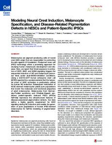
Fig. 1. Electron microscope micrograph. No nuclear or cytoplasmic abnormalities were detected after a treatment of 24h with lectin. Bar = 0-5./xm. (N, nucleus; G, Golgi apparatus; me, melanin granule; m, mitochondria; nm, nuclear membrane; np, nuclear pores). The exclusion test with trypan blue indicated a similar % of dead cells between treated and control batches(