Targeted gene expression in transgenic Xenopus using the binary Gal4-UAS system Katharine O. Hartley*†, Stephen L. Nutt*†‡, and Enrique Amaya*†§ *Wellcome兾CRC Institute, Tennis Court Road, Cambridge CB2 1QR, United Kingdom; and †Department of Zoology, University of Cambridge, Cambridge CB2 3EJ, United Kingdom Communicated by John C. Gerhart, University of California, Berkeley, CA, December 4, 2001 (received for review October 18, 2001)
he Gal4-UAS binary system has become the system of choice for targeted misexpression experiments in Drosophila (1–3). This system has also been adapted for misesxpression studies in other organisms, such as zebrafish and mouse (4–7). We reasoned that the same system would be beneficial for studying gene function in Xenopus, as it would overcome a number of the limitations of the transgenic and mRNA misexpression strategies currently being utilized. Here we show that the Gal4-UAS binary system is applicable to Xenopus. Furthermore, we use this system to characterize the neural and mesodermal defects caused by early misexpression of Xvent-2, a transcriptional target of bone morphogenetic protein 4 (BMP4) signaling. Materials and Methods
Generation of Transgenic Lines. Generation of transgenic Xenopus
laevis embryos was carried out essentially according to Kroll and Amaya (8). In each reaction, 100 ng of linearized DNA (100 ng) was used.
In Situ Hybridization and Antibody Staining. In situ hybridization was performed following the protocol of Harland (9). Whole-mount immunocytochemistry with monoclonal antibody MZ15 specific for notochord (10) was performed as described (11). Probes were generated from the following plasmids, after linearization with the indicated restriction enzymes and transcribed with the indicated RNA polymerase: CSGFP3 (BamHI, T7; ref. 12); XBF-1 (ClaI, T7; ref. 13); Xotx2 (NotI, T7; ref. 14); Xrx1 (BamHI, T7; ref. 15); Pax-3 (BglII, SP6; ref. 16); CsGal4(KH) (HindIII, T7); Cardiac Actin (Ac100) (PvuII, SP6; ref. 33); and En-2 (XbaI, T3; ref. 17). www.pnas.org兾cgi兾doi兾10.1073兾pnas.022646899
individual embryos collected at stage 35, by proteinase K treatment. PCR was performed as follows: 94°C for 2 min, 94°C for 30 s, 57–59°C for 30 s, 72°C for 1 min (for 30 cycles), and 72°C for 10 min. PCR products were run on a 1% agarose gel with ethidium bromide and visualized and photographed under UV illumination. Primers used were as follows: Gal4, 5⬘-GCTGTCTCAATGTTAGAGGC3⬘; UAS, 5⬘-CAAGCGCAGCTGAACAAG-3⬘; Xvent-2, 5⬘GTCTCCAGGGGTTGAGGTATTAA-3⬘; and Pax-6, 5⬘CAAACCAGTGAACCAATG-3⬘.
mRNA Synthesis and Embryo Microinjection. After fertilization, embryos at the two-cell stage were transferred to 1% agarose dishes containing 1⫻ MMR with 6% Ficoll. mRNA (100 pg) was injected into the marginal zone of one cell of two-cell stage embryos. Gal4 mRNA was prepared in vitro by using the SP6 Ambion mMessage mMachine in vitro transcription kit, after linearization of CsGal4(KH) with NotI.
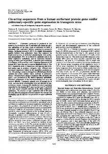
Results and Discussion To adapt the Gal4-UAS system to Xenopus, we first generated stable transgenic lines containing ‘‘activator’’ and ‘‘effector’’ constructs (Fig. 1a). Activator constructs contained the yeast transcriptional activator Gal4 under the control of the strong ubiquitous simian cytomegalovirus promoter (18) (CMVGal4) or the tissue specific Pax-6 promoter (Pax-6Gal4; ref. 19). Effector constructs contained the five tandem repeats of the Gal4-binding motif (UAS) along with the hsp70 minimal promoter, from pUAST (gift from A. Brand; ref. 2), linked to green fluorescent protein (UASGFP) or Xvent-2 (UASXvent-2). On reaching maturity, founders containing effector constructs were tested for germ-line transmission of the transgene and for responsiveness to Gal4. Gal4 mRNA (100 pg) was injected into one cell of two-cell stage F1 embryos generated after a mating between the UASGFP lines and wild-type frogs. Transactivation of the effector gene, GFP, was observed by fluorescence microscopy in 50% to 75% of resulting embryos at late-neurula stages in four of five lines containing UASGFP (Fig. 1b). In the absence of Gal4 RNA injection, none of the embryos expressed GFP, suggesting that the UASGFP transgene remains transcriptionally silent, unless transactivated specifically by Gal4. We next tested whether transactivation by Gal4 could be accomplished in a temporally and spatially controlled fashion on cross-fertilization of effector lines with lines containing activator constructs expressing Gal4. We observed ubiquitous GFP expression in 28% and 31% of F1 embryos resulting from matings Abbreviations: GFP, green fluorescent protein; CMV, cytomegalovirus; UAS, upstream activating sequence Gal4-binding motif; BMP4, bone morphogenetic protein 4; MMR, Marc’s modified Ringers. ‡Present
address: Division of Immunology, The Walter and Eliza Hall Institute, The Royal Melbourne Hospital, Melbourne 3050, Australia.
§To
whom reprint requests should be addressed. E-mail:
[email protected].
The publication costs of this article were defrayed in part by page charge payment. This article must therefore be hereby marked “advertisement” in accordance with 18 U.S.C. §1734 solely to indicate this fact.
PNAS 兩 February 5, 2002 兩 vol. 99 兩 no. 3 兩 1377–1382
BIOLOGY
T
PCR Analysis of Genomic DNA. Genomic DNA was extracted from
DEVELOPMENTAL
The transgenic technique in Xenopus allows one to misexpress genes in a temporally and spatially controlled manner. However, this system suffers from two experimental limitations. First, the restriction enzyme-mediated integration procedure relies on chromosomal damage, resulting in a percentage of embryos failing to develop normally. Second, every transgenic embryo has unique sites of integration and unique transgene copy number, resulting in variable transgene expression levels and variable phenotypes. For these reasons, we have adapted the Gal4-UAS method for targeted gene expression to Xenopus. This technique relies on the generation of transgenic lines that carry ‘‘activator’’ or ‘‘effector’’ constructs. Activator lines express the yeast transcription factor, Gal4, under the control of a desired promoter, whereas effector lines contain DNA-binding motifs for Gal4-(UAS) linked to the gene of interest. We show that on intercrossing of these lines, the effector gene is transcribed in the temporal and spatial manner of the activator’s promoter. Furthermore, we use the Gal4-UAS system to misexpress Xvent-2, a transcriptional target of bone morphogenetic protein 4 (BMP4) signaling during early embryogenesis. Embryos inheriting both the Gal4 activator and Xvent-2 effector transgenes display a consistent microcephalic phenotype. Finally, we exploit this system to characterize the neural and mesodermal defects obtained from early misexpression of Xvent-2. These results emphasize the potential of this system for the controlled analyses of gene function in Xenopus.
Fig. 1. (a) Schematic diagram showing the activator and effector constructs used in this study. Activator constructs contain the yeast transcriptional activator Gal4 under the control of the ubiquitously expressed CMV promoter or the tissue-specific Pax-6 promoter. Effector constructs contain five repeat concatemers of the consensus binding site for Gal4 (UAS), linked to the hsp70 minimal promoter and the gene of interest, green fluorescent protein (GFP) or Xvent-2. (b) F1 progeny from effector lines were tested for the transmission of the transgene and transactivation by Gal4. Embryos were injected into one cell at the two-cell stage with 100 pg of Gal4 mRNA. Two stage 19 sibling F1 embryos from GUASGFP line 2 injected with Gal4 mRNA (arrow). GFP fluorescence is observed in the right embryo on its right side, and that embryo is deemed to have inherited the UASGFP transgene, which has been transactivated by Gal4. The embryo on the left shows no GFP fluorescence and is assumed not to have inherited the transgene.
between two independent UASGFP female founder frogs (lines 6 and 9) and a CMVGal4 male frog (CMVGal4 line1), respectively (Fig. 2 e and g). GFP fluorescence was observed from mid-late neurula stages, suggesting a slight time delay between the accumulation of Gal4 protein and its transactivation of GFP.
Fig. 2. GFP is transactivated by Gal4 in temporally and spatially restricted manners on cross-fertilization of founder activator and effector lines. (a–c) Pax-6Gal4 line 2 ⫻ UASGFP line 2. (a) GFP fluorescence is restricted to the eye field in a stage 25 F1 embryo. GFP RNA (b) or Gal4 RNA (c) as detected by in situ hybridization is expressed in the anterior neural plate and presumptive hindbrain and spinal cord in stage 19 F1 embryos. (d) Stage 19 F0 embryo transgenic for Pax-6GFP showing the expression of GFP RNA driven by the Pax-6 promoter. (e–g) CMVGal4 line 1⫻ UASGFP line 6. (e) Stage 25 F1 embryo expressing GFP ubiquitously. ( f) Sibling stage 25, F1 embryo not expressing GFP. (g) Stage 40, F1 tadpole expressing GFP ubiquitously. 1378 兩 www.pnas.org兾cgi兾doi兾10.1073兾pnas.022646899
Moreover, tissue-specific expression of GFP was obtained in 51.5% of F1 embryos from a cross between a female Pax6Gal4 founder (line 2) (carrying two separate transgene integrations) and a male transgenic for UASGFP (line 2; also carrying two separate transgene integrations; Fig. 2 a–c). GFP fluorescence was observed at stage 25, predominately in the eye primordia (Fig. 2a), whereas GFP mRNA was detected at stage 20 by in situ hybridization in the eye field and regions fated to become hindbrain and spinal cord (Fig. 2b). Importantly, the activation of GFP RNA correlated with the expression of Gal4 RNA as observed by in situ hybridization to F1 embryos from the same mating (Fig. 2 b and c) and with the expression of GFP in embryos transgenic for the Pax-6 promoter driving GFP (Fig. 2d). Therefore we can conclude that the Gal4 system is applicable to Xenopus, resulting in spatially and temporally controlled activation of the marker gene GFP. To test the utility of these transgenic activator lines for the study of gene function during Xenopus development, we asked whether we could regulate the expression of a developmentally important gene by means of Gal4 transactivation in the frog. For these investigations, we chose the ventralizing homeobox gene, Xvent-2, a direct downstream target of BMP signaling, otherwise known as Vox, Xom, and Xbr-1 (20 –24). We crossed the CMVGal4 male with a female containing the responsive transgene, UASXvent-2 (line 5). Until the early tailbud stages, there was no discernible phenotype in the embryos. However, by stage 23, 156 of 667 (23%) embryos developed a striking and consistent phenotype, a failure of the head to form. By stage 30, this microcephalic phenotype was clearly distinguishable from the normally developing sibling embryos (Fig. 3a) and mimicked the predicted phenotype obtained by the overexpression of Xvent-2 by mRNA injection into the two- to four-cell-stage embryo (20 –22). To confirm that the microcephalic phenotype resulted from the transactivation of Xvent-2 by Gal4, genomic DNA from six embryos exhibiting this phenotype was individually extracted, and PCR analysis was carried out to determine whether each embryo carried the parental transgenes. For comparison, the genomic DNA from six phenotypically normal sibling embryos was also extracted and tested by PCR to determine whether they had inherited the transgenes. All six embryos with the microcephalic phenotype carried the activator, CMVGal4, and effector, UASXvent-2 transgenes (Fig. 3a, lanes 1– 6), whereas none of the phenotypically normal sibling embryos inherited both the maternal and paternal transgenes (Fig. 3a, lanes 7–12). Additional crosses between the male Pax-6Gal4 (line 1) and two further responsive lines containing UASXvent-2 (lines 4 and 6) revealed strikingly similar and consistent results to those obtained above. In the first cross, 16% (10 of 62) of the F1 embryos exhibited severe ventralization or ‘‘bauchstuck’’ phenotype by stage 28 (Fig. 3b). The dorsal axis of the embryos involved was shortened and all head structures were absent. Again PCR analysis revealed that every severely ventralized embryo contained both activator, Pax-6Gal4, and effector, UASXvent-2, transgenes (Fig. 3b, lanes 1– 6), whereas none of the normal siblings tested had inherited both parental transgenes (Fig. 3b, lanes 7–12). A second cross also revealed a microcephalic phenotype in 13% (52 of 406) of the F1 progeny (Fig. 3c) which again absolutely correlated with the presence of both transgenes (Fig. 3c, lanes 1–5 vs. 6 –10). Therefore, we can conclude that, in three independent crosses, the phenotype is a result of the overexpression of the effector gene, Xvent-2, by means of transactivation by Gal4. It was, however, surprising that the phenotypes of activating Xvent-2 with the ubiquitous activator CMVGal4 or by a tissue-specific activator Pax6Gal4 were broadly similar. To investigate the underlying cause of this result, we performed in situ hybridizations to Gal4 mRNA in gastrula stage embryos. Unexpectedly, this PaxHartley et al.
6Gal4 line was found to have an early ubiquitous burst of Gal4 expression at around stage 10 (Fig. 3d), which would explain the similarity in the observed phenotypes. Indeed, when this Pax-6Gal4 male was crossed with the GUASGFP female lines 6 and 9, the progeny inheriting both transgenes expressed GFP Hartley et al.
ubiquitously. As mentioned before, our other Pax-6Gal4 line expressed Gal-4 in a pattern more reminiscent of the endogenous Pax-6 expression pattern. However, because this founder animal was female, we were unable to cross this line with the GUASXvent2 founders, which were also female. The PNAS 兩 February 5, 2002 兩 vol. 99 兩 no. 3 兩 1379
BIOLOGY
DEVELOPMENTAL
Fig. 3. Microcephalic and ventralized phenotypes result from transactivation of Xvent-2 by Gal4 in three independent crosses. (a) CMVGal4 line 1 ⫻ UASXvent-2 line 5. (b) Pax-6Gal4 line 1 ⫻ UASXvent-2 line 4. (c) Pax-6Gal4 line 1 ⫻ UASXvent-2 line 6. (a–c Left) F1 stage 30 embryos showing the characteristic microcephalic or ventralized phenotype (Lower) when compared to an apparently normal sibling (Upper). (Right) PCR genotyping of individual embryos with the indicated phenotype. PCR primer combinations are indicated. ⫺ve, PCR control without DNA. (d and e) Half of F1 progeny from Pax-6Gal4 line 1 ⫻ wild type express Gal4 as revealed by in situ hybridization to Gal4 RNA. (d) At stage 10, Gal4 is expressed throughout the marginal zone of the embryo. (e) At stage 19, Gal4 expression is restricted to the eye field in the anterior neural plate.
Fig. 4. Analysis of the microcephalic phenotype at stage 3. F1 progeny from CMVGal4 line 1 ⫻ UASXvent-2 line 5. (a–f and i) In situ hybridization to the indicated marker genes. Representative sibling F1 embryos that are phenotypically normal (Upper) or microcephalic (Lower) are shown for each marker. (g and j) Transverse sections through the dorsal region of embryos stained for Pax-3 and cardiac actin, respectively, with the section of the microcephalic embryo on the right. (h) MZ15 monoclonal antibody staining of notochordal tissue for normal (Upper) and microcephalic (Lower) F1 embryos.
difference in expression between the two Pax6-Gal4 founder animals is probably caused by the positional effect of the transgene, and emphasizes the need to screen through both 1380 兩 www.pnas.org兾cgi兾doi兾10.1073兾pnas.022646899
activator and effector lines for correct expression and response. In addition it is also interesting to note that the two different UASXvent-2 lines resulted in a consistent phenotype Hartley et al.
within each mating but of differing severity between crosses. We have also recorded similar variations on crossing different GFP effector lines to the same activator line (data not shown) and surmise that effector lines vary in their response to Gal4 by transactivating the responsive gene to various degrees. We decided to extend our understanding of the microcephalic phenotype resulting from the cross between the CMVGal4 male and a UASXvent-2 female line 5. Xvent-2 is an immediate early target of BMP signaling (20, 22, 24), which is thought to act as a transcriptional repressor to ventralize mesoderm and inhibit neural differentiation (20–22, 25, 26). The loss of the most dorsal mesoderm, the notochord, has been noted, but the extent of neural defects has not been extensively catalogued. The phenotype obtained in our Gal4 crosses suggested that Xvent-2 appeared to selectively inhibit anterior structures, a phenotype similar to the one observed when BMP4 is misexpressed in the anterior neural plate after gastrulation (19). In situ hybridization to a range of neural markers showed that the structures of the forebrain, midbrain, and retinal markers are missing, up to and including the midbrain– hindbrain border (Fig. 4 a–d). In contrast, the dorsal hindbrain and spinal cord marker, Pax-3, was expressed, suggesting that the posterior neural tube can be induced in embryos ectopically expressing Xvent-2 (Fig. 4 e and f ). One potential model that may account for the loss of dorso-anterior structures in embryos ectopically expressing Xvent-2 is that Xvent-2 functions to temporarily inhibit the Spemann organizer. Indeed, Xvent-2 has been shown to repress the organizer genes goosecoid and chordin (26, 27), whereas in the chick the expression of Pax-3, is induced by late-stage chick Henson node and not early-stage nodes (28). Sectional analysis of embryos stained for Pax-3 showed that the expression domain of this dorsal neural tube marker is expanded ventrally in the spinal cord of embryos exhibiting the microcephalic phenotype (Fig. 4g). The expansion of Pax-3 could be the direct result of the ectopic expression of Xvent-2 in the neural structures of these embryos, as the expression of Pax-3 in the dorsal neural tube is maintained by BMP signaling and Xvent-2 is normally expressed in the dorsal neural tube. Thus, it is possible that Xvent-2 is the BMP-responsive gene that acts to maintain expression of Pax-3 in this region and that the ectopic expression of Xvent-2 causes a similar upregulation of Pax-3 in the ventral neural tube. However, we also noted that the microcephalic embryos lack notochord tissue (Fig. 4h). Therefore, the ventralization of the neural tube could be caused by the loss of notochord, which normally produces signals, such as sonic hedgehog, that ventralize the neural tube (29 –32).
We thank Andrea Brand and Aaron Zorn for helpful discussions and Roz Friday for comments on the manuscript. This work was supported by a Medical Research Council studentship (to K.O.H.), a Human Frontier Science Program long-term fellowship (to S.L.N.), and National Institutes of Health Grant RR13221 and a Wellcome Trust Senior Research Fellowship (to E.A.).
1. Fischer, J. A., Giniger, E., Maniatis, T. & Ptashne, M. (1988) Nature (London) 332, 853–856. 2. Brand, A. H. & Perrimon, N. (1993) Development (Cambridge, U.K.) 118, 401–415. 3. Phelps, C. B. & Brand, A. H. (1998) Methods 14, 367–379. 4. Rowitch, D. H., St-Jacques, B., Lee, S. M., Flax, J. D., Snyder, E. Y. & McMahon, A. P. (1999) J. Neurosci. 19, 8954–8965. 5. Scheer, N. & Camnos-Ortega, J. A. (1999) Mech. Dev. 80, 153–158. 6. Scheer, N., Groth, A., Hans, S. & Campos-Ortega, J. A. (2001) Development (Cambridge, U.K.) 128, 1099–1107. 7. Koster, R. W. & Fraser, S. E. (2001) Dev. Biol. 233, 329–346. 8. Kroll, K. L. & Amaya, E. (1996) Development (Cambridge, U.K.) 122, 3173– 3183. 9. Harland, R. M. (1991) Methods Cell Biol. 36, 685–695. 10. Smith, J. C. & Watt, F. M. (1985) Differentiation 29, 109–115. 11. Smith, J. C. (1993) in Cellular Interactions in Development: A Practical Approach, ed. Hartley, D. A. (Oxford Univ. Press, Oxford), pp. 181–204. 12. Bronchain, O. J., Hartley, K. O. & Amaya, E. (1999) Curr. Biol. 9, 1195– 1198. 13. Bourguignon, C., Li, J. & Papalopulu, N. (1998) Development (Cambridge, U.K.) 125, 4889–4900.
14. Lamb, T. M., Knecht, A. K., Smith, W. C., Stachel, S. E., Economides, A. N., Stahl, N., Yancopolous, G. D. & Harland, R. M. (1993) Science 262, 713–718. 15. Andreazzoli, M., Gestri, G., Angeloni, D., Menna, E. & Barsacchi, G. (1999) Development (Cambridge, U.K.) 126, 2451–2460. 16. Espeseth, A., Johnson, E. & Kintner, C. (1995) Mol. Cell. Neurosci. 6, 199–211. 17. Hemmati-Brivanlou, A., de la Torre, J. R., Holt, C. & Harland, R. M. (1991) Development (Cambridge, U.K.) 111, 715–724. 18. Turner, D. L. & Weintraub, H. (1994) Genes Dev. 8, 1434–1447. 19. Hartley, K. O., Hardcastle, Z., Friday, R. V., Amaya, E. & Papalopulu, N. (2001) Dev. Biol. 238, 168–184. 20. Onichtchouk, D., Gawantka, V., Dosch, R., Delius, H., Hirschfeld, K., Blumenstock, C. & Niehrs, C. (1996) Development (Cambridge, U.K.) 122, 3045–3053. 21. Schmidt, J. E., von Dassow, G. & Kimelman, D. (1996) Development (Cambridge, U.K.) 122, 1711–1721. 22. Ladher, R., Mohun, T. J., Smith, J. C. & Snape, A. M. (1996) Development (Cambridge, U.K.) 122, 2385–2394. 23. Papalopulu, N. & Kintner, C. (1996) Dev. Biol. 174, 104–114. 24. Rastegar, S., Friedle, H., Frommer, G. & Knochel, W. (1999) Mech. Dev. 81, 139–149.
Hartley et al.
PNAS 兩 February 5, 2002 兩 vol. 99 兩 no. 3 兩 1381
BIOLOGY
DEVELOPMENTAL
Finally, the embryos were analyzed for the appearance of somitic tissue with the marker cardiac actin (Fig. 4 i and j). Both microcephalic and wild-type embryos stained strongly for this marker. However, analysis of sections revealed that the somites of the microcephalic embryos were fused in the midline underlying the neural tube (Fig. 4j Right), whereas in wild-type embryos, the somites form two separate blocks on either side of the neural tube and notochord (Fig. 4j Left). Together these data show that the Gal4-UAS system can transactivate the expression of a responsive gene in a temporally and spatially restricted manner during Xenopus embryogenesis. As with other organisms where the Gal4 system has been applied, we have found that it is essential to screen through activator and effector lines to find the experimentally appropriate expression profiles. Variations in expression of Gal4 occur between activator lines containing the same construct as well as variations in the response to Gal4 by effector lines. These differences are most likely to be caused by the effect of the site of integration on the transgene, variations in copy number of transgenes between lines, or partial deletions of the transgene. However, once the lines are identified, these can be bred to homozygosity and utilized in potentially endless combinations of crosses to activate genes at specific locations. We particularly advocate this system for gain-of-function experiments, as exemplified by the Xvent-2 misexpression studies presented here. However, we would be cautious about suggesting using the Gal4 system for dominant-negative experiments, which require massive overexpression, as we have found it difficult to achieve the high levels of expression required for such studies. Perhaps the most attractive advantage of this method is that it is possible to generate hundreds of embryos, expressing a gene of interest in controlled domains of space and time after a simple cross-fertilization of appropriate transgenic lines. As indicated here, the phenotypes arising from these crosses are strikingly consistent, as every resulting experimental embryo has the same number of copies of each transgene, at the same site of integration. Hence, any phenotype observed is the result of the expression of the gene of interest. With the future generation of a range of tissue-specific promoters, this system will be of great benefit for the study of gene function at late stages in development.
25. Onichtchouk, D., Glinka, A. & Niehrs, C. (1998) Development (Cambridge, U.K.) 125, 1447–1456. 26. Melby, A. E., Clements, W. K. & Kimelman, D. (1999) Dev. Biol. 211, 293–305. 27. Trindade, M., Tada, M. & Smith, J. C. (1999) Dev. Biol. 216, 442–456. 28. Bang, A. G., Papalopulu, N., Kintner, C. & Goulding, M. D. (1997) Development (Cambridge, U.K.) 124, 2075–2085. 29. Chiang, C., Litingtung, Y., Lee, E., Young, K. E., Corden, J. L., Westphal, H.
1382 兩 www.pnas.org兾cgi兾doi兾10.1073兾pnas.022646899
& Beachy, P. A. (1996) Nature (London) 383, 407–413. 30. Liem, K. F., Jr., Tremml, G., Roelink, H. & Jessell, T. M. (1995) Cell 82, 969–979. 31. Goulding, M. D., Lumsden, A. & Gruss, P. (1993) Development (Cambridge, U.K.) 117, 1001–1016. 32. Dietrich, S., Schubert, F. R. & Gruss, P. (1993) Mech. Dev. 44, 189–207. 33. Hemmati-Brivanluv, D., Frank, D., Bolce, M. E., Brown, B. D., Sive, H. L. & Harland, R. M. (1990) Development (Cambridge, U.K.) 110, 325–330.
Hartley et al.