TCR Triggering by the pMHC Complex: Valency, Affinity, and Dynamics Rajat Varma (13 May 2008) Science Signaling 1 (19), pe21. [DOI: 10.1126/stke.119pe21]
The following resources related to this article are available online at http://stke.sciencemag.org. This information is current as of 14 May 2008.
Article Tools Related Content
References Glossary
The editors suggest related resources on Science's sites: http://stke.sciencemag.org/cgi/content/abstract/sigtrans;1/12/pt2 http://stke.sciencemag.org/cgi/content/abstract/sigtrans;2002/122/pe10 This article cites 37 articles, 10 of which can be accessed for free: http://stke.sciencemag.org/cgi/content/full/sigtrans;1/19/pe21#otherarticles Look up definitions for abbreviations and terms found in this article: http://stke.sciencemag.org/glossary/ Obtain information about reproducing this article: http://www.sciencemag.org/about/permissions.dtl
Science Signaling (ISSN 1937-9145) is published weekly, except the last week in December, by the American Association for the Advancement of Science, 1200 New York Avenue, NW, Washington, DC 20005. Copyright 2008 by the American Association for the Advancement of Science; all rights reserved.
Downloaded from stke.sciencemag.org on May 14, 2008
Permissions
Visit the online version of this article to access the personalization and article tools: http://stke.sciencemag.org/cgi/content/full/sigtrans;1/19/pe21
PERSPECTIVE RECEPTORS
TCR Triggering by the pMHC Complex: Valency, Affinity, and Dynamics Rajat Varma Published 13 May 2008
T cells constitute one important arm of the immune system and are of two types: those with the cell surface marker CD4 (helpers) and those with CD8 (killers). [There is another class of T cells called γδ T cells, but these are not included in this discussion, because they interact with their ligands in a peptide-independent manner (1).] Both CD4+ and CD8+ T cells have the same kind of TCR, but they interact with different kinds of ligands. CD4+ T cells interact with class II pMHCs, and CD8+ T cells interact with class I pMHCs. Each T cell has a distinctive TCR and is selected in the thymus such that the TCR does not have a high affinity for peptides derived from self proteins, thus avoiding autoreactivity. The TCR complex comprises the ligand-binding heterodimeric α and β chains (TCRαβ) and the signal-transducing CD3 chains: δ, ε, γ, and ζ. TCR triggering occurs when the TCR binds to the pMHC, which is the first step in TCR signaling. Several models exist to explain how the TCR-pMHC interaction leads to phosphorylation of CD3 chains (2, 3). These models take into account such factors as conformational change, kinetic segregation, and receptor aggregation. However, each of these models has differLaboratory of Cellular and Molecular Immunology, National Institute of Allergy and Infectious Diseases, National Institutes of Health, 4 Center Drive, Bethesda, MD 20892, USA. E-mail,
[email protected]
ent valency requirements for pMHCs (valency being def ined as the number of pMHC molecules required to trigger the TCR). This has resulted in a controversy over how many pMHC molecules are required to trigger the TCR (4). Activation of T cells occurs as a consequence of TCR signaling initiated after triggering of the TCR by the pMHC. T cell activation results in various measurable responses, including phosphorylation of the CD3 chains, recruitment of kinases to signaling complexes, Ca2+ flux, cell proliferation, or cytokine production. The idea that conformational change of the TCR might be a mechanism for TCR triggering was based on the observation that the binding of the cytoplasmic noncatalytic region of the tyrosine kinase adaptor protein Nck to a proline-rich sequence (PRS) of CD3ε only occurred upon TCR engagement (5). However, f indings from a study of knock-in mice—expressing a form of CD3ε from which the PRS was deleted—demonstrated that it only plays a role in controlling the abundance of the TCR at the cell surface during development and has no impact on the response of peripheral T cells to stimulation by the pMHC (6). This study also demonstrated that Nck binds to the PRS even in the absence of engagement of the TCR by the pMHC. Soluble monomers of agonist peptide–loaded pMHC fail to activate T cells (7–11). This finding, along with some structural data that fail to show any conformational change in TCRαβ upon ligand binding,
www.stke.org/cgi/content/full/1/19/pe21
Page 1
Downloaded from stke.sciencemag.org on May 14, 2008
The interaction between the T cell receptor (TCR) and a peptide-loaded major histocompatibility complex (pMHC) is one of the most-studied interactions in immunology, and yet the precise mechanism by which this system operates is still not fully understood. One key issue is whether TCR triggering minimally requires monomeric pMHC complexes or higher-order multimers (two or more pMHCs). Any model of TCR triggering must explain the high sensitivity, specificity, and dynamic range of ligand responsiveness that this receptor system exhibits. Most models of TCR triggering have not fully appreciated the dynamic aspects of TCR triggering. TCR triggering happens very quickly, and the properties of sensitivity and specificity can be explained by a model that accounts for the interaction dynamics of such a receptor system. In this paper, it is proposed that the important parameter in TCR triggering is the immobilization of the TCR-pMHC complex in the plasma membrane. Whether this involves monomeric or multimeric pMHCs may depend on the affinity of the TCR for the pMHC.
has largely been used as evidence against conformational change as the mechanism for TCR triggering (12). From what is known about the architecture of the TCRCD3 complex, dimerization of the receptor is not required for signaling. Cytoplasmic kinases are recruited to CD3 chains even if the TCR complex is monomeric. However, soluble dimers of agonist pMHCs can activate T cells (10, 11, 13), although it is controversial whether surface-bound monomeric pMHCs can activate T cells (4). Artificial substrates that present pMHCs have contributed toward the understanding of TCR triggering and signaling (14). These substrates can be as simple as pMHCs adsorbed to plastic (15, 16), coated on transwell membranes (17, 18), or attached to glass-supported planar bilayers (16, 19–21). These artificial surfaces offer a reductionist approach to understanding signaling, in which the components and their surface densities can be experimentally controlled. The antigen-specific interaction between a T cell and an antigen-presenting cell (APC) is known as the immunological synapse (19, 22). Apart from containing TCR-pMHC interactions, this region is the site of interactions—some of which are costimulatory, whereas others are coinhibitory—between accessory molecules on the T cells and APCs (Fig. 1). Hence, attributing a specific phenotype to TCR-pMHC interactions becomes difficult in the context of T cell interactions with APCs. Proteins incorporated in a glass-supported planar bilayer are free to diffuse. This property brings this system a step closer to the context of the APC than does having proteins bound to solid supports, but the interaction of membrane proteins with the APC cytoskeleton cannot be reconstructed (23). Very few pMHCs on the surface of an APC are required for the activation of a T cell (24–26), and this holds true when pMHCs are presented on artificial substrates (13, 19, 27). Because such substrates are easily amenable to high-resolution microscopy, they are a good system to investigate the requirements for TCR triggering by pMHCs (14). Using a substratepresenting immobile photoactivable pMHC, Davis and co-workers showed that TCR triggering happens at incredible speeds (15). After ligand engagement, it takes 4 s for the phosphorylation of a key TCR adaptor protein linker of activated T cells to occur and 6 to 7 s for diacylglycerol production and calcium flux to begin. Hence, if TCRs need to dimerize in order to signal, then these dimers need to persist at the cell
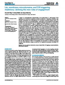
PERSPECTIVE Fig. 1. Formation of the immunological synapse. Initially, receptors and coreceptors in the plasma membranes of the APC (upper cell in top panel) and T cell (lower cell in top panel) are randomly arranged. TCR triggering occurs as a result of the binding of the pMHC complex to the TCR. Interactions between the adhesion molecules intercellular adhesion molecule–1 (ICAM-1) and leukocyte function-associated antigen–1 (LFA-1) bring the APC and T cell in contact with each other, leading to the formation of the immunological synapse. At the immunological synapse, TCR-pMHC and B7-CD28 complexes form clusters at the center, while adhesion molecules interact at the sides. Not shown are the CD4 and CD8 T cell coreceptors, which interact with the pMHC.
pMHC
ICAM-1
B7
LFA-1 TCR CD28
Downstream signaling leading to T cell activation
acting TCRs and pMHCs in the plasma membrane. Diffusion trapping is the immobilization of the interaction pair in the plasma membrane. Parameters governing diffusion trapping of TCR-pMHC interactions include the aff inity of the TCR for the pMHC, the valency of the TCR-pMHC interaction (whether the pMHC is presented as a monomer or a dimer), and the diffusion coefficient of pMHC on the surface of the APC. TCR-pMHC complexes with different binding affinities have different valency requirements for diffusion trapping (Fig. 2). Formation of TCR-pMHC complexes in which the TCR has a low affinity for the pMHC is predicted to require pMHC dimers; complexes with TCRs of intermediate affinity for the pMHC involve pseudodimers, whereas those complexes in which the TCR has a high affinity for the pMHC require only monomeric pMHCs. This model is largely inspired by our own
www.stke.org/cgi/content/full/1/19/pe21
observations and a theoretical proposal of Weng and Delisi (31). In experiments with T cells that have a relatively high-affinity TCR, we have observed the trapping of very few agonist pMHCs in lipid bilayers, which translocate from the periphery of the immune synapse to the center and hence are diffusion trapped because their motion is guided by the motion of TCR. Diffusion trapping is most likely caused by the interaction of activated receptors with the actin cytoskeleton in the T cell. Delisi and colleagues first proposed diffusion trapping as a mechanism to explain signaling by the complement receptor C1r (32) and later extended this analysis to the TCR (31). Assuming that dimerization of the TCR is important for triggering, they propose that the diffusion coeff icient of a specif ic pMHC in the APC membrane may be critical in causing diffusion trapping and triggering of the TCR. One could, in principle,
Page 2
Downloaded from stke.sciencemag.org on May 14, 2008
TCR triggering
surface for only a short period of time. T cells are extremely sensitive to very few agonist pMHCs in the presence of a milieu of self antigen–loaded pMHCs (28, 29). To explain this sensitivity, researchers proposed a pseudodimer model of T cell activation (4, 13). Soluble dimers of pMHCs, in which one of the MHC molecules is loaded with an agonist peptide and the other is loaded with an endogenous peptide, activate T cells. These dimers require an intact CD4-binding site on the agonist pMHC. (It is thought that a similar mechanism occurs with CD8 + T cells, though this has not been explicitly demonstrated.) A similar contribution of self peptides to agonist-mediated stimulation was observed with planar membranes and Chinese hamster ovary cells. Based on these observations, it was proposed that a basic unit of signaling is a pseudodimer of TCRs, in which one TCR interacts with an agonist pMHC while the other interacts with an endogenous pMHC. The CD4 associated with the TCR that interacts with endogenous pMHC also interacts with and binds to the agonist pMHC in trans (13). A recent report contradicts this finding and the ability of self peptides to enhance the response by the agonist pMHC (16). At the heart of this contradiction could be the presence of B7-1 in the bilayer used by Davis et al. (13), which was not present in the studies of Finkel and colleagues (16). B7-1 is a costimulatory ligand expressed on APCs that is known to enhance the sensitivity of naïve T cells by its interaction with the costimulatory molecule CD28 found on the surface of T cells (30). I propose that the first step in triggering the TCR is the diffusion trapping of inter-
sites of active signaling because they recruit signaling molecules, such as Src family tyrosine kinases, and ζ chain–associated protein kinase of 70 kD (ZAP-70) and exclude phosphatases, such as CD45 (21, 23, 27). We cannot say with certainty whether microcluster formation is an early or late event in TCR triggering. It is conceivable that conformational changes in the TCR precede or follow clustering and interaction with the actin cytoskeleton. There is a plethora of models to explain how TCRpMHC interactions occur. It is possible that the ideas presented in each of these models explain different steps in TCR triggering and hence are all true, although individually they may be temporally distinct. Advances in spectroscopic and imaging methods have the potential to unravel these mysteries and unify these models. Although we may have a unified model for TCR triggering in a few years, it will perhaps be decades before we can exploit this model for therapeutic purposes.
Agonist monomers
Pseudo dimers
Agonist dimers
0
Low
Moderate
High 100
100-fold range of TCR-pMHC affinities selected in the thymus
Fig. 2. Model depicting the valency requirement for TCR triggering that depends on the affinity of the TCR for the pMHC. The x axis represents an arbitrary 100-fold range of TCR-pMHC interaction affinities selected in the thymus. The affinity ranges for TCR-pMHC interactions are arbitrarily divided into low, medium, and high categories. Lowaffinity interactions would minimally require dimerization of agonist pMHCs for TCR triggering, medium-affinity interactions would occur with a pseudodimer of pMHC, and high-affinity interactions would occur with pMHC monomers. Not shown in the figure are the influence of the diffusion coefficient of pMHC on the valency requirements for TCR triggering and the function of coreceptors. The prediction is that slower diffusion of pMHC would reduce the valency requirements such that medium-affinity interactions that trigger TCRs may occur with pMHC monomers, and so on. Considering the speed of TCR triggering, the association of the coreceptors CD4 and CD8 need only be transient.
References and Notes 1. Y. H. Chien, Y. Konigshofer, Antigen recognition by γδ T cells. Immunol. Rev. 215, 46–58 (2007). 2. K. Choudhuri, P. A. van der Merwe, Molecular mechanisms involved in T cell receptor triggering. Semin. Immunol. 19, 255–261 (2007). 3. M. S. Kuhns, M. M. Davis, K. C. Garcia, Deconstructing the form and function of the TCR/CD3 complex. Immunity 24, 133–139 (2006). 4. M. Krogsgaard, M. M. Davis, How T cells “see” antigen. Nat. Immunol. 6, 239–245 (2005). 5. D. Gil, W. W. Schamel, M. Montoya, F. SanchezMadrid, B. Alarcon, Recruitment of Nck by CD3ε reveals a ligand-induced conformational change essential for T cell receptor signaling and synapse formation. Cell 109, 901–912 (2002). 6. M. Mingueneau, A. Sansoni, C. Gregoire, R. Roncagalli, E. Aguado, A. Weiss, M. Malissen, B. Malissen, The proline-rich sequence of CD3ε controls T cell antigen receptor expression on and signaling potency in preselection CD4+CD8+ thymocytes. Nat. Immunol. 9, 522–532 (2008). 7. J. P. Abastado, Y. C. Lone, A. Casrouge, G. Boulot, P. Kourilsky, Dimerization of soluble major histocompatibility complex-peptide complexes is sufficient for activation of T cell hybridoma and induction of unresponsiveness. J. Exp. Med. 182, 439–447 (1995). 8. S. Casares, C. S. Zong, D. L. Radu, A. Miller, C. A. Bona, T. D. Brumeanu, Antigen-specific signaling
www.stke.org/cgi/content/full/1/19/pe21
9.
10.
11.
12.
13.
14.
by a soluble, dimeric peptide/major histocompatibility complex class II/Fc chimera leading to T helper cell type 2 differentiation. J. Exp. Med. 190, 543–553 (1999). J. J. Boniface, J. D. Rabinowitz, C. Wulfing, J. Hampl, Z. Reich, J. D. Altman, R. M. Kantor, C. Beeson, H. M. McConnell, M. M. Davis, Initiation of signal transduction through the T cell receptor requires the multivalent engagement of peptide/MHC ligands. Immunity 9, 459–466 (1998). J. R. Cochran, T. O. Cameron, L. J. Stern, The relationship of MHC-peptide binding and T cell activation probed using chemically defined MHC class II oligomers. Immunity 12, 241–250 (2000). J. D. Stone, L. J. Stern, CD8 T cells, like CD4 T cells, are triggered by multivalent engagement of TCRs by MHC-peptide ligands but not by monovalent engagement. J. Immunol. 176, 1498–1505 (2006). M. G. Rudolph, R. L. Stanfield, I. A. Wilson, How TCRs bind MHCs, peptides, and coreceptors. Annu. Rev. Immunol. 24, 419–466 (2006). M. Krogsgaard, Q. J. Li, C. Sumen, J. B. Huppa, M. Huse, M. M. Davis, Agonist/endogenous peptide-MHC heterodimers drive T cell activation and sensitivity. Nature 434, 238–243 (2005). J. T. Groves, M. L. Dustin, Supported planar bilayers in studies on immune cell adhesion and communication. J. Immunol. Methods 278, 19–32 (2003).
Page 3
Downloaded from stke.sciencemag.org on May 14, 2008
come up with a mathematical model that accounts for the diffusion coefficient of the pMHC and the affinity of the TCR for the pMHC in order to predict the valency required for triggering of the TCR. Experiments involving a high-aff inity TCR showed that a very low density of plasticadsorbed or bilayer-attached pMHC activated T cells, which suggests that monomers of pMHC are sufficient (16). In this study, plastic-adsorbed pMHC monomers may have activated the T cells because of their high affinity for the TCR and their slow diffusion rates, whereas in the case of lipid bilayers, one cannot rule out transient dimerization events. The conf iguration of kinases that is achieved by slowing the diffusion rates of TCRs is not known. One possibility is that there is an equilibrium interaction among kinases, phosphatases, and CD3 immunoreceptor tyrosine-based activation motifs (ITAMs) that is critically dependent on the rate of diffusion of the TCR in the plasma membrane. When TCRs diffuse more slowly, this equilibrium may somehow favor phosphorylation of CD3 ITAMs by promoting the exclusion of phosphatases (2, 27). The other possibility is that TCR triggering is a two-step process: the first step being its immobilization and the second step being a conformational change. This conformational change may also change the affinity of the TCR for pMHC, because it has been difficult to compete for TCR-pMHC interactions with pMHC-blocking antibodies (27). Actin polymerization could facilitate TCR clustering and could also provide the force needed to cause a conformational change in the TCR, as has been suggested in the receptor deformation model (33). TCR signaling results in the polymerization of actin (34), which may be connected to the TCR complex directly or through adaptor proteins (35–37). Hence, actin polymerization has the potential to apply a force to the TCR complex (33). However, it will be important to determine whether actin polymerization occurs parallel or perpendicular to the plasma membrane. If even a small component of actin polymerization occurs parallel to the plasma membrane, then the force transmitted to the TCR may be reduced if pMHCs are free to diffuse, and thus this force may vary depending on the diffusion coefficient of the pMHC. We have shown that very few pMHC complexes are needed to generate microclusters of TCRs (27). These microclusters are
Minimally required valency of pMHC to trigger TCR
PERSPECTIVE
PERSPECTIVE
23.
24.
25.
26.
27.
28.
29. 30.
31.
32.
supramolecular activation clusters in T cells. Nature 395, 82–86 (1998). G. Campi, R. Varma, M. L. Dustin, Actin and agonist MHC-peptide complex-dependent T cell receptor microclusters as scaffolds for signaling. J. Exp. Med. 202, 1031–1036 (2005). C. V. Harding, E. R. Unanue, Quantitation of antigen-presenting cell MHC class II/peptide complexes necessary for T-cell stimulation. Nature 346, 574–576 (1990). K. Kimachi, M. Croft, H. M. Grey, The minimal number of antigen-major histocompatibility complex class II complexes required for activation of naive and primed T cells. Eur. J. Immunol. 27, 3310–3317 (1997) P. A. Reay, K. Matsui, K. Haase, C. Wulfing, Y. H. Chien, M. M. Davis, Determination of the relationship between T cell responsiveness and the number of MHC-peptide complexes using specific monoclonal antibodies. J. Immunol. 164, 5626–5634 (2000). R. Varma, G. Campi, T. Yokosuka, T. Saito, M. L. Dustin, T cell receptor-proximal signals are sustained in peripheral microclusters and terminated in the central supramolecular activation cluster. Immunity 25, 117–127 (2006). D. J. Irvine, M. A. Purbhoo, M. Krogsgaard, M. M. Davis, Direct observation of ligand recognition by T cells. Nature 419, 845–849 (2002). R. N. Germain, T-cell activation: The power of one. Curr. Biol. 13, R137–R139 (2003). R. J. Greenwald, G. J. Freeman, A. H. Sharpe, The B7 family revisited. Annu. Rev. Immunol. 23, 515–548 (2005). Z. Weng, C. Delisi, Toward a predictive understanding of molecular recognition. Immunol. Rev. 163, 251–266 (1998). C. Delisi, F. W. Wiegel, Membrane fluidity and the
www.stke.org/cgi/content/full/1/19/pe21
33.
34.
35.
36.
37.
38.
probability of complement fixation. J. Theor. Biol. 102, 307–322 (1983). Z. Ma, P. A. Janmey, T. H. Finkel, The receptor deformation model of TCR triggering. FASEB J. 22, 1002–1008 (2008). M. L. Dustin, J. A. Cooper, The immunological synapse and the actin cytoskeleton: Molecular hardware for T cell signaling. Nat. Immunol. 1, 23–29 (2000). M. Barda-Saad, A. Braiman, R. Titerence, S. C. Bunnell, V. A. Barr, L. E. Samelson, Dynamic molecular interactions linking the T cell antigen receptor to the actin cytoskeleton. Nat. Immunol. 6, 80–89 (2005). S. Caplan, M. Baniyash, Normal T cells express two T cell antigen receptor populations, one of which is linked to the cytoskeleton via ζ chain and displays a unique activation-dependent phosphorylation pattern. J. Biol. Chem. 271, 20705–20712 (1996). M. M. Rozdzial, B. Malissen, T. H. Finkel, Tyrosine-phosphorylated T cell receptor ζ chain associates with the actin cytoskeleton upon activation of mature T lymphocytes. Immunity 3, 623–633 (1995). I thank R. Schwartz, P. Matzinger, R. Germain, N. Singh, and K. Laky for helpful and inspiring discussions and K. Laky for critically reading the manuscript. I also apologize to all those authors whose work could not be cited or acknowledged as a result of space limitations. 10.1126/stke119pe21
Citation: R. Varma, TCR triggering by the pMHC complex: Valency, affinity, and dynamics. Sci. Signal. 1, pe21 (2008).
Page 4
Downloaded from stke.sciencemag.org on May 14, 2008
15. M. Huse, L. O. Klein, A. T. Girvin, J. M. Faraj, Q. J. Li, M. S. Kuhns, M. M. Davis, Spatial and temporal dynamics of T cell receptor signaling with a photoactivatable agonist. Immunity 27, 76–88 (2007). 16. Z. Ma, K. A. Sharp, P. A. Janmey, T. H. Finkel, Surface-anchored monomeric agonist pMHCs alone trigger TCR with high sensitivity. PLoS Biol. 6, e43 (2008). 17. S. K. Bromley, D. A. Peterson, M. D. Gunn, M. L. Dustin, Cutting edge: Hierarchy of chemokine receptor and TCR signals regulating T cell migration and proliferation. J. Immunol. 165, 15–19 (2000). 18. D. Skokos, G. Shakhar, R. Varma, J. C. Waite, T. O. Cameron, R. L. Lindquist, T. Schwickert, M. C. Nussenzweig, M. L. Dustin, Peptide-MHC potency governs dynamic interactions between T cells and dendritic cells in lymph nodes. Nat. Immunol. 8, 835–844 (2007). 19. A. Grakoui, S. K. Bromley, C. Sumen, M. M. Davis, A. S. Shaw, P. M. Allen, M. L. Dustin, The immunological synapse: A molecular machine controlling T cell activation. Science 285, 221–227 (1999). 20. C. Sumen, M. L. Dustin, M. M. Davis, T cell receptor antagonism interferes with MHC clustering and integrin patterning during immunological synapse formation. J. Cell Biol. 166, 579–590 (2004). 21. T. Yokosuka, K. Sakata-Sogawa, W. Kobayashi, M. Hiroshima, A. Hashimoto-Tane, M. Tokunaga, M. L. Dustin, T. Saito, Newly generated T cell receptor microclusters initiate and sustain T cell activation by recruitment of Zap70 and SLP-76. Nat. Immunol. 6, 1253–1262 (2005). 22. C. R. Monks, B. A. Freiberg, H. Kupfer, N. Sciaky, A. Kupfer, Three-dimensional segregation of