Cellular & Molecular Immunology
9
Review
The Classical and Regulatory Functions of C1q in Immunity and Autoimmunity Jinhua Lu1, 3, Boon King Teh1, Linda Wang1, Yinan Wang1, Yen Seah Tan1, Min Chern Lai1 and Kenneth B. M. Reid2
A classical function of C1q is to bind immune complexes and initiate complement activation producing membrane lytic complexes, opsonins and anaphylatoxins. This classical pathway of complement activation is also elicited when C1q binds some other ligands. Besides complement activation, C1q also regulates cell differentiation, adhesion, migration, activation and survival. C1q deficiency is associated with autoimmunity as well as increased susceptibility to infections. In this article, we discuss the basic properties of C1q, its expression, and classical and regulatory functions. Cellular & Molecular Immunology. 2008;5(1):9-21.
Key Words: C1q, macrophage, dendritic cell, apoptotic cell, autoimmunity
alternative pathway can function independently and can also amplify the other two pathways.
Introduction Complement is a major arm of the host innate immune system providing critical protection against microbial infections. It can be activated through 3 different pathways, i.e., the classical, lectin and alternative pathways (1, 2). The classical pathway is mainly elicited by immune complexesinvolving binding of C1q, which forms a pentameric complex with the C1r2C1s2 serine protease tetramer, to the Fc regions of antibodies (1, 3). Complement is also activated by other C1q ligands besides immune complexes. Ultimately, complement activation generates membrane-lytic complexes, opsonins and inflammatory anaphylatoxins (1). Complement activation through the lectin pathway is initiated upon binding of the mannan-binding lectin (MBL) or ficolin, which complex with C1s-related serine proteases known as MBL-associated serine proteases (MASPs), to common carbohydrate structures on microorganisms (2, 4-6). The alternative pathway is activated when low “tick-over” of C3 activation is amplified on microbial surfaces (1). The 1 Department of Microbiology, Yong Loo Lin School of Medicine and 2NUS Immunology Program, National University of Singapore, Blk MD4, 5 Science Drive 2, Singapore 117597; 2
MRC Immunochemistry Unit, Department of Biochemistry, University of Oxford, South Parks Road, Oxford OX1 3QU, UK; 3
Corresponding to: Dr. Jinhua Lu, Department of Microbiology, Yong Loo Lin School of Medicine and National University of Singapore, Blk MD4, 5 Science Drive 2, Singapore 117597. Tel: +65-6516-3277, Fax: +65-67766872, E-mail:
[email protected] Received Dec 10, 2007. Accepted Jan 30, 2008.
C1q and MBL protects host against infections In line with the role of C1q in complement activation, its deficiency causes increase in host susceptibility to microbial infections, e.g., Salmonella infections (7), malaria reinfection (8), and polymicrobial peritonitis (9). MBL deficiency is associated with increased susceptibility to childhood infections (10). The latter is due to low complement activation on microbial surfaces and hence suboptimal opsonization with C3b and iC3b and insufficient phagocytosis (11). MBL-/- mice show increased susceptibility to infections (12).
C1q deficiency causes autoimmunity While C1q deficiency is anticipated to render hosts more susceptible to microbial infections, the consistent association of C1q deficiency, in human and mice, with excessive inflammation and systemic lupus erythematosus (SLE)-like autoimmunity is not directly explained by its classical role in complement activation (13, 14). Instead, it suggests immunosuppressive or tolerogenic roles for C1q. In this respect, the ability of the complement system to enhance apoptotic cell (AC) phagocytosis or clearance is relevant (15). Excessive
Abbreviations: DC, dendritic cell; SLE, systemic lupus erythematosus; MBL, mannan-binding lectin; AC, apoptotic cell; CRP, C-reactive protein; LPS, lipopolysaccharide; APC, antigen-presenting cell; CR, complement receptor.
Copyright © 2008 by The Chinese Society of Immunology Volume 5
Number 1
February 2008
10
C1q in Immunity and Autoimmunity
AC can cause autoimmunity (16). C1q-/- mice have increased tissue apoptotic bodies (14).
C1q can directly opsonize AC for phagocytosis C1q may enhance AC phagocytosis through several mechanisms. In vitro, C1q binds to surface blebs on AC but not normal host cells (17). C1q binds to calreticulin (CRT) in AC blebs and opsonizes AC for enhanced phagocytosis through C1q interaction with CD91, which is also known as the 2-macroglobulin receptor or low-density lipoprotein receptor-related protein 1 (LRP-1), on phagocytes (18). CRT is a ubiquitously expressed acidic Ca2+-binding protein in the endoplasmic reticulum (ER) (19). Despite the finding of CRT expression within the ER it has also been reported as a receptor for C1q (cC1qR) and the collectins including MBL (20, 21). CRT apparently moves to the surface on AC (18). In this mechanism of C1q-mediated AC clearance, C1q directly opsonizes AC without eliciting complement activation.
C1q can indirectly opsonize AC through complement activation C1q can also cause AC opsonization through complement activation (15). AC can bind polyclonal IgM antibodies which then recruits C1q and activates the complement classical pathway (22-24). IgM probably binds late but not early AC (22). C3 deposition on AC enhances phagocytosis and, in this regard, complement receptor 3 (CR3) plays a critical role (15, 23, 25). The other mechanism involves C1q binding to its ligand C-reactive protein (CRP) (26). Like IgM, CRP also binds to phosphorylcholine on AC and then recruits C1q and elicits complement activation (27). Binding of CRP to phosphorylcholine on bacteria also elicit C1qmediated complement activation (28). Compared with direct C1q opsonization, C1q-mediated C3 deposition on AC is likely to lead to more effective AC phagocytosis as a single C1q bound to AC can cause the deposition of many C3 molecules (1). This is provided that the C3 activation does not cause excessive tissue damages and inflammation. In this connection, it must be noted that C3 activation was not required in C1q-mediated protection against glomerulonephritis (29).
Deficiencies in the classical pathway are associated with autoimmunity Autoimmunity is associated with elevated immune complex formation and tissue deposition which activate complement as well as leukocytes (30). Complement activation can cause tissue inflammation and damages and contribute to disease (31). Then it is challenging to reconcile that deficiency in this pathway is prominently associated with increased autoimmunity (13, 32, 33). Autoimmunity is mainly associated with deficiency of the early components of this pathway, especially C1q and C4, and association with deficiency of Volume 5
Number 1
late complement components (i.e., C5-C9) has not been reported (33). While C1q protection against autoimmunity may attribute to its ability to opsonize AC for phagocytosis, the counter argument is that MBL also binds AC and enhances AC phagocytosis through CD91 like C1q (18), but its deficiency is not associated with autoimmunity (10). MBL-/- mice show increased tissue AC but, unlike the C1q-/mice, these MBL-/- mice showed no obvious autoimmunity (34). This raises the question whether AC opsonization by C1q for enhanced phagocytosis represents a key host protective mechanism against autoimmunity.
The role of opsonic iC3b beyond AC phagocytosis A possible difference between C1q and MBL, in response to AC, is the lack of report that MBL binding to AC also activates complement. MBL binding to AC surface CRT or cC1qR is expected to involve its collagenous domain and there is no evidence that this induces complement activation (18). While C1q similarly binds to AC, it also binds IgM- and CRP-opsonized AC, through its globular domains, which can activate complement on AC causing deposition of iC3b (25, 27). Besides phagocytosis, the opsonic iC3b potentially promotes immune suppression or tolerance in phagocytes through stimulating CR3-simultaneous CR3 ligation inhibits LPS induction of IL-12 from macrophages (35, 36). IL-12 is produced by antigen-presenting cells (APCs), like macrophages and DCs, and plays a major role in host T differentiation into IFN-γ producing Th1 cells (37). TNF-α is a major inflammatory cytokine causing multiple inflammatory diseases (38). Its production is also inhibited by CR3 signaling as a mechanism of immune evasion by fungal pathogen (39). CR3-mediated AC phagocytosis indeed rendered DC tolerogenic (25). In this connection, a defective complement classical pathway could lead to increased autoimmunity (13, 32, 33).
C1q is synthesized by macrophages Most complement proteins are synthesized in the liver by hepatocytes (40). However, macrophages can synthesize early complement components of both the classical and alternative pathways and can provide sufficient local tissue complement for opsonization without recruiting plasma complement (40). C1q is unusual, for a complement protein, that it is synthesized by tissue macrophages but not hepatocytes (41-43). While there are also earlier indications that C1q is synthesized by other cell types, Petry et al. (2001) showed clearly the hemapoietic origin of C1q in vivo (44). They showed that, when wild type mouse bone marrow was transferred to irradiated C1q-/- mice, C1q production was restored in the recipient mice and, when irradiated wild type mice received bone marrow from C1q-/- mice, C1q production was not detected (44). Tissue macrophages originate from the monocyte precursors in blood circulation (45). C1q was not detectable in monocytes at protein or February 2008
Cellular & Molecular Immunology
11
Table 1. Regulation of C1q production Regulatory agent TLR ligands Stimuli that up-regulate C1q IL-6 expression SIV Tumor LPS IFN-γ Dexamethasone Prednisone Thioglycollate Non-steroidal drugs
Stimuli that inhibit C1q expression
Hydrocortisone C3b-opsonized zymosan Immune complex
Mechanisms of regulation Induce C1q and PTX3 from immature DCs Increases C1q expression by pMac and THP-1 Increases C1q expression by microglia Increases C1q, IL-10 and TGF-β expression Induces C1q from THP-1; transiently increases C1q expression by macrophages Induces C1q from THP-1; transiently increases C1q expression by macrophages Induces C1q from THP-1; increases C1q expression by KC, pMac, MDM Induces C1q from THP-1 Increases C1q expression by pMac Increase C1q production from resident pMac (but inhibit C1q production from resident pMac) Increases C1q production from pMac Transiently increases C1q expression from macrophages Transiently increases C1q expression by macrophages
Ref 53 54, 55 56 57 55, 58 55, 58-60 55, 61 55 62 62 62 58 58
Non-steroidal drugs IFN-γ IL-1 PMA Tacrine LPS Thioglycollate
Inhibit C1q production from thioglycollate-treated pMac Inhibits C1q production by pMac, KC, MDM and microglia Inhibits C1q expression by pMac Inhibits C1q production from THP-1 Inhibits IFN-γ induction of C1q from THP-1 Inhibits C1q expression from KC, pMac, MDM and DC Inhibits C1q production by pMac
62 61, 63, 64 63 55 55 52, 61 61
Reports on the induction and regulation of C1q production by TLR ligands, live virus, tumor, immune complex, and drugs are listed under the general name of ‘regulatory agents’. The mechanism of regulation and reference articles (Ref) are also listed. The following abbreviations are used: SIV, simian immunodeficiency virus; pMac, peritoneal macrophage; LPS, lipopolysaccharide; KC, Kupffer cell; MDM, in vitro monocyte-derived macrophage; PMA, phorbol 12-myristate 13-acetate; ‘TLR ligands’ in the specified study include LPS, peptidoglycan (PGN), and lipoteichoic acid (LTA).
mRNA levels (46, 47). However, monocytes synthesize C1q upon differentiation into macrophages (46).
C1q is synthesized by monocyte-derived DCs Besides macrophages, monocytes can also differentiate into DCs and, like macrophages, DCs also populate peripheral tissues (48, 49). How DCs synthesize complement proteins is unclear. A recent report shows that C3-/- DCs preferentially stimulate CD4 T cells into Th2 and regulatory T cells (50). The first report that DCs synthesize C1q came from immunohistochemical staining of spleen, in which C1q was detected in interdigitating cells (51). DCs are major APCs that exhibit two distinct maturation stages (49). Peripheral tissue DCs are mostly immature DCs (imDCs) which are potent phagocytes and, upon activation, DCs down-regulate these properties and migrate to draining lymph nodes as mature DCs (mDCs). imDCs synthesize C1q but there are conflicting reports as to whether, or not, mDCs synthesize C1q (47, 52). The detection of C1q in interdigitating cells in the spleen also suggests C1q synthesis by mDCs (51). Macrophages are abundant in tissues and are probably the major source of blood C1q. It is unclear how DCs contribute to blood C1q. Nonetheless, there is growing evidence that C1q can regulate DC differentiation and activation affecting DC stimulation of T cells. Volume 5
Number 1
Regulation of C1q production While macrophages and DCs constitutively produce C1q, it is also regulated by factors of microbial and host origins (Table 1). Toll-like receptor (TLR) ligands such as lipopolysaccharide (LPS), peptidoglycan (PGN), lipoteichoic acid (LTA) and zymosan up-regulate C1q production by macrophages or DCs but LPS has also been reported to inhibit C1q production. Inflammatory cytokines also regulate C1q expression. Limited data show that IL-6 increases but IL-1 inhibits C1q production from macrophages. IFN-γ has been found to increase or inhibit C1q expression from different studies. Simian immunodeficiency virus (SIV) infection increased C1q production by macrophages. Immune complexes also up-regulate C1q expression. The up-regulation of C1q by microbial and inflammatory stimuli appears consistent with the classical role of C1q in complement activation against microbial infections. The down-regulation of C1q expression by IFN-γ, IL-1 and LPS is less understood. It should be noted that C1q up-regulation by some of these factors could be transient which is followed by down-regulation (58). Drugs like phorbol 12-myristate 13-acetate (PMA) and tacrine inhibit C1q production. PMA inhibits C1q production by THP-1 cells and tacrine inhibits C1q up-regulation by IFN-γ (55). Some non-steroidal drugs inhibit C1q production from thioglycollate-treated macroFebruary 2008
12
C1q in Immunity and Autoimmunity
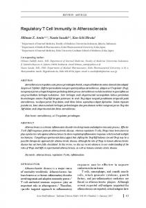
Figure 1. Assembly of the 18 polypeptide C1q molecule. C1q is assembled in macrophages and DCs from three types of chains, i.e., A-chain, B-chain and C-chain. Each chain has a collagenous N-terminal half and a non-collagenous C-terminal half which form globules. A and B chains dimerize through a disulphide bond at the N-terminal end and two C chains form homodimers through similar disulphide bonding. An A-B dimer and a single C-chain form a triple helix and the other C-chain in a C-C dimer trimerizes with another A-B dimer forming to triple helices linked by the disulphide between the two C-chains. Three such structures form a C1q molecule through N-terminal association (65).
phages but the same drugs up-regulated C1q production from resident macrophages (62). It is unclear whether these drugs affect the development of autoimmune conditions. It is interesting to note that C1q expression is generally up-regulated under immunosuppressive conditions. For example, a microarray study revealed that tumor-associated macrophages expressed elevated levels of C1q and two major immunosuppressive cytokines IL-10 and TGF-β (57). Immunosuppresive drugs like dexamethasone, prednisone and hydrocortisone all promoted C1q production from macrophages (Table 1).
Biochemical aspects of C1q The biochemical characterization of C1q was mostly completed in 1970s and, as a result, a model was proposed for C1q (65). A C1q molecule consists of 18 polypeptide chains of three different types (6 A-chains, 6 B-chains, and 6 Volume 5
Number 1
C-chains) (Figure 1). Each polypeptide consists of a collagenous N-terminal half and a globular C-terminal half with the collagenous region from each of the three chain types forming a triple helix bringing the 3 C-terminal globules together. A C1q consists of 6 such triple helical structures which are held together through inter-chain disulphide bonds at the N-terminal ends (Figure 1). It is viewed as a “bundle-of-tulips” under the electron microscope (66). Earlier work has established conditions for the selective degradation of the collagenous or globular regions of C1q to evaluate the functions of each region in the absence of the other (67, 68). The overall theoretical isoelectric point (pI) of the three C1q chains is 9.03 showing that it is a highly basic protein (http://au.expasy.org/tools/pi_tool.html). Collagens are characterized by Gly-Xaa-Yaa repeating sequences where Xaa and Yaa are often proline residues and Yaa is often hydroxylproline or hydroxyllysine residues (69). This is also true for the collagenous halves of the C1q polypeptides (67). Post-translational hydroxylation of proline and lysine residues is critical to the expression of C1q and other collagen-containing proteins. Two inhibitors of this process, i.e., 2,2’-dipyrodyl (DP) and 3,4-dehydro-DL-proline (DHP), have been used to block C1q synthesis in macrophages to generate C1q-deficient macrophages before C1q-/- macrophages became available from knockout mice (41, 70). However, macrophages generated with these inhibitors are also expected to lack collagen expression.
C1q binds pentraxins Many molecules or structures bind to C1q but the consequences of the binding vary substantially (Table 2). The C1r2C1s2 tetramer associates with the collagenous region of C1q and the C1r and C1s proenzymes are activated upon C1q binding to the Fc regions of antibodies through its globular domains (1). This triggers the complement classical pathway. Another well-studied C1q-binding molecule is CRP. C1q binds to CRP and similarly activates the complement classical pathway (27, 28). The difference between CRP and immune complexes as C1q ligands is that CRP but not immune complexes also binds factor H (71). CRP also up-regulates complement inhibitory proteins such as CD59, decay-accelerating protein (DAF) and membrane co-factor protein (MCP) on target cells (72). These mechanisms act to limit complement activation within the early components of the cascade which causes effective opsonization with C3 and C4 fragments for phagocytosis while minimizing the formation of anaphylatoxins and membrane-lytic complement complexes and therefore minimizing tissue inflammation and damage (71-73). CRP and another pentraxin serum amyloid P component (SAP) both bind to chromatin on AC and recruit C1q to elicit complement activation (27, 73). C1q also binds to another pentraxin (PTX3) which inhibits C1q deposition on AC (53). PTX3 was also reported to binds AC, but it binds to domains on AC that are distinct from the C1q-binding domains. Therefore, PTX3 apparently inhibits C1q binding to AC by blocking the AC-binding site on C1q (53). Both PTX3 February 2008
Cellular & Molecular Immunology
13
Table 2. C1q-binding proteins C1q ligands and receptors (pI value) CRP (5.44)
SAP (5.83)
PTX3 (4.86) CD35 (complement receptor 1 or CR1) SIGN-R1 cC1qR (calreticulin or CRT) gC1qR
CD91 CD93 α2β1 integrin
Effects 1) CRP binds to phosphorylcholine on mocrobes and host cells 2) CRP binds to chromatin 3) C1q binding to CRP-bound AC elicits complement activation and enhances phagocytosis 4) C1q binding to bacteria-bound CRP activates complement and protect mice from pneumococcal infection 1) SAP binds to DNA and chromotins in a Ca2+-dependent manner 2) C1q binds to SAP and activates complement 3) SAP-/- mice show autoimmunity as for C1q-/- mice PTX3 binds to C1q to prevent C1q deposition on ACs No known effect
Ref 26-28, 108
91, 109, 110 53 83
C1q binds to SIGN-R1, which the bacteria Streptococcus pneumoniae also bind, to activate complement on the 87 bacteria surface causing C3 deposition and bacterial killing on macrophages 1) CRT is an ER luminal protein but it is surface-exposed on ACs 18 2) C1q binds CRT on ACs and enhance AC phagocytosis through CD91 on phagocytes 1) It is an acidic mitochondria protein without a transmembrane domain 74-78 2) It binds to C1q 3) Its cell surface expression is not consistently reported 1) It is also known as low-density lipoprotein receptor-related protein 1 (LRP1) or α2-macroglobulin receptor 18 2) C1q binds to ACs and mediates AC phagocytosis through CD91 1) It is a large single unit receptor containing a C-type lectin and 5 EGF-like domains 79-82 2) Its role in mediating imC1q-elicited enhancement of FcγR- and CR-mediated phagocytosis is controversial 1) Mediates mast cell adhesion to C1q 107 2) Enhances mast cell activation by antibody-opsonized listeria monocytogenes in a C1q-dependent manner
Listed are proteins that have been reported to bind C1q. Some are soluble secreted proteins (CRP, SAP and PTX3), some are typical transmembrane receptors (CD35, SIGN-R1, CD91, CD93 and integrin α2β1), and some are cellular proteins without transmembrane domains which have been reported on the cell surface (cC1qR and gC1qR). cC1qR is also known as an endoplasmic reticulum protein calreticulin (CRT). gC1qR is also known as the mitochondria protein p33. CD91 is also known as α2-macroglobulin receptor or low-density lipoprotein receptor-related protein 1 (LRP-1). CD35 is complement receptor 1 (CR1). CD93 is also known as C1qRp. SAP, serum amloid P component; CRP, C-reactive protein; AC, apoptotic cell. NA, not determined. The isoelectric points (pIs) of each C1q-binding protein are indicated which were theoretical and were derived from the amino acid sequences of each mature peptide (http://au.expasy.org/tools/pi_tool.html). Sources of data are indicated by the numbered reference articles (Ref).
and C1q are produced by activated DCs. These pentraxins can apparently co-operate to regulate C1q-mediated complement activation on AC. Besides their structural similarities, these three pentraxins also have the common property of being acidic with low pI values (Table 2). This is in contrast to the high pI value of C1q. In fact, most other C1q-binding molecules are also acidic (Table 2).
Cell-associated C1q ligands or receptors A number of C1q-binding proteins are cellular proteins. CRT was first identified as a receptor for C1q that binds to the collagenous region of C1q (20, 21). It is a ubiquitous Ca2+-binding protein which is naturally retained in the ER lumen and lacks a transmembrane domain (19). On AC, it is found in blebs (18). Its role as a C1q receptor (cC1qR) to modulate C1q-opsonized phagocytosis or signaling is doubtable, but its ability to tag AC for C1q recognition and phagocytosis through CD91 renders the C1q-CRT interaction a valid mechanism (18). CRT is an acidic protein with a pI value of 4.29. CD91 is a type I receptor which also has an Volume 5
Number 1
acidic extracellular domain (pI, 5.16). gC1qR is another acidic cellular protein (pI, 4.74) that binds to C1q; it binds to the globular, rather than collagenous domains of C1q (74). It is naturally expressed in the mitochondria lacking a transmembrane domain and its expression on the cell surface is not consistently reported (75, 76). Nonetheless, treatment of macrophages and DCs with anti-gC1qR antibodies, aiming at gC1qR ligation, was shown to activate phosphatidylinositol 3-kinase (PI3K) and inhibit LPS induction of IL-12 from these cells (77). While it is difficult to reconcile between mitochondrial expression of gC1qR and its response to extracellular molecules, the report of gC1qR expression on DCs upon maturation bridges these differences (78).
CD93 and CD35 Besides CD91, another two cell surface transmembrane proteins bind to C1q. CD93 was identified for its ability to recognize immobilized C1q and promote macrophage phagocytosis through Fc and complement receptors (79-81). February 2008
14
C1q in Immunity and Autoimmunity
Figure 2. C1q production and its regulatory functions. The classical function of C1q in complement classical pathway activation is not highlighted in this illustration. The production of C1q by macrophages and DCs are up-regulated (+) or down-regulated (-) by various stimuli. C1q produced by macrophages and DCs can directly or indirectly stimulate various cell types for the listed effector functions which are detailed in Table 3.
It recognizes the collagenous region of C1q and is also known as C1qRp (79, 80). It is a type I receptor that contains a C-type carbohydrate recognition domain (CRD) and 5 EGF-like domains in its extracellular domain (80, 81). Like most other C1q-binding proteins, the extracellular domain of CD93 is also acidic (pI, 4.95). With regard to AC phagocytosis, CD93 appears to function independently of C1q C1q opsonization which is not required in CD93-mediated phagocytosis of AC (82). CD35 or complement receptor 1 (CR1) is the least acidic C1q-binding protein known so far (PI, 6.57) (83).
C1q regulates multiple macrophage activities Through interactions with soluble and cellular molecules, C1q modulates a range of macrophage interaction with microbial pathogens (Figure 2). C1q binds to Listeria monocytogenes and directly opsonizes the bacteria for enhanced macrophage uptake (84). C1q opsonization of L. monocytogenes enhances IFN-γ induced superoxide (O2-) and nitric oxide (NO) production from macrophages leading to increased cytotoxicity (85). Without obvious requirement for ligands, soluble C1q can stimulate macrophage production of TNF-α and up-regulate C3 (86). In a recent study, another mechanism was described by which C1q mediates complement activation on Streptococcus (S.) pneumoniae without binding to the bacteria (87). Both C1q and S. pneumoniae Volume 5
Number 1
bind to the lectin receptor SIGN-R1 on a subpopulation of spleen macrophages (87). C1q binding to SIGN-R1 elicits complement activation which apparently “spills” over to the surface of the bacteria leading to bacterial killing and clearance (87). A mechanism by which soluble C1q enhances macrophage cytotoxicity is its increased expression by IFN-γ and its autocrine up-regulation of TNF receptor expression on macrophages (88, 89). As a result, the elevated TNF receptor enhances autocrine macrophage stimulation by TNF-α which is induced by lipid A and these collectively lead potent NO production (88, 89). In one report, C1q was shown to inhibit alveolar macrophage phagocytosis of serum-opsonized yeast (90). C1q can directly and indirectly opsonize AC for phagocytosis by macrophages. C1q can directly bind AC and mediate AC phagocytosis through CD91 or LRP-1 on macrophages (18). It can also bind indirectly, through CRP and IgM, to cause complement activation and C3 deposition (24, 27, 73). SAP binds to chromatins in AC blebs and C1q binds to SAP to activate complement (91). Interestingly, like C1q-/- mice, SAP-/- mice also exhibit autoimmunity (91). Phagocytosis of C1q-opsonized AC through CD91, also known as LRP-1, induced ERK activation in a manner which requires the expression of ATP-binding cassette member A7 (ABCA7) (92). ERK activation promotes IL-10 but suppresses IL-12 production (93). However, the extent to which this mechanism contributes to AC induction of tolerogenic APCs has not been evaluated. February 2008
Cellular & Molecular Immunology
15
Table 3. Classical and other effector functions of C1q C1q/receptor agonists sC1q binding to AC
Cells
Effector functions
Ref
DCs
1) Endogenous C1q promotes AC phagocytosis by DCs 2) Endogenous C1q increases TLR4/AC induction of IL-12 from DCs 1) Enhance AC uptake by DCs 2) C1q-opsonized AC induces IL-6, IL-10 and TNF-α, but not IL-12p70, from DCs 1) Inhibits LPS-induced NF-κB, p38, c-Jun and ERK activation 2) Inhibits CpG-indced IL-12p40 and TNF-α production 3) No effect on CD40 and CD86 expression 1) Promotes gC1qR- and cC1qR-mediated DC chemotaxis 1) Binding through IgM, CRP or SAP causes C3 deposition and enhanced phagocytosis 2) C1q-binding to CRP-opsonized AC causes limited complement activation without significant activation of late components
53 94 95 99 23-29, 73, 18, 109
sC1q
DCs
1) Induces monocyte differenctiation into tolerogenic DCs
98
Anti-gC1qR
DCs
77
sC1q binding to AC
Mac
1) PI3-kinase activation 2) Inhibition of LPS-induced IL-12 production 1) Opsonizes AC for phagocytosis through CD91/LRP1 2) Induces ERK activation in a ABCA7-dependent manner 1) CRP and polyclonal IgM bind to phosphorylcholine on AC 2) C1q binds to both CRP and IgM to promote Ac phagocytosis 3) AC phagocytosis inhibits IL-12p40 production by macrophages 4) Necrotic cells induce IL-12p40 5) IgM-/- and C1q-/- additively reduce AC clearance by pMac 1) C1q binding to CRP-opsonized ACs activates early but not late complement component and promotes phagocytosis 2) It also induces TGF-β from Mac 1) Both Streptococcus pneumoniae and C1q bind to SIGN-R1 on macrophages 2) Bound C1q activates complement resulting complement activation on the bacteria and formation of C3 convertase and bacteria killing 1) C1q binding to Listeria monocytogenes enhances IFN-γ induction of O2- and NO 2) Directly opsonizes the bacteria for uptake 1) Induces TNF-α and C3 production 1) Inhibits suspension aMac phagocytosis of serum-opsonized yeast
87
Regulation of Mac Mac interaction with pathogens by sC1q
sC1q
Mo Neu
Fib
Mast imC1q
DC
Mac Mo Neu T
18, 92 22-25 53, 94, 95
71-73
84, 85 86 90
1) C1q promotes monocyte but not aMac chemotaxis 1) Up-regulates CR3 2) Opsonizes Staphylococci for respiratory burst induction 3) Opsonizes immune complexes for uptake 4) Chemotaxis 1) p38 activation in proliferating fibroblasts 2) Induction of fibroblast apoptosis 3) Partially replicated by cross-linking of surface cC1qR (CRT) 4) Ca2+-activated K+ channels and chemotaxis 1) Chemotaxis, chemokinesis 2) IL-6 induction by Listeria monocytogenes
100 103, 106
1) Increases MHC II, CD80, CD83, CD86 and CCR7 expression 2) Enhances IL-12, IL-10 and TNF-α secretion 3) Induces NF-κB activation 4) Promotes DC stimulation of T cell production of IFN-γ 1) Enhances FcγR- and CR-mediated phagocytosis through its collagen domain 2) No enhancement of FcR- and CR-mediated AC phagocytosis 3) CD93-/- mice show reduced AC phagocytosis which is C1q-independent 1) Inhibits phagocytosis of SP-A-opsonized bacteria 1) Induces respiratory burst through Ca2+ signaling 2) Co-operates with ICAM-1 and CD18 to induce respiratory burst 3) Induces respiratory burst through lipid raft 1) Regulates IFN-γ production by CD4 T cells
96, 97
114, 115
102, 107
79-82 44 105, 111, 112 113
The effects of soluble C1q (sC1q) and immobilized C1q (imC1q) on the various cell types are separately grouped. The C1q effector mechanisms are further grouped based on specific cell types. Mac, macrophage; Mo, monocyte; Neu, neutrophil; Fib, fibroblast; O2-, superoxide; NO, nitric oxide; MHC, major histocompatibility complex. The reference articles from which data were obtained are listed. Volume 5
Number 1
February 2008
16
C1q in Immunity and Autoimmunity
As C1q deposits around tissue macrophages (47), understanding macrophage response to tissue-deposited or in vitro immobilized C1q is physiologically relevant. In this regard, immobilized C1q has been reported to engage CD93 on macrophages to enhance phagocytosis through Fc and complement receptors (79-81). It should be noted that, while CD93 is involved in AC phagocytosis and clearance, there is no evidence that C1q was involved in this process (82).
C1q regulates DC differentiation, activation and antigen presentation C1q regulates several aspects of DC functions (Figure 2). It has been consistently reported to opsonize AC for DC phagocytosis (23, 53, 94, 95). C1q-opsonized AC also regulates DC production of cytokines, but data from different studies are not entirely consistent. In one study, C1q was shown to increase IL-12 production by DCs when the cells were co-stimulated with LPS and AC (53). In another study, C1q-opsonized AC was shown to induce IL-6, IL-10 and TNF-α from DCs but not IL-12 (94). C1q-/- mice have higher serum IL-12p40 than wild type mice suggesting inhibition of IL-12p40 production by normal C1q production (95). When DCs were derived from the bone marrow of C1q-/- mice, these cells showed reduced NF-κB, p38, c-Jun and ERK activation and decreased IL-12p40 and TNF-α upon LPS stimulation (95). However, these C1q-/- DCs showed normal CD40 and CD86 expression. As stated above, ligation of gC1qR on DCs inhibited LPS-induction of IL-12 through PI3K activation (77). Apparently, most studies report C1q inhibition of IL-12 production from DCs and suggest a tolerogenic property of C1q which is consistent to the development of autoimmunity at C1q deficiency (14). C1q deposits in extracellular matrix around DCs (47). DCs have also been cultured on immobilized C1q (96, 97). Immobilized C1q was shown to activate DCs -- it up-regulated co-stimulatory molecules and induced cytokines including IL-10, IL-12 and TNF-α (96, 97). These C1q-activated DCs also stimulated T cell proliferation and the production of IFN-γ (96, 97). In contrast, DCs cultured from monocytes in the presence of soluble C1q exhibited tolerogenic or immunosuppressive properties (98). These C1q-DCs expressed reduced co-stimulatory molecules and less cytokines and are also poorer in stimulating T cell proliferation and IFN-γ production (98). C1q has been reported to stimulate immature DC chemotaxis through gC1qR and gC1qR (99).
C1q regulates monocytes Monocytes are blood precursors of tissue macrophages and DCs which are constitutively under the stimulation of plasma C1q. Nonetheless, C1q has been reported to stimulate monocyte chemotaxis (100). However, C1q does not stimulate alveolar macrophage chemotaxis (100). While immobilized C1q was shown to enhance FcR- and CRVolume 5
Number 1
mediated phagocytosis by macrophages, it was shown to inhibit monocyte phagocytosis of surfactant protein A (SP-A)-opsonized bacteria (101). This was not observed with alveolar macrophages (101). Like MBL, SP-A is a member of the collectin family (6). The mechanism of this inhibition is unclear (101).
C1q regulates granulocytes, mast cells and fibroblasts Soluble C1q stimulates mast cell, neutrophil and eosinophil chemotaxis (102-104) (Figure 2). In view that C1q is predominantly found in the plasma, the significance of C1q in chemotaxis is unclear. However, this is clearly significant for newly synthesized C1q in peripheral tissues (47). If C1q secreted from macrophages and DCs in peripheral tissues translocates to the blood circulation, it is likely to form radial C1q gradients under steady state originating from tissue macrophages and DCs. Immobilized C1q induces respiratory burst in neutrophils causing the production of O2- (105). This involves the activation of Ca2+ signaling in neutrophils (105). Unlike macrophages, neutrophils do not exhibit FcRmediated phagocytosis upon stimulation on immobilized C1q (105). Immobilized C1q also failed to induce the release of primary and secondary granules from neutrophils (105). C1q, especially when it binds to bacteria Staphylococci, stimulates neutrophil respiratory burst and up-regulates CR3 expression (106). In a recent study, it was shown that IL-6 induction from mast cells, by antibody-opsonized bacteria L. monocytogenes, is dependent on the presence of C1q and integrin α2β1 expression on mast cells (107). Antibody-opsonized L. monocytogenes failed to induce IL-6 from mast cells when either C1q-/- mouse serum or α2-/- mast cells were used. It suggests that bacteria-bound C1q engages integrin α2β1 to provide a co-stimulatory signal through α2β1 signaling which synergizes with other receptors, i.e., FcR and CR, to induce IL-6 from mast cells (107). Mast cells adhere to immobilized C1q through the α2β1 integrin (107). C1q can regulate fibroblast migration and apoptosis. It stimulates fibroblast intracellular Ca2+ rise leading to K+ channel activation and fibroblast chemotaxis (114). An independent study showed that proliferating fibroblasts enter mitotic arrest and apoptosis upon stimulation with C1q or its collagenous region and this is associated with p38 MAPK phosphorylation in fibroblasts (115). Inhibition of p38 abrogated fibroblast response to C1q. Pre-incubation of fibroblasts with anti-CRT antibody inhibited C1q stimulation of p38 phosphorylation (115). This raises another regulatory role of tissue C1q in tissue remodeling where it is produced by macrophages and DCs.
C1q in immunity and tolerance The central conduit of C1q functions is its association with the C1r2C1s2 to form the complement C1 complex. Variations February 2008
Cellular & Molecular Immunology
17
of its function arise from its selective ligand recognition which targets this complex to various patho- physiologically relevant entities. Its ability to elicit lethal attack by producing opsonins, anaphylatoxins and lytic complexes is well understood. When the targets are host entities, this is tightly regulated through multiple inhibitory mechanisms including C1 inhibitor, C4-binding protein (C4BP), factor I, factor H, DAF, MCP, CR1 and CD59 (1). Even the targets are microbial pathogens, some of these control mechanisms are also in place to limit the scale of complement consumption and consequent tissue damages. For example, factor I and factor H cleave C4b and C3b on microorganisms as well as on host cells to reduce complement activation. However, DAF, MCP, CR1 and CD59 are largely restricted to host cells for the control of complement activation. AC has been increasingly recognized as a target of C1qmediated complement activation which has distinct or opposite consequences from its activation on microorganisms. AC-loaded APCs appear to be tolerogenic, or immunosuppressive, and this is largely consistently reported for C1q-opsonized AC. A number of mechanisms may contribute the immunosuppressive property of C1q-opsonized AC. Firstly, phagocytosis of C1q-opsonized AC mediated by CD91 is likely to activate ERK or PI3K in APCs (18, 77, 92). ERK activation favours IL-10 production (93). ACstimulated macrophages indeed produce more IL-10 and less IL-12 (116). PI3K activation, which can be potentially elicited through gC1qR, is known to inhibit IL-12 production (117). Secondly, when complement is elicited on AC, this is likely to be limited by various inhibitory mechanisms leading to the deposition of C3 or C4 opsonins without generating significant C5a anaphylatoxin and lytic complexes. Under steady state, macrophages synthesize early but not late complement components such as C5-C9 (40). It is unclear what other complement components are produced in peripheral tissues by other cells. If C5-C9 is absent or scarce in peripheral tissues, complement activation on AC naturally reduces or ceases after C3 opsonin deposition and this is not expected to cause significant inflammation and tissue damages associated with anaphylatoxins and lytic complexes. Besides membrane destruction, activated late complement components (C5b-C9) also activate DC production of IL-12 and TNF-α and such DCs promote immunity rather than tolerance (118). When AC is bound by CRP, the activation of late complement components can be inhibited through two additional mechanisms. CRP can limit the activation of late complement components on AC by binding to factor H which enhances C3b cleavage into iC3b (71). It also up-regulates surface expression of DAF, MCP and CD59 on host cells although whether this occurs on AC is unclear (72). CRP is an acute phase protein and is likely to be significant under inflammatory conditions when plasma complement infiltrates peripheral tissues (119). It is also unclear how C1q-mediated complement activation on polyclonal IgM-opsonized AC is controlled under inflammatory conditions. The report that SAP-/- mice developed autoimmunity, like C1q-/- mice, is Volume 5
Number 1
consistent with its binding to AC and triggering of C1q-mediated complement activation (14, 91). The exact mechanism by which SAP control complement activation on AC is unclear. Thirdly, C3 deposition on AC, especially iC3b, can effectively regulate phagocyte activation. Ligation of the iC3b receptor CR3 on macrophages inhibits IL-12 production (35, 36). Phagocytosis of iC3b-opsonized AC by DCs renders these cells tolerogenic (25). The ability of C1q to elicit iC3b deposition on AC without inducing inflammation and tissue damages can result in suppression of phagocyte activation or render APCs tolerogenic while promoting AC uptake.
Summary The structure of C1q, as a major component of the complement system, has been extensively investigated providing an elegant model for the investigation of its functions. While its primary role in the complement system is the formation of C1 complex with the C1r2C1s2 proteases and its ability to bind different ligands directs complement activation on different targets, growing evidence suggests a broader regulatory role for C1q. It remains to be investigated why C1q expression is restricted to monocyte-derived macrophages and dendritic cells. Since these are major antigen-presenting cells, this mode of C1q synthesis may regulate antigen presentation and adaptive immunity. It is also not fully understood how C1q-deficiency is associated with autoimmunity. C1q is synthesized from three distinct types of chains. In the absence of one or two C1q chains, can the remaining chains form a C1q-like molecule? The expression of recombinant C1q may explain some of these questions. In addition, although isolated collagenous and globular domains of C1q have been widely used to locate its binding sites for various ligands or effector molecules, specific C1q binding sites for these molecules also require the expression of recombinant C1q.
Acknowledgements JH would like to thank Ken Reid and Michael Loos for guidance and support for many years. This project is funded by the Singapore National Medical Research Council Grant NMRC/0950/2005.
References 1. Reid KBM. Activation and control of the complement system. Essays Biochem. 1986;22:27-68. 2. Fujita T, Matsushita M, Endo Y. The lectin-complement pathway-its role in innate immunity and evolution. Immunol Rev. 2004;198:185-202. 3. Duncan AR, Winter G. The binding site for C1q on IgG. Nature. 1988;332:738-740. 4. Matsushita M, Fujita T. Activation of the classical complement pathway by mannose-binding protein in association with a novel C1s-like serine protease. J Exp Med. 1992;176:1497-1502. February 2008
18
C1q in Immunity and Autoimmunity
5. Thiel S, Vorup-Jensen T, Stover CM, et al. A second serine protease associated with mannan-binding lectin that activates complement. Nature. 1997;386:506-510. 6. Lu J, Teh C, Kishore U, Reid KBM. Collectins and ficolins: sugar pattern recognition molecules of the mammalian innate immune system. Biochim Biophys Acta. 2002;1572:387-400. 7. Warren J, Mastroeni P, Dougan G, et al. Increased susceptibility of C1q-deficient mice to Salmonella enterica serovar Typhimurium infection. Infect Immun. 2002;70:551-557. 8. Taylor PR, Seixas E, Walport MJ, Langhorne J, Botto M. Complement contributes to protective immunity against reinfection by Plasmodium chabaudi chabaudi parasites. Infect Immun. 2001;69:3853-3859. 9. Celik I, Stover C, Botto M, et al. Role of the classical pathway of complement activation in experimentally induced polymicrobial peritonitis. Infect Immun. 2001;69:7304-7309. 10. Super M, Thiel S, Lu J, Levinsky RJ, Turner MW. Association of low levels of mannan-binding protein with a common defect of opsonisation. Lancet. 1989;2:1236-1239. 11. Turner MW, Grant C, Seymour ND, et al. Evaluation of C3b/C3bi opsonization and chemiluminescence with selected yeasts and bacteria using sera of different opsonic potential. Immunology. 1986;58:111-115. 12. Shi L, Takahashi K, Dundee J, et al. Mannose-binding lectindeficient mice are susceptible to infection with Staphylococcus aureus. J Exp Med. 2004;199:1379-1390. 13. Kölble K, Reid KBM. Genetic deficiencies of the complement system and association with disease--early components. Int Rev Immunol. 1993;10:17-36. 14. Botto M, Dell'Agnola C, Bygrave AE, et al. Homozygous C1q deficiency causes glomerulonephritis associated with multiple apoptotic bodies. Nat Genet. 1998;19:56-59. 15. Mevorach D, Mascarenhas JO, Gershov D, Elkon KB. Complement-dependent clearance of apoptotic cells by human macrophages. J Exp Med. 1998;188:2313-2320. 16. Mevorach D, Zhou JL, Song X, Elkon KB. Systemic exposure to irradiated apoptotic cells induces autoantibody production. J Exp Med. 1998;188:387-392. 17. Korb LC, Ahearn JM. C1q binds directly and specifically to surface blebs of apoptotic human keratinocytes: complement deficiency and systemic lupus erythematosus revisited. J Immunol. 1997;158:4525-4528. 18. Ogden CA, deCathelineau A, Hoffmann PR, et al. C1q and mannose binding lectin engagement of cell surface calreticulin and CD91 initiates macropinocytosis and uptake of apoptotic cells. J Exp Med. 2001;194:781-795. 19. Michalak M, Corbett EF, Mesaeli N, Nakamura K, Opas M. Calreticulin: one protein, one gene, many functions. Biochem J. 1999;344:281-292. 20. Malhotra R, Willis AC, Jensenius JC, et al. Structure and homology of human C1q receptor (collectin receptor). Immunology. 1993;78:341-348. 21. Malhotra R, Thiel S, Reid KBM, Sim RB. Human leukocyte C1q receptor binds other soluble proteins with collagen domains. J Exp Med. 1990;172:955-959. 22. Zwart B, Ciurana C, Rensink I, et al. Complement activation by apoptotic cells occurs predominantly via IgM and is limited to late apoptotic (secondary necrotic) cells. Autoimmunity. 2004; 37:95-102. 23. Quartier P, Potter PK, Ehrenstein MR, Walport MJ, Botto M. Predominant role of IgM-dependent activation of the classical pathway in the clearance of dying cells by murine bone marrow-derived macrophages in vitro. Eur J Immunol. 2005;35: 252-260. Volume 5
Number 1
24. Ogden CA, Kowalewski R, Peng Y, Montenegro V, Elkon KB. IgM is required for efficient complement mediated phagocytosis of apoptotic cells in vivo. Autoimmunity. 2005;38:259-264. 25. Skoberne M, Somersan S, Almodovar W, et al. The apoptotic-cell receptor CR3, but not αvβ5, is a regulator of human dendritic-cell immunostimulatory function. Blood. 2006;108:947-955. 26. Richards RL, Gewurz H, Osmand AP, Alving CR. Interactions of C-reactive protein and complement with liposomes. Proc Natl Acad Sci U S A. 1977;74:5672-5676. 27. Kim SJ, Gershov D, Ma X, Brot N, Elkon KB. Opsonization of apoptotic cells and its effect on macrophage and T cell immune responses. Ann N Y Acad Sci. 2003;987:68-78. 28. Suresh MV, Singh SK, Ferguson DA Jr, Agrawal A. Role of the property of C-reactive protein to activate the classical pathway of complement in protecting mice from pneumococcal infection. J Immunol. 2006;176:4369-4374. 29. Mitchell DA, Taylor PR, Cook HT, et al. Cutting edge: C1q protects against the development of glomerulonephritis independently of C3 activation. J Immunol. 1999;162:56765679. 30. Jancar S, Sánchez Crespo M. Immune complex-mediated tissue injury: a multistep paradigm. Trends Immunol. 2005;26:48-55. 31. Karp DR. Complement and systemic lupus erythematosus. Curr Opin Rheumatol. 2005;17:538-542. 32. Morgan BP, Walport MJ. Complement deficiency and disease. Immunol Today. 1991;12:301-306. 33. Sturfelt G, Truedsson L. Complement and its breakdown products in SLE. Rheumatology (Oxford). 2005;44:1227-1232. 34. Stuart LM, Takahashi K, Shi L, et al. Mannose-binding lectin-deficient mice display defective apoptotic cell clearance but no autoimmune phenotype. J Immunol. 2005;174:32203226. 35. Marth T, Kelsall BL. Regulation of interleukin-12 by complement receptor 3 signaling. J Exp Med. 1997;185:19871995. 36. Sutterwala FS, Noel GJ, Clynes R, Mosser DM. Selective suppression of interleukin-12 induction after macrophage receptor ligation. J Exp Med. 1997;185:1977-1985. 37. Trinchieri G. Interleukin-12 and the regulation of innate resistance and adaptive immunity. Nat Rev Immunol. 2003;3: 133-146. 38. Tracey KJ, Cerami A. Tumor necrosis factor: a pleiotropic cytokine and therapeutic target. Annu Rev Med. 1994;45:491503. 39. Brandhorst TT, Wüthrich M, Finkel-Jimenez B, Warner T, Klein BS. Exploiting type 3 complement receptor for TNF-α suppression, immune evasion, and progressive pulmonary fungal infection. J Immunol. 2004;173:7444-7453. 40. Colten HR, Strunk RC, Perlmutter DH, Cole FS. Regulation of complement protein biosynthesis in mononuclear phagocytes. Ciba Found Symp. 1986;118:141-154. 41. Müller W, Hanauske-Abel H, Loos M. Biosynthesis of the first component of complement by human and guinea pig peritoneal macrophages: evidence for an independent production of the C1 subunits. J Immunol. 1978;121:1578-1584. 42. Loos M, Martin H, Petry F. The biosynthesis of C1q, the collagen-like and Fc-recognizing molecule of the complement system. Behring Inst Mitt. 1989;84:32-41. 43. Gulati P, Lemercier C, Guc D, Lappin D, Whaley K. Regulation of the synthesis of C1 subcomponents and C1-inhibitor. Behring Inst Mitt. 1993;93:196-203. 44. Petry F, Botto M, Holtappels R, Walport MJ, Loos M. Reconstitution of the complement function in C1q-deficient February 2008
Cellular & Molecular Immunology
45. 46.
47. 48.
49. 50.
51.
52. 53. 54.
55. 56.
57.
58.
59.
60.
61.
19
(C1qa-/-) mice with wild-type bone marrow cells. J Immunol. 2001;167:4033-4037. Gordon S. The macrophage. Bioessays. 1995;17:977-986. Lu J, Le Y, Kon OL, Chan J, Lee SH. Biosynthesis of human ficolin, an Escherichia coli-binding protein, by monocytes: comparison with the synthesis of two macrophage-specific proteins, C1q and the mannose receptor. Immunology. 1996; 89:289-294. Cao W, Bobryshev YV, Lord RS, et al. Dendritic cells in the arterial wall express C1q: potential significance in atherogenesis. Cardiovasc Res. 2003;60:175-186. Sallusto F, Cella M, Danieli C, Lanzavecchia A. Dendritic cells use macropinocytosis and the mannose receptor to concentrate macromolecules in the major histocompatibility complex class II compartment: downregulation by cytokines and bacterial products. J Exp Med. 1995;182:389-400. Banchereau J, Briere F, Caux C, et al. Immunobiology of dendritic cells. Annu Rev Immunol. 2000;18:767-811. Peng Q, Li K, Patel H, Sacks SH, Zhou W. Dendritic cell synthesis of C3 is required for full T cell activation and development of a Th1 phenotype. J Immunol. 2006;176:33303341. Schwaeble W, Schäfer MK, Petry F, et al. Follicular dendritic cells, interdigitating cells, and cells of the monocytemacrophage lineage are the C1q-producing sources in the spleen. Identification of specific cell types by in situ hybridization and immunohistochemical analysis. J Immunol. 1995;155:4971-4978. Castellano G, Woltman AM, Nauta AJ, et al. Maturation of dendritic cells abrogates C1q production in vivo and in vitro. Blood. 2004;103:3813-3820. Baruah P, Dumitriu IE, Peri G, et al. The tissue pentraxin PTX3 limits C1q-mediated complement activation and phagocytosis of apoptotic cells by dendritic cells. J Leukoc Biol. 2006;80:87-95. Faust D, Loos M. In vitro modulation of C1q mRNA expression and secretion by interleukin-1, interleukin-6, and interferon-γ in resident and stimulated murine peritoneal macrophages. Immunobiology. 2002;206:368-376. Walker DG. Expression and regulation of complement C1q by human THP-1-derived macrophages. Mol Chem Neuropathol. 1998;34:197-218. Depboylu C, Schäfer MK, Schwaeble WJ, et al. Increase of C1q biosynthesis in brain microglia and macrophages during lentivirus infection in the rhesus macaque is sensitive to antiretroviral treatment with 6-chloro-2', 3'-dideoxyguanosine. Neurobiol Dis. 2005;20:12-26. Biswas SK, Gangi L, Paul S, et al. A distinct and unique transcriptional program expressed by tumor-associated macrophages (defective NF-κB and enhanced IRF-3/STAT1 activation). Blood. 2006;107:2112-2122. Zhou AQ, Herriott MJ, Leu RW. Kinetics of the biosynthesis of complement subcomponent C1q by murine macrophages: LPS, immune complexes, and zymosan alone and in combination with interferon-γ. J Leukoc Biol. 1991;50:453-463. Leu RW, Zhou AQ, Rummage J, Fast DJ, Shannon BJ. Reconstitution of a deficiency of AKR mouse macrophages for their response to lipid A activation for tumor cytotoxicity by complement subcomponent C1q: role of IFN-γ. J Immunol. 1991;147:1315-1321. Zhou AQ, Herriott MJ, Leu RW. Kinetics of the biosynthesis of complement subcomponent C1q by murine macrophages: effects of stimulation by interferon-γ. J Interferon Res. 1991;11: 111-119. Armbrust T, Nordmann B, Kreissig M, Ramadori G. C1q Volume 5
Number 1
62.
63.
64.
65. 66. 67. 68. 69. 70.
71. 72. 73.
74. 75. 76. 77.
78.
synthesis by tissue mononuclear phagocytes from normal and from damaged rat liver: up-regulation by dexamethasone, down-regulation by interferon γ, and lipopolysaccharide. Hepatology. 1997;26:98-106. Trinder PK, Faust D, Petry F, Loos M. Modulation of mRNA expression and secretion of C1q in mouse macrophages by anti-inflammatory drugs and cAMP: evidence for the partial involvement of a pathway that includes cyclooxygenase, prostaglandin E2 and adenylate cyclase. Immunology. 1995;84: 638-644. Ohmi K, Greenberg DS, Rajavel KS, Ryazantsev S, Li HH, Neufeld EF. Activated microglia in cortex of mouse models of mucopolysaccharidoses I and IIIB. Proc Natl Acad Sci U S A. 2003;100:1902-1907. Haga S, Aizawa T, Ishii T, Ikeda K. Complement gene expression in mouse microglia and astrocytes in culture: comparisons with mouse peritoneal macrophages. Neurosci Lett. 1996;216:191-194. Reid KBM, Porter RR. Subunit composition and structure of subcomponent C1q of the first component of human complement. Biochem J. 1976;155:19-23. Knobel HR, Villiger W, Isliker H. Chemical analysis and electron microscopy studies of human C1q prepared by different methods. Eur J Immunol. 1975;5:78-82. Reid KBM. A collagen-like amino acid sequence in a polypeptide chain of human C1q (a subcomponent of the first component of complement). Biochem J. 1974;141:189-203. Reid KBM. Isolation, by partial pepsin digestion, of the three collagen-like regions present in subcomponent Clq of the first component of human complement. Biochem J. 1976;155:5-17. Myllyharju J. Prolyl 4-hydroxylases, the key enzymes of collagen biosynthesis. Matrix Biol. 2003;22:15-24. Mocharla R, Mocharla H, Leu RW. Effects of inhibitors of C1q biosynthesis on macrophage Fc receptor subclass-mediated antibody-dependent cellular cytotoxicity and phagocytosis. Cell Immunol. 1987;105:127-135. Mold C, Gewurz H, Du Clos TW. Regulation of complement activation by C-reactive protein. Immunopharmacology. 1999;42:23-30. Li SH, Szmitko PE, Weisel RD, et al. C-reactive protein upregulates complement-inhibitory factors in endothelial cells. Circulation. 2004;109:833-836. Gershov D, Kim S, Brot N, Elkon KB. C-Reactive protein binds to apoptotic cells, protects the cells from assembly of the terminal complement components, and sustains an antiinflammatory innate immune response: implications for systemic autoimmunity. J Exp Med. 2000;192:1353-1364. Peerschke EI, Reid KBM, Ghebrehiwet B. Identification of a novel 33-kDa C1q-binding site on human blood platelets. J Immunol. 1994;152:5896-5901. Dedio J, Jahnen-Dechent W, Bachmann M, Müller-Esterl W. The multiligand-binding protein gC1qR, putative C1q receptor, is a mitochondrial protein. J Immunol. 1998;160:3534-3342. van Leeuwen HC, O' Hare P. Retargeting of the mitochondrial protein p32/gC1Qr to a cytoplasmic compartment and the cell surface. J Cell Sci. 2001;114:2115-2123. Waggoner SN, Cruise MW, Kassel R, Hahn YS. gC1q receptor ligation selectively down-regulates human IL-12 production through activation of the phosphoinositide 3-kinase pathway. J Immunol. 2005;175:4706-4714. Vegh Z, Goyarts EC, Rozengarten K, et al. Maturationdependent expression of C1q-binding proteins on the cell surface of human monocyte-derived dendritic cells. Int Immunopharmacol. 2003;3:345-357.
February 2008
20
C1q in Immunity and Autoimmunity
79. Tenner AJ, Robinson SL, Borchelt J, Wright JR. Human pulmonary surfactant protein (SP-A), a protein structurally homologous to C1q, can enhance FcR- and CR1-mediated phagocytosis. J Biol Chem. 1989;264:13923-13928. 80. Nepomuceno RR, Henschen-Edman AH, Burgess WH, Tenner AJ. cDNA cloning and primary structure analysis of C1qR(P), the human C1q/MBL/SPA receptor that mediates enhanced phagocytosis in vitro. Immunity. 1997;6:119-129. 81. Steinberger P, Szekeres A, Wille S, et al. Identification of human CD93 as the phagocytic C1q receptor (C1qRp) by expression cloning. J Leukoc Biol. 2002;71:133-140. 82. Norsworthy PJ, Fossati-Jimack L, Cortes-Hernandez J, et al. Murine CD93 (C1qRp) contributes to the removal of apoptotic cells in vivo but is not required for C1q-mediated enhancement of phagocytosis. J Immunol. 2004;172:3406-3414. 83. Klickstein LB, Barbashov SF, Liu T, et al. Complement receptor type 1 (CR1, CD35) is a receptor for C1q. Immunity. 1997;7:345-355. 84. Alvarez-Dominguez C, Carrasco-Marin E, Leyva-Cobian F. Role of complement component C1q in phagocytosis of Listeria monocytogenes by murine macrophage-like cell lines. Infect Immun. 1993;61:3664-3672. 85. Alvarez-Domínguez C, Carrasco-Marín E, López-Mato P, Leyva-Cobián F. The contribution of both oxygen and nitrogen intermediates to the intracellular killing mechanisms of C1q-opsonized Listeria monocytogenes by the macrophage-like IC-21 cell line. Immunology. 2000;101:83-89. 86. Bajtay Z, Józsi M, Bánki Z, Thiel S, Thielens N, Erdei A. Mannan-binding lectin and C1q bind to distinct structures and exert differential effects on macrophages. Eur J Immunol. 2000;30:1706-1713. 87. Kang YS, Do Y, Lee HK, et al. A dominant complement fixation pathway for pneumococcal polysaccharides initiated by SIGN-R1 interacting with C1q. Cell. 2006;125:47-58. 88. Jiang H, Rummage JA, Stewart CA, et al. Evidence for endogenous C1q modulates TNF-α receptor synthesis and autocrine binding of TNF-α associated with lipid A activation of murine macrophages for nitric oxide production. Cell Immunol. 1996;170:34-40. 89. Jiang H, Rummage JA, Zhou A, et al. IFN-αβ reconstitutes the deficiency in lipid A-activated AKR macrophages for nitric oxide synthase. J Immunol. 1996;157:305-312. 90. Rosseau S, Guenther A, Seeger W, Lohmeyer J. Phagocytosis of viable Candida albicans by alveolar macrophages: lack of opsonin function of surfactant protein A. J Infect Dis. 1997; 175:421-428 91. Bickerstaff MC, Botto M, Hutchinson WL, et al. Serum amyloid P component controls chromatin degradation and prevents antinuclear autoimmunity. Nat Med. 1999;5:694-697. 92. Jehle AW, Gardai SJ, Li S, et al. ATP-binding cassette transporter A7 enhances phagocytosis of apoptotic cells and associated ERK signaling in macrophages. J Cell Biol. 2006; 174:547-556. 93. Agrawal A, Dillon S, Denning TL, Pulendran B. ERK1-/- mice exhibit Th1 cell polarization and increased susceptibility to experimental autoimmune encephalomyelitis. J Immunol. 2006; 176:5788-5796. 94. Nauta AJ, Castellano G, Xu W, et al. Opsonization with C1q and mannose-binding lectin targets apoptotic cells to dendritic cells. J Immunol. 2004;173:3044-3050. 95. Yamada M, Oritani K, Kaisho T, et al. Complement C1q regulates LPS-induced cytokine production in bone marrowderived dendritic cells. Eur J Immunol. 2004;34:221-230. 96. Fonseca MI, Carpenter PM, Park M, et al. C1qR(P), a myeloid Volume 5
97. 98. 99.
100. 101. 102.
103.
104. 105.
106. 107. 108.
109. 110.
111.
112. 113. 114.
Number 1
cell receptor in blood, is predominantly expressed on endothelial cells in human tissue. J Leukoc Biol. 2001;70:793800. Csomor E, Bajtay Z, Sándor N, et al. Complement protein C1q induces maturation of human dendritic cells. Mol Immunol. 2007;44:3389-3397. Castellano G, Woltman AM, Schlagwein N, et al. Immune modulation of human dendritic cells by complement. Eur J Immunol. 2007;37:2803-2811. Vegh Z, Kew RR, Gruber BL, Ghebrehiwet B. Chemotaxis of human monocyte-derived dendritic cells to complement component C1q is mediated by the receptors gC1qR and cC1qR. Mol Immunol. 2006;43:1402-1407. Tino MJ, Wright JR. Surfactant proteins A and D specifically stimulate directed actin-based responses in alveolar macrophages. Am J Physiol. 1999;276:L164-174. Tino MJ, Wright JR. Surfactant protein A stimulates phagocytosis of specific pulmonary pathogens by alveolar macrophages. Am J Physiol. 1996;270:L677-688. Ghebrehiwet B, Kew RR, Gruber BL, et al. Murine mast cells express two types of C1q receptors that are involved in the induction of chemotaxis and chemokinesis. J Immunol. 1995;155:2614-2619. Leigh LE, Ghebrehiwet B, Perera TP, et al. C1q-mediated chemotaxis by human neutrophils: involvement of gClqR and G-protein signalling mechanisms. Biochem J. 1998;330:247254. Kuna P, Iyer M, Peerschke EI, Kaplan AP, Reid KBM, Ghebrehiwet B. Human C1q induces eosinophil migration. Clin Immunol Immunopathol. 1996;81:48-54. Goodman EB, Tenner AJ. Signal transduction mechanisms of C1q-mediated superoxide production. Evidence for the involvement of temporally distinct staurosporine-insensitive and sensitive pathways. J Immunol. 1992;148:3920-3928. Eggleton P, Ghebrehiwet B, Coburn JP, et al. Characterization of the human neutrophil C1q receptor and functional effects of free ligand on activated neutrophils. Blood. 1994;84:1640-1649. Edelson BT, Stricker TP, Li Z, et al. Novel collectin/C1q receptor mediates mast cell activation and innate immunity. Blood. 2006;107:143-150. Robey FA, Jones KD, Tanaka T, Liu TY. Binding of C-reactive protein to chromatin and nucleosome core particles. A possible physiological role of C-reactive protein. J Biol Chem. 1984; 259:7311-7316. Bristow CL, Boackle RJ. Evidence for the binding of human serum amyloid P component to Clq and Fab γ. Mol Immunol. 1986;23:1045-1052. Hicks PS, Saunero-Nava L, Du Clos TW, Mold C. Serum amyloid P component binds to histones and activates the classical complement pathway. J Immunol. 1992;149:36893694. Tyagi S, Nicholson-Weller A, Barbashov SF, et al. Intercellular adhesion molecule 1 and β2 integrins in C1q-stimulated superoxide production by human neutrophils: an example of a general regulatory mechanism governing acute inflammation. Arthritis Rheum. 2000;43:2248-2259. Otabor I, Tyagi S, Beurskens FJ, et al. A role for lipid rafts in C1q-triggered O2- generation by human neutrophils. Mol Immunol. 2004;41:185-190. Lu J, Wu X, Teh BK. The regulatory roles of C1q. Immunobiology. 2007;212:245-252. Oiki S, Okada Y. C1q induces chemotaxis and K+ conductance activation coupled to increased cytosolic Ca2+ in mouse fibroblasts. J Immunol. 1988;141:3177-3185
February 2008
Cellular & Molecular Immunology
21
115. Bordin S, Whitfield D. Cutting edge: proliferating fibroblasts respond to collagenous C1q with phosphorylation of p38 mitogen-activated protein kinase and apoptotic features. J Immunol. 2003;170:667-671. 116. Chung EY, Kim SJ, Ma XJ. Regulation of cytokine production during phagocytosis of apoptotic cells. Cell Res. 2006;16:154161. 117. Fukao T, Koyasu S. PI3K and negative regulation of TLR
Volume 5
signaling. Trends Immunol. 2003;24:358-363. 118. Chen Y, Yang C, Jin N, et al. Terminal complement complex C5b-9-treated human monocyte-derived dendritic cells undergo maturation and induce Th1 polarization. Eur J Immunol. 2007; 37:167-176. 119. Young B, Gleeson M, Cripps AW. C-reactive protein: a critical review. Pathology. 1991;23:118-124.
Number 1
February 2008