Mar. Drugs 2015, 13, 1647-1665; doi:10.3390/md13041647 OPEN ACCESS
marine drugs ISSN 1660-3397 www.mdpi.com/journal/marinedrugs Article
The Effect of Cholesterol on the Long-Range Network of Interactions Established among Sea Anemone Sticholysin II Residues at the Water-Membrane Interface Sara Garcí a-Linares 1, Ida Alm 2, Terhi Maula 2, JoséG. Gavilanes 1,*, Johan Peter Slotte 2 and Álvaro Martínez-del-Pozo 1,* 1
2
Department of Biochemistry and Molecular Biology I, Complutense University, 28040 Madrid, Spain; E-Mail:
[email protected] Biochemistry, Department of Biosciences, Åbo Akademi University, 20520 Turku, Finland; E-Mails:
[email protected] (I.A.);
[email protected] (T.M.);
[email protected] (J.P.S.)
* Authors to whom correspondence should be addressed; E-Mails:
[email protected] (J.G.G.);
[email protected] (Á.M.-P.); Tel.: +34-91-394-4158; Fax: +34-91-394-4159. Academic Editor: Keith Glaser Received: 23 January 2015 / Accepted: 16 March 2015 / Published: 25 March 2015
Abstract: Actinoporins are α-pore forming proteins with therapeutic potential, produced by sea anemones. Sticholysin II (StnII) from Stichodactyla helianthus is one of its most extensively characterized members. These proteins remain stably folded in water, but upon interaction with lipid bilayers, they oligomerize to form a pore. This event is triggered by the presence of sphingomyelin (SM), but cholesterol (Chol) facilitates pore formation. Membrane attachment and pore formation require changes involving long-distance rearrangements of residues located at the protein-membrane interface. The influence of Chol on membrane recognition, oligomerization, and/or pore formation is now studied using StnII variants, which are characterized in terms of their ability to interact with model membranes in the presence or absence of Chol. The results obtained frame Chol not only as an important partner for SM for functional membrane recognition but also as a molecule which significantly reduces the structural requirements for the mentioned conformational rearrangements to occur. However, given that the DOPC:SM:Chol vesicles employed display phase coexistence and have domain boundaries, the observed effects could be also due to the presence of these different phases on the membrane. In addition, it is also shown that the Arg51 guanidinium group is strictly required for membrane recognition, independently of the presence of Chol.
Mar. Drugs 2015, 13
1648
Keywords: actinoporin; equinatoxin; sphingomyelin; pore-forming toxin; sphingomyelin
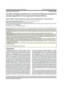
1. Introduction Sea anemones are a group of benthic marine animals which secrete various toxins [1] including a group of small and basic α-pore forming proteins known as actinoporins [2]. These actinoporins form cation-selective pores on the cell membranes, causing colloid-osmotic shock that leads to cells death [3–5]. They are believed to participate in anemone functions such as predation, defense, and digestion, and have been shown to be lethal to small crustaceans, mollusks, fish [6], and parasites [7]. All known actinoporins display high sequence identity and appear as multigene families [8,9]. However, only four of them have been characterized in deep detail: Equinatoxin II (EqtII) from Actinia equina [8], Sticholysins I and II (StnI and StnII) from Stichodactyla helianthus [10,11], and Fragaceatoxin C (Fra C) from Actinia fragacea [12]. Like many other marine toxins, actinoporins show some therapeutic potential, including different pharmacological effects, presumable anticancer activities, and use in the construction of specific immunotoxins [1,7,13–18]. In addition to their potential as therapeutic drugs, actinoporins have gained remarkable attention because they show a singular behavior at the water–lipid membrane interface. In aqueous solution they remain stably folded, but they become integral membrane structures upon interaction with lipid bilayers, oligomerizing to form pores [10,11,19]. It is widely accepted that the bilayers targeted must contain sphingomyelin (SM) and/or display phase coexistence [20–25]. In fact, the effect of not only SM but also Chol on the membrane pore-forming ability of StnII has been thoroughly studied [21,22,25–29]. According to those results, it is now quite clear that the presence of Chol eases the formation of pores by StnII, a conclusion which is in agreement with the coexistence of Chol and SM in biological membranes [27,30–33]. However, what still remains poorly studied is the nature of the protein determinants which explain this effect. To answer this question, we have studied a battery of different StnII mutants affecting different protein regions presumably involved in pore formation. The water-soluble structure of StnII is known in detail [34]. It folds as a β-sandwich motif composed of 10 β-strands flanked by two α-helices which interact with both sides of the β-sandwich (Figure 1). One of these helices (α1) is located near the N-terminal end. In fact, the first 30 residues appear to be able to adopt alternative conformations without disrupting the fold of the β-sandwich [35]. This feature, altogether with the amphipathic character of this stretch, seems to be extremely important for the final functionality of the pore, since the α1 helix has been proposed to extend and further insert into the membrane to form the pore walls [17,36,37]. The most recent model explaining the mechanism of actinoporins’ pore formation [17,37–39] assumes a toroidal protein-lipid structure without a well-defined fixed stoichiometry [17,38–40], although an alternative model has been proposed for Fra C [41]. Nevertheless, it is reasonably well-proven that insertion of the N-terminus into the membrane takes place in anon-coordinate way, shortly after the binding of the toxin and before their oligomerization into the final pore [37]. In addition to this N-terminal α-helix, three more regions of the structure seem to be especially important from a functional point of view: a phosphocholine (POC) binding site, a cluster of aromatic residues, and an array of basic amino acids [11].
Mar. Drugs 2015, 13
1649
Figure 1. Diagram of the three-dimensional structure of StnII indicating the location of the eight mutated positions: Ala10, Arg29, Arg51, Phe106, Tyr111, Tyr135, Tyr136, and Gly142. The different elements of ordered secondary structure, as well as the N- and C-terminal ends, are also indicated. The dotted line is a representation of the membrane surface. The diagram was constructed from the atomic coordinates deposited to the PDB (Protein Data Bank, reference 1GWY for StnII). The image was generated by the MolMol Program [42]. The work presented here studies the effect of Chol on the membrane-interacting behavior of eight different StnII mutants affecting the stretch of the 30 first residues (A10P and R29Q), the aromatic cluster (F106L, Y111N, Y135F, and Y136F), the POC binding site (R51Q, Y111N, Y135F, and Y136F), and a residue involved in maintaining the pore-competent state of protein oligomerization (G142A). 2. Results 2.1. Protein Purification and Characterization All proteins used in the study were purified to homogeneity according to their behavior in SDS-PAGE. Their amino acid compositions were consistent with the introduced mutations. The calculated E0.1% (280 nm, 1 cm) values were also in good agreement with the amino acid changes made (Table 1). All far-UV CD spectra of the individual mutants were indistinguishable from that corresponding to the wild-type StnII (data not shown). In summary, all eight mutants retained the overall native water-soluble conformation.
Mar. Drugs 2015, 13
1650
The hemolytic activity was diminished in all the mutants studied (Table 1). This may not be a surprise, given that all the residues changed are presumably involved in essential steps for the formation of the final functional pore [10,11,37]. This effect was especially evident for mutants R29Q, Y111N, and G142A (Table 1) [29,43,44]. Stability analyses also revealed the importance of some of these residues (Arg29 and Gly142) in maintaining the protein conformation as deduced from the large decrease of the Tm values for R29Q and G142A mutant variants (Table 1). It is also remarkable that substitution of Tyr111 by Asn and Tyr136 by Phe produced mutant proteins with higher Tm values (Table 1). All mutants studied showed values high above the temperatures used along the study and, therefore, the results described below should not be attributed to thermal denaturing effects. Table 1. Structural and functional parameters of the proteins used in the study. StnII Variant Wild-type A10P R29Q R51Q F106L Y111N Y135F Y136F G142A a
E0.1% (280 nm, 1 cm) 2.54 a 2.69 a 2.54 a 2.38 2.62 a 2.58 a 2.47 2.66 2.30 b
Tm (°C) 67 a 66 a 60 a 67 66 a 70 a 66 69 61 b
Relative Hemolytic Activity c 1.00 0.26 a