International Journal of
Molecular Sciences Article
The Long Non-Coding RNA RHPN1-AS1 Promotes Uveal Melanoma Progression Linna Lu † , Xiaoyu Yu † , Leilei Zhang † , Xia Ding, Hui Pan, Xuyang Wen, Shiqiong Xu, Yue Xing, Jiayan Fan, Shengfang Ge, He Zhang, Renbing Jia * and Xianqun Fan * Department of Ophthalmology, Ninth People’s Hospital, Shanghai Jiao Tong University School of Medicine, Shanghai 200025, China;
[email protected] (L.L.);
[email protected] (X.Y.);
[email protected] (L.Z.);
[email protected] (X.D.);
[email protected] (H.P.);
[email protected] (X.W.);
[email protected] (S.X.);
[email protected] (Y.X.);
[email protected] (J.F.);
[email protected] (S.G.);
[email protected] (H.Z.) * Correspondence:
[email protected] (R.J.);
[email protected] (X.F.); Tel./Fax: +86-138-1856-7505 (R.J.); +86-21-6313-5606 (X.F.) † These authors contributed equally to this work. Academic Editor: Martin Pichler Received: 30 December 2016; Accepted: 16 January 2017; Published: 23 January 2017
Abstract: Increasing evidence suggests that aberrant long non-coding RNAs (lncRNAs) are significantly correlated with the pathogenesis, development and metastasis of cancers. RHPN1 antisense RNA 1 (RHPN1-AS1) is a 2030-bp transcript originating from human chromosome 8q24. However, the role of RHPN1-AS1 in uveal melanoma (UM) remains to be clarified. In this study, we aimed to elucidate the molecular function of RHPN1-AS1 in UM. The RNA levels of RHPN1-AS1 in UM cell lines were examined using the quantitative real-time polymerase chain reaction (qRT-PCR). Short interfering RNAs (siRNAs) were designed to quench RHPN1-AS1 expression, and UM cells stably expressing short hairpin (sh) RHPN1-AS1 were established. Next, the cell proliferation and migration abilities were determined using a colony formation assay and a transwell migration/invasion assay. A tumor xenograft model in nude mice was established to confirm the function of RHPN1-AS1 in vivo. RHPN1-AS1 was significantly upregulated in a number of UM cell lines compared with the normal human retinal pigment epithelium (RPE) cell line. RHPN1-AS1 knockdown significantly inhibited UM cell proliferation and migration in vitro and in vivo. Our data suggest that RHPN1-AS1 could be an oncoRNA in UM, which may serve as a candidate prognostic biomarker and target for new therapies in malignant UM. Keywords: lncRNA; RHPN1-AS1; uveal melanoma; migration
1. Introduction Uveal melanoma (UM) is the most common primary intraocular tumor in adults, which arises from uveal melanocytes and has a strong propensity to metastasize [1]. The most frequent metastatic site is the liver, followed by the lung and soft tissues [2]. Although optimal treatments (surgery or radiation) have been developed for primary tumors, there are no effective therapies for metastatic UM. In the Collaborative Ocular Melanoma Study, the prognosis for metastatic UM was found to be poor, with a one-year overall mortality rate of 80%–87% [3,4]. Highly metastatic UM tumors are usually caused by the loss of one copy of chromosome 3 and the gain of an additional 8q [5]. Recent studies have shown that somatic mutations occur in a mutually exclusive pattern in Guanine Nucleotide-Binding Protein α-Q (GNAQ) or Guanine Nucleotide-Binding Protein G α-11 (GNA11) in ~83% of UM cases [6], and inactivating somatic mutations of BRCA associated protein-1 (BAP1) occur in ~84% of metastasizing tumors [7].
Int. J. Mol. Sci. 2017, 18, 226; doi:10.3390/ijms18010226
www.mdpi.com/journal/ijms
Int. J. Mol. Sci. 2017, 18, 226
2 of 13
Research on mammalian transcriptomes suggests that only 1.5% of the human genome encodes protein-coding genes [8]. However, recent data from the Encyclopedia of DNA Elements (ENCODE) consortium indicate that around 70% of human genome is actively transcribed, generating a vast range of non-coding RNAs (ncRNAs) [9]. Based on the transcript length, ncRNAs are classified into small ncRNAs (200 nt). Although lncRNAs share common features with mRNAs, as many of them are transcribed by RNA polymerase II, spliced, and 50 -capped [9], lncRNAs also have several distinct features. Some lncRNAs are evolutionarily conserved, implying that they are functionally important [10]. Genome screening studies indicate that lncRNAs are often expressed in a tissue-, developmental stage- or disease-specific pattern [11]. In addition, there is evidence indicating that lncRNAs are important regulatory molecules at various levels, including involvement in chromatin modification, transcription, and post-transcriptional processing [8]. Previous studies have shown that the lncRNA ROR occupies and activates the tescalcin (TESC) promoter and promotes metastasis [12]. However, the function of lncRNAs in UM is not well understood. Aberrant expression of lncRNAs has been shown to contribute to tumorigenesis in cancers such as prostate cancer, gastric cancer and leukemia [13–15], and we previously conducted a study of cDNA microarrays in UM samples and normal tissues (data unpublished). We found that RHPN1 Antisense RNA 1 (RHPN1-AS1) was highly expressed in UM cancerous tissues compared to normal tissues. Down regulating RHPN1-AS1 in a variety of UM cells inhibitedcolony formation, migration and invasionin vitroand tumor growthin vivo. Our results show, for the first time, that RHPN1-AS1 plays a potential role in the progression of UM. Thus, this lncRNA might be an attractive biomarker and therapeutic target in UM. 2. Results 2.1. RHPN1-AS1 Is a Cytoplasmic lncRNA That Is Upregulated in Uveal Melanoma (UM) To investigate the expression profiling and the role of lncRNAs in UM, microarrays containing probes targeting 12,784 lncRNAs were used in uveal melanoma samples and noncancerous tissues (unpublished data). We found that the expression level of RHPN1-AS1 was upregulated in UM tissues compared with normal tissues. Furthermore, the expression level of RHPN1-AS1 was detected by qRT-PCR in a variety of UM cell lines, including OCM1 and OM431. RPE cells served as controls. Compared with the normal RPE cells, RHPN1-AS1 was significantly overexpressed in UM cells (Figure 1A). The non-coding nature of RHPN1-AS1 was confirmed by coding-potential analysis (Figure S1). Prediction of putative proteins encoded by RHPN1-AS1 using Open Reading Frame Finder and the condon substitution frequency scores (CSF) of RHPN1-AS1 indicated that RHPN1-AS1 lacks protein coding potential. Next, to determine the subcellular localization of RHPN1-AS1, we performed RNA fluorescence in situ hybridization (FISH) analyses using cy3-labeled probes that recognize RHPN1-AS1. We found that fluorescent signals (red) appeared in cytoplasm (Figure 1B), suggesting that RHPN1-AS1 is located in the cytoplasm. This was further confirmed by nuclear/cytoplasmic RNA fractionation, which revealed that RHPN1-AS1 is associated with the cytoplasmic fractions (Figure 1C). 2.2. RHPN1-AS1 Knockdown Cell Lines To decipher the potential role of RHPN1-AS1 in UM, three siRNAs were designed to knockdown RHPN1-AS1 expression. RHPN1-AS1 expression was significantly knocked down after transfection with these siRNAs in OM431 and OCM1 cells. The highest interference rate was about 70% in OM431 cells and 60% in OCM1 cells (p < 0.05 vs. controls and Mock, Figure 1D,E). RHPN1-AS1-si1 and RHPN1-AS1-si2 were was selected for use in subsequent experiments.
Int. J. Mol. Sci. 2017, 18, 226 Int. J. Mol. Sci. 2017, 18, 226
3 of 13 3 of 13
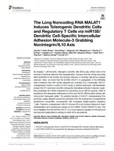
Figure 1. RHPN1-AS1, aa cytoplasmic expressed in in UM cellcell lines. (A)(A) RHPN1-AS1 Figure cytoplasmiclncRNA, lncRNA,isisaberrantly aberrantly expressed UM lines. RHPN1expression was was measured by real-time PCRPCR in different UMUM cellscells andand normal cell cell (RPE). RHPN1-AS1 AS1 expression measured by real-time in different normal (RPE). RHPN1presented higherhigher expression in melanoma cell linescell OCM1 OM431 than RPE cells; RHPN1-AS1 AS1 presented expression in melanoma linesand OCM1 and OM431 than(B) RPE cells; (B) is cytoplasmically distributed. RNA fluorescence in situ hybridization (FISH) (red) was performed RHPN1-AS1 is cytoplasmically distributed. RNA fluorescence in situ hybridization (FISH) (red) with was cy3-labeledwith probes that recognizing RHPN1-AS1. The scale bars represent µm; (C) RHPN1-AS1 is performed cy3-labeled probes that recognizing RHPN1-AS1. The scale 20 bars represent 20 µm; (C) associated with cytoplasmic Total RNAsfractions. from OCM1 cellsRNAs were separated into cytoplasmic RHPN1-AS1 is the associated withfractions. the cytoplasmic Total from OCM1 cells were and nuclear fractions. U1, GAPDH were used as controls; (D,E)used The as interference rate The was separated intosoluble cytoplasmic and nuclear soluble fractions.U1, GAPDH were controls; (D,E) detected 48 h after RHPN1-AS1 siRNAs transfection in OCM1 and OM431 cells. Triplicate assays interference rate was detected 48 h after RHPN1-AS1siRNAs transfection in OCM1 and OM431 cells. were performed eachperformed sample andfor the each relative level of RHPN1-AS1 waslevel normalized to the GAPDH. Triplicate assaysfor were sample and the relative of RHPN1-AS1 was (* p < 0.05). normalized to the GAPDH. (* p < 0.05).
The pGIPZ-shRNAvectors vectors empty pGIPZ an enhanced green The pGIPZ-shRNA andand empty pGIPZ controlcontrol vectorsvectors with an with enhanced green fluorescent fluorescent protein (EGFP) maker was packaged into lenti-viruses andhuman transfected human OCM1 cells, and protein (EGFP) maker was packaged into lenti-viruses and transfected OCM1 and OM431 OM431 cells, namedRHPN1-AS1-sh1, RHPN1-AS1-sh2 and mock, respectively. Transfection named RHPN1-AS1-sh1, RHPN1-AS1-sh2 and mock, respectively. Transfection efficiency was efficiency was by GFP seen in the mock, RHPN1-AS1determined bydetermined GFP expression. GFPexpression. expressionGFP was expression seen in the was mock, RHPN1-AS1-sh1 and -sh2 cells sh1 and 2A). -sh2cells 2A). The knockdown efficiencyby was measured by qRT-PCR. RHPN1-AS1 (Figure The (Figure knockdown efficiency was measured qRT-PCR. RHPN1-AS1 expression was expression was significantly decreased in RHPN1-AS1-sh1 and RHPN1-AS1-sh2 transfected cells significantly decreased in RHPN1-AS1-sh1 and RHPN1-AS1-sh2 transfected cells (Figure 2B). (Figure 2B). 2.3. Down-Regulation of RHPN1-AS1 Inhibited Cell Proliferation, Migrationand Invasion In Vitro 2.3. Down-Regulation of RHPN1-AS1 Inhibited Cell Proliferation, Migrationand In Vitro Next, we investigated whether the characteristics of the tumor cells wereInvasion altered after RHPN1-AS1 knockdown. first examined thethecolony formation of shRHPN1-AS1-OCM1 and Next, we We investigated whether characteristics of ability the tumor cells were altered aftercells RHPN1shRHPN1-AS1-OM431 cells compared the control and mock using the colony formation AS1 knockdown. We first examined thetocolony formation abilitygroups of shRHPN1-AS1-OCM1 cells and assay. The number of colonies was significantly decreased aftergroups RHPN1-AS1 knockdown (p < 0.05, shRHPN1-AS1-OM431 cells compared to the control and mock using the colony formation FigureThe 3A).number We alsoofexamined of RHPN1-AS1 knockdown on the migration and(p invasion assay. colonies the waseffect significantly decreased after RHPN1-AS1 knockdown < 0.05, ability 3A). of UM resultsthe demonstrated that the RHPN1-AS1 knockdown inhibited UM cell Figure Wecells. also The examined effect of RHPN1-AS1 knockdown on the migration and invasion migration by ~55% OCM1 cellsdemonstrated and by ~50% in OM431 cells, respectively (p < 0.05, Figure 3B) ability of UM cells.inThe results that the RHPN1-AS1 knockdown inhibited UMusing cell trans-well by assay. The matrigel invasion showed thatrespectively RHPN1-AS1(pknockdown in3B) UMusing cells migration ~55% in OCM1 cells and byassay ~50%also in OM431 cells, < 0.05, Figure caused a significant cell invasion < 0.05, Figurethat 3C).RHPN1-AS1 These data indicate that RHPN1-AS1 trans-well assay. Thedecrease matrigelininvasion assay(palso showed knockdown in UM cells plays a aregulatory in tumor progression. caused significantrole decrease in cell invasion (p < 0.05, Figure 3C). These data indicate that RHPN1AS1 plays a regulatory role in tumor progression.
Int. J.J. Mol. Mol. Sci. Sci.2017, 2017,18, 18,226 226 Int.
of 13 13 44 of
Figure 2. 2. RHPN1-AS1 RHPN1-AS1 knockdown knockdown by by two two shRNAs: shRNAs: (A) (A) EGFP EGFP was was used used to to track track the the expression expression of of Figure RHPN1-AS1 shRNAs and control vectors in OCM1 and OM431 cells. The scale bars represent 100 µm; RHPN1-AS1shRNAs and control vectors in OCM1 and OM431 cells. The scale bars represent 100 µm; and (B) (B) detection detection of of RHPN1-AS1 RHPN1-AS1 mRNA mRNA level level in in OCM1 OCM1 and and OM431 OM431 cells cells after and after shRNA-mediated shRNA-mediated knockdown of RHPN1-AS1 by qRT-PCR. knockdown of RHPN1-AS1 by qRT-PCR.
2.4. Down-Regulation Down-Regulation of of RHPN1-AS1 RHPN1-AS1 Decreased Decreased Xenograft Xenograft Growth Growth In In Vivo Vivo 2.4. To examine examine the the biological biological significance significance of of RHPN1-AS1 RHPN1-AS1 on on tumor tumor growth growth in in vivo, vivo, we we established established To 66 of the control OCM1 or RHPN1-AS1-sh1OCM1 cells were a xenograft model in nude mice; 1 × 10 a xenograft model in nude mice; 1 × 10 of the control OCM1 orRHPN1-AS1-sh1OCM1 cells were subcutaneously injected injected into into the the right right flanks flanks of of mice. mice. The The tumor tumor volume volume was was measured measured once once every every subcutaneously 3–4 days. Fourteen days after injection, the tumor growth of RHPN1-AS1-sh1OCM1 cells was significant 3–4 days. Fourteen days after injection, the tumor growth of RHPN1-AS1-sh1OCM1 cells was slower thanslower that of than control cells < 0.05,cells Figure After 28 days, mice28 were sacrificed, and the significant that of (p control (p 4A). < 0.05, Figure 4A).the After days, the mice were tumors were removed and analyzed (Figureand 4B).analyzed The average tumor weight of RHPN1-AS1-sh1OCM1 sacrificed, and the tumors were removed (Figure 4B). The average tumor weight of cells was significantly lower that of the control < 0.05, Figure 4C). These that RHPN1-AS1-sh1OCM1 cells than was significantly lowercells than(pthat of the control cells (p