Use of a Capture-Based Pathogen Transcript Enrichment Strategy for RNA-Seq Analysis of the Francisella Tularensis LVS Transcriptome during Infection of Murine Macrophages Zachary W. Bent*, David M. Brazel, Mary B. Tran-Gyamfi, Rachelle Y. Hamblin, Victoria A. VanderNoot, Steven S. Branda Sandia National Laboratories, Livermore, California, United States of America
Abstract Francisella tularensis is a zoonotic intracellular pathogen that is capable of causing potentially fatal human infections. Like all successful bacterial pathogens, F. tularensis rapidly responds to changes in its environment during infection of host cells, and upon encountering different microenvironments within those cells. This ability to appropriately respond to the challenges of infection requires rapid and global shifts in gene expression patterns. In this study, we use a novel pathogen transcript enrichment strategy and whole transcriptome sequencing (RNA-Seq) to perform a detailed characterization of the rapid and global shifts in F. tularensis LVS gene expression during infection of murine macrophages. We performed differential gene expression analysis on all bacterial genes at two key stages of infection: phagosomal escape, and cytosolic replication. By comparing the F. tularensis transcriptome at these two stages of infection to that of the bacteria grown in culture, we were able to identify sets of genes that are differentially expressed over the course of infection. This analysis revealed the temporally dynamic expression of a number of known and putative transcriptional regulators and virulence factors, providing insight into their role during infection. In addition, we identified several F. tularensis genes that are significantly up-regulated during infection but had not been previously identified as virulence factors. These unknown genes may make attractive therapeutic or vaccine targets. Citation: Bent ZW, Brazel DM, Tran-Gyamfi MB, Hamblin RY, VanderNoot VA, et al. (2013) Use of a Capture-Based Pathogen Transcript Enrichment Strategy for RNA-Seq Analysis of the Francisella Tularensis LVS Transcriptome during Infection of Murine Macrophages. PLoS ONE 8(10): e77834. doi: 10.1371/journal.pone.0077834 Editor: Yung-Fu Chang, Cornell University, United States of America Received July 3, 2013; Accepted September 9, 2013; Published October 14, 2013 This is an open-access article, free of all copyright, and may be freely reproduced, distributed, transmitted, modified, built upon, or otherwise used by anyone for any lawful purpose. The work is made available under the Creative Commons CC0 public domain dedication. Funding: Sandia National Laboratories is a multi-program laboratory managed and operated by Sandia Corporation, a wholly owned subsidiary of Lockheed Martin Corporation, for the U.S. Department of Energy's National Nuclear Security Administration under contract DE-AC04-94AL85000. The funders had no role in study design, data collection and analysis, decision to publish, or preparation of the manuscript. Competing interests: The authors have declared that no competing interests exist. * E-mail:
[email protected]
Introduction
infectious dose, F. tularensis has long been feared for its potential as a biological weapon and has been designated a Category A Select Agent [5,6]. The species F. tularensis is comprised of two sub-species types, with type A strains endemic to North America and type B strains endemic to Europe and Asia [2]. A live vaccine strain (LVS) derived from a type B strain was created in the former Soviet Union over 50 years ago; due to safety concerns, however, it is not currently licensed for human use [7]. Although the F. tularensis LVS strain does not cause illness in humans, it is lethal to mice, causing a disease that very closely mimics human tularemia [8]. These features have made the F. tularensis LVS murine infection model an ideal and well-established system for study of F. tularensis pathogenesis [9].
Francisella tularensis, the causative agent of tularemia, is a Gram-negative facultative intracellular pathogen that is capable of infecting a wide variety of hosts, including mammals, birds, amphibians, fish, and insects [1]. Originally isolated in 1911 in Tulare County, California during a plague-like outbreak in the rodent population, F. tularensis was subsequently found to be endemic in most of the northern hemisphere [2]. Human infections most commonly occur upon contact with infected animals or from the bite of an infected tick, leading to cutaneous ulceroglandular tularemia [3]. A pneumonic infection can result from inhalation of as few as 10 bacteria, leading to severe and often fatal disease [4]. Because of the seriousness of its disease, ability to be aerosolized, and extremely low
PLOS ONE | www.plosone.org
1
October 2013 | Volume 8 | Issue 10 | e77834
F. tularensis LVS Transcriptome during Infection
Results
As an intracellular pathogen, F. tularensis must adapt to multiple environments throughout the course of an infection. The bacteria enter host cells via phagocytosis, escape the phagosome, replicate within the host cell cytosol, and at later stages of infection are found within a double membranous compartment that resembles an autophagosome [10]. The bacteria infect a variety of cell types in a variety of locations throughout the body, each presenting different stresses and challenges to bacterial survival [7,11]. The bacteria must appropriately respond to each of these microenvironments for the infection to proceed. To accomplish this, F. tularensis must rapidly alter transcription of numerous genes in a coordinated manner as it moves from site to site within the host, as well as within the compartments of individual host cells. Typically, pathogens respond to infection-associated stresses through up-regulation of virulence factors - genes that have been demonstrated by mutational or genetic analysis to play a critical role in the bacteria’s ability to cause disease. These genes encode a wide range of products, including secretion systems, adhesins, invasins, iron uptake pathways, and toxins. During infection pathogens also down-regulate expression of transcripts that are no longer necessary, or are potentially detrimental, in a given microenvironment. These changes in gene expression are ultimately responsible for the success of the pathogen in evading or subverting the immune response and surviving within its host. Previous work to characterize F. tularensis transcriptome dynamics during infection has focused on the type A strain SCHU S4. Wehrly and colleagues used microarray analysis to track the transcriptional profiles of the bacteria during infection of murine bone marrow-derived macrophages (BMDM) [12]. Walters and colleagues used RNA-Seq to investigate the transcriptome of the bacteria at late time points in the lungs of infected mice [13]. Both studies revealed up-regulation of known and previously unknown virulence factors, demonstrating that distinct stages of F. tularensis infection are accompanied by global changes in transcriptional profile. Given the importance of these global transcriptional shifts to the virulence and persistence of the bacteria within the host, it is critical to understand when and how these shifts occur. Using RNA-Seq to address this issue presents a technical challenge, because the vast majority of transcripts present in infected cells are derived from the host, rather than from the bacteria of interest. In this study, we use a newly developed capture-based bacterial transcript enrichment strategy [14] to obtain enough pathogen reads , despite the host-dominated background, to analyze the complete transcriptome of F. tularensis LVS during phagosomal escape and cytosolic replication within murine macrophages. Comparison of the transcriptional profiles of the bacteria at these two distinct time points, relative to that of the bacteria in culture, revealed upregulation of numerous known virulence factors as well as many genes with unknown function that play, as yet, undetermined roles in F. tularensis virulence. Further, by analyzing the expression of the known and putative transcriptional regulators encoded by the F. tularensis genome, we were able to identify pathways and products that are important at each stage of infection.
PLOS ONE | www.plosone.org
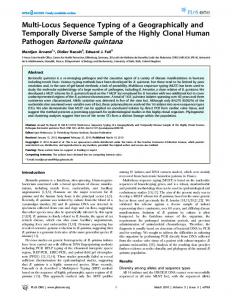
F. tularensis transcriptional changes during infection F. tularensis bacteria grown to exponential phase in a rich medium, such as the one used in this study, are highly invested in replication, and therefore would be expected to express genes associated with metabolic functions and cell division. Switching from culture in a rich medium to an active infection of host cells presents a specific series of challenges to the bacteria that are addressed through global transcriptional changes. The first phase of F. tularensis infection requires that the bacteria be taken up into the host cell by phagocytosis. This is followed by escape from the phagosome, and establishment of a replicating population within the cytosol of the host cell. The exact timing of these events varies in different F. tularensis strains, host cell types, and infection protocols. However, it is well documented that in the murine macrophage cell line J774A.1, as well as in murine bone marrow derived macrophages (BMDM), after 4 hours of infection the bacteria are in a transition state in which some are still within phagosomes while others have managed to escape into the cytosol [10,15-18]. After 8 hours the vast majority of bacteria are located within the cytosol [10,12,15,18-20]. Work by Mack and colleagues [21] as well as Edwards and colleagues [22] has demonstrated, through direct comparisons, that F. tularensis infections show an essentially identical disease progression in J774A.1 cells as P388D1 cells, the murine macrophage line used in this study. To characterize the F. tularensis transcriptome dynamics associated with transition from growth in culture to infection of host cells, as well as the transition from phagosome to the cytosol, we performed a differential gene expression analysis of F. tularensis LVS before infection and after 4 or 8 hours of infection. Table S1 presents the biological duplicate FPKM (fragments per kilobase of transcript per million mapped reads) values and differential expression results for all F. tularensis genes, comparing the transcriptome of the culture grown inoculum to the transcriptome after 4 and 8 hours of infection. As expected, after 4 hours of infection we observed downregulation of genes that are involved in protein synthesis, protein fate, and central intermediary metabolism (Figure 1); these results are consistent with slowing of replication during the transition from culture to infection. Genes that encode mobile elements, such as transposases, also showed downregulated expression after 4 hours of infection. In contrast, genes encoding virulence determinants (e.g., transport proteins) and components of virulence-related biochemical pathways (e.g., biosynthesis of amino acids and cofactors) were up-regulated after 4 hours of infection. These upregulated genes include many located within the Francisella pathogenicity island (FPI), which is known to play a critical role in F. tularensis virulence [23]. These trends in gene expression were still apparent after 8 hours of infection, with the exception of mobile element expression, which was up-regulated at the later time point. In both cases, and particularly at the later time point, many genes of unknown function showed differential expression. There are 21 differentially expressed genes that produce products of unknown function after 4 hours of infection
2
October 2013 | Volume 8 | Issue 10 | e77834
F. tularensis LVS Transcriptome during Infection
Figure 1. Number of genes up- and down-regulated, by functional category. All differentially expressed F. tularensis LVS genes were categorized by function, and the number of genes in each category were plotted according to whether their expression increased or decreased at 4 hours (left) and 8 hours (right) after infection. doi: 10.1371/journal.pone.0077834.g001
whose regulation is sensitive to the environmental changes associated with transition from culture to infection. Of the genes consistently up-regulated during infection (Table S4), ~20% are located within the FPI. Additionally, ~18% of these genes were categorized as transport and binding proteins, a group of proteins that include several genes implicated in virulence such as siderophore synthesis, and transmembrane peptide transport. Of the genes consistently down-regulated during infection (Table S5), ~49% are involved in protein synthesis and fate, and several others in biosynthesis of enzyme cofactors such as riboflavin, cyanophycin, and anthranilate.
and 60 genes after 8 hours (Tables S2 and S3, respectively). The highly up-regulated unknown genes at each time-point are especially interesting because they are likely to be important in specific stages of pathogenesis and yet have not been previously identified as virulence factors. Table 1 lists the F. tularensis genes showing the largest changes in expression during transition from culture to infection. Strikingly, 30-40% of the most strongly up-regulated genes are of unknown function. Among the genes of known function, those that were most strongly up-regulated are involved in purine and amino acid biosynthesis, peptide transport, and competence. The genes most strongly downregulated are involved in protein synthesis and central metabolism - functions predicted to play diminished roles during infection, as compared to exponential growth in culture. Interestingly, although the genes differentially expressed after 4 hours versus 8 hours of infection are closely related with respect to their annotated functions, only 39 (20.1%) of the upregulated genes, and 46 (21.8%) of the down-regulated genes, were differentially expressed at both time points (Figure 2). The consistency with which these genes were differentially expressed suggests that they represent a core set of genes
PLOS ONE | www.plosone.org
Genomic localization of genes differentially expressed during infection To determine whether the genes differentially expressed during infection are located in particular regions of the F. tularensis genome, we identified the genes showing the largest changes in expression (> 4 fold) after 4 hours and 8 hours of infection, and mapped their locations within the genome. This analysis revealed that most of the differentially expressed genes are broadly distributed throughout the F. tularensis
3
October 2013 | Volume 8 | Issue 10 | e77834
F. tularensis LVS Transcriptome during Infection
Table 1. Genes showing the largest changes in expression during infection.
Gene ID
Name/Function
4hr Fold Change
Adj P-Value
Gene ID
Name/Function
8hr Fold Change
Adj P-Value
FTL_0721
DedA family protein*
9.69
0.016
FTL_0815
PRC-barrel protein*
28.51