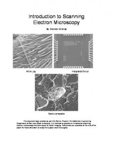
Abstract: Frozen hydrated specimens of Pratylenchus agilis and dauer larvae of Steinernema carpo- capsae were observed with low-temperature field emission ...
Journal of Nematology 25(2):214-226. 1993.
Use of Low Temperature Scanning Electron Microscopy to Observe Frozen Hydrated Specimens of Nematodes WILLIAM P. WERGIN, 1 RICHARD M . SAYRE, 2 AND ERIC F. ERBE 1
Abstract: Frozen hydrated specimens of Pratylenchus agilis and dauer larvae of Steinernema carpocapsae were observed with low-temperature field emission scanning electron microscopy. This new technique provides information about the surface features of nematodes and also allows specimens to be fractured to reveal their internal structure. Furthermore, both halves of fractured specimens can be retained, examined, and photographed either as two-dimensional micrographs or as threedimensional images for stereo observation (stereology) or quantitative measurements (stereometry). This technique avoids artifacts normally associated with procedures required to prepare nematodes for examination in the transmission and scanning electron microscopes, such as chemical fixation, dehydration, and sectioning or critical point drying. Key words: Complementary imaging, low-temperature SEM, nematode, Pratylenchus agilis, SEM, Steinernema carpocapsae, stereology, technique, three-dimensional imaging.
During the last 25 years, electron microcopy (EM) greatly expanded our knowledge and understanding of plant-parasitic nematodes. T h e transmission electron microscope (TEM) revealed considerable structural information about most internal organs and tissues (4,34), thus enabling nematologists to relate ultrastructural details to physiological functions. Alternatively, the scanning electron microscope (SEM) revealed detailed structural features on the nematode surface (7,20). This EM knowledge, which complemented and extended that from light microscopic investigations, has enabled taxonomists to describe species more thoroughly and in some cases to differentiate races (20). Both the T E M and the SEM have provided structural information that has helped to visualize and postulate the nature of hostparasite interactions involving nematodes and many agriculturally important crop plants (11,20). Although EM studies are invaluable, the nematode and plant materials examined with these techniques are subjected to chemical fixation, dehydration, embedReceived for publication 29 October 1992. i Electron Microscopy Laboratory, USDA ARS, Building 177B, BARC-East, Beltsville, MD 20705. 2 Nematology Laboratory, USDA ARS, Building 011A, BARC-West, Beltsville, MD 20705. We thank Christopher Pooley for photographic assistance in preparing the final plates, Oxford Instruments for the use of the CT 1500 Cryotrans System, and Biosys for the cultures of Steinernemacarpocapsae.
214
ding or critical point drying, and staining or coating. Unfortunately, all o f these p r e p a r a t i o n p r o c e d u r e s can p r o d u c e structural artifacts (21,24,32). For example, chemical fixatives can extrude materials onto the nematode surface, solvent dehydration may extract cellular constituents, critical point drying can shrink and distort tissues, and sputter coating with 20-30 nm of gold-palladium may obliterate fine-structural details. Although examination of unfixed hydrated tissues would avoid these problems, the high vacuum required for conventional TEM and SEM imaging quickly collapses biological samples, which contain as much as 90% water. To avoid tissue collapse, Echlin et al. (6) developed a procedure to examine frozen, fully hydrated specimens in a low-temperature SEM. This procedure has two obvious advantages. First, although freezing rates vary depending on the size and nature of a bio~6gical specimen, the time necessary to f r e e z e - f i x t h e s p e c i m e n by quenching in liquid nitrogen is shorter by several orders of magnitude than that required for chemical fixation (2). Second, the frozen specimen will not collapse because it is observed on a cryostage at temperatures below - 130 C, where the vapor pressure of water is insignificant. Therefore, sublimation does not occur at a detectable rate. F u r t h e r m o r e , pure water does not recrystallize at temperatures below - 130 C (3). Consequently, as long as
Low Temperature SEM: Wergin et al. 215 the stage remains cold, stable frozen samples can be observed for several hours (28). With these advantages in mind, a study was undertaken to devise procedures that utilized a standard SEM cryostage and a field emission SEM to observe frozen hydrated tissues from intact and fractured nematodes. The technique allowed observation of the external surface of frozen hydrated nematodes as well as the internal structure of frozen, fractured nematode specimens. F u r t h e r m o r e , b o t h halves (complementary images) of the fractured nematodes were retained, observed, and p h o t o g r a p h e d to make two-dimensional and three-dimensional (stereo) comparisons of the opposing faces. MATERIALS AND METHODS
An Oxford CT 1500 Cryotrans System was m o u n t e d on a Hitachi S-4000 field emission SEM to p e r f o r m low-tempera t u r e manipulations and observations. Samples consisted o f a suspension o f a mixed population o f Pratylenchus agilis T h o r n e & Malek recovered from root explant cultures of Zea mays (9) or dauer larvae of Steinernema carpocapsae Weiser strain All, which were obtained as the commercial product "Bit-Safe" from Biosys (Palo Alto, CA). The samples were not repeatedly washed to remove salts or particulate debris from the suspension. T h e specimens were placed on the surface of a flat specimen holder or in a 1-mm 3 vertical chamber f o r m e d by slots traversing the two halves of a closed, hinged 24k gold holder (Fig. 1). The specimen holders containing t h e s u s p e n s i o n s w e r e r a p i d l y p l u n g e - f r o z e n in liquid n i t r o g e n . Although use of the small gold holder reduced freezing times, no attempts were made to address the relationship of freezing rates to cellular damage caused by ice crystal formation. As many as six hinged gold specimen holders containing samples were mounted onto a Denton complementary freeze-etch specimen cap (Fig. 2) that was submerged in liquid nitrogen. The cap, which had a
threaded hole in the base, was affixed to the matching threads of a vertical screw that had been soldered to a modified Oxf o r d stub. T h e stub was oriented and mounted on the Oxford carrier with a set screw. The entire assembly was then inserted into the O x f o r d nitrogen slush chamber, evacuated, and withdrawn into a cryo-transfer arm for transfer to the prechamber. A pre-cooled pick in the prechamber was then used to fracture the samples by lifting and rotating 180 degrees the fracture arm of the complementary cap. This action, similar to opening a book, separated the two halves of each of the six hinged holders by flipping the u p p e r halves of the holders 180 degrees to expose the complementary areas o f each f r e e z e - f r a c t u r e d sample (Fig. 3). T h e clamping action of the cap retained the lower half of the holder while the hinge provided thermal continuity to the u p p e r half. The standard flat holder containing the specimens could be mounted directly on the cryo-transfer arm for transfer to the prechamber. The specimens were then either sputter coated with platinum in the prechamber and inserted onto the cryostage of the microscope or etched and coated in the prechamber and moved to the cryostage for observation. Locating and matching the two complementary areas of the fractured sample were facilitated by using the raster rotation feature of the microscope to rotate the beam until the longitudinal axis o f the open gold holder was parallel or perpendicular to the horizontal axis of the viewing cathode ray tube. This orientation expedited the measurement or estimation of the distance between the hinge pin of the holder (or some conspicuous feature of the sample) and the desired area on the fractured specimen. After an area was viewed and photographed, the holder was systematically moved to the opposite half, a similar distance from the pin (or sample feature), to view the complementary surface. Accelerating voltages o f 10 kV were used to observe and record images onto Polaroid Type 55 P/N film. T o obtain ste-
216
Journal of Nematology, Volume25, No. 2,June 1993
1
2
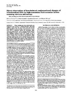
FIGS. 1--3. Specimen holders used to fracture samples and retain complementary faces. 1) Drawing (8× actual size) of a hinged gold holder. About 1 p,1 of a nematode suspension is placed in the slot (arrow) of this holder. 2) Denton complementary freeze-etch cap mounted on an Oxford specimen carrier (1.5 × actual size). The cap accommodates up to six hinged gold holders that retain both halves of a fractured sample. The frozen samples are fractured when a cryo-pick in the Oxford prechamber is used to flip and rotate the fracture arm of the cap (arrows). 3) Low-temperature field emission SEM of an opened, hinged specimen holder containing the complementary halves of a freeze-fractured sample. Arrows indicate complementary fractured protrusion of the frozen sample. Bar = 1 mm.
r e o pairs, t h e first i m a g e was r e c o r d e d , the stage was tilted 5 to 10 d e g r e e s , t h e specim e n was r e c e n t e r e d , a n d a s e c o n d i m a g e was r e c o r d e d . T o g e t h e r t h e s e two i m a g e s c o n t a i n e d t h e p a r a l l a x i n f o r m a t i o n necessary f o r s t e r e o viewing. T o a c h i e v e s t e r e o p s i s (a t h r e e - d i m e n sional view) o f t h e s t e r e o pairs, t h e f i g u r e
m u s t be viewed so t h a t t h e left m e m b e r o f t h e pair is seen o n l y with t h e left eye a n d r i g h t m e m b e r with t h e o t h e r eye. T h i s c a n be a c c o m p l i s h e d e i t h e r b y e m p l o y i n g a simple s t e r e o viewer m a d e f o r this p u r p o s e o r by u s i n g a file c a r d o r s h e e t o f p a p e r to e n t r a i n t h e line o f vision o n t h e left a n d r i g h t m e m b e r s o f t h e pair. T h e r e a d e r
Low Temperature SEM: Wergin et al. 217 should place the card vertically, midway between the members of the pair, with the reader's head positioned about 30 cm (12 inches) from the page. The upper edge o f the card should be positioned midway between the eyes, parallel to the nose, and extend from the face toward the midpoint of the figure. In this position, the card acts as a barrier that prevents the left eye from seeing the right image and vice versa. When the eyes are relaxed, the two images will move horizontally until they overlap. When this occurs, three images are perceived. T h e Center one will be the superimposed 3-D image of the micrographs; the two lateral images will appear flat and should be ignored. After practice and experience with this procedure, stereo pairs can be easily viewed without the assistance of a viewer or a card. RESULTS
When the temperature of the flat specimen holder containing a suspension of P. agilis was raised to - 90 to - 95 C to etch or sublime the surface water-ice that covered the specimens, the surface of the cuticle was exposed (Figs. 4,5). However, because of the three-dimensional nature of the nematodes and their random orientation in the suspension, portions generally remained embedded in the water-ice matrix. The shape of the nematodes did not appear distorted; they were generally sinuous, similar to living specimens examined with the light microscope. An amorphous material, probably representing the residual compounds from the growth medium, usually occurred on the surface o f the etched juveniles. T h e cuticular annulations a p p e a r e d s m o o t h a n d r e g u l a r l y spaced, and the lateral fields did not reveal any "dimpling" or irregularities frequently found on specimens that are air dried, critical point dried, or dried by other methods. Occasionally, small cracks, referred to as prefractures, occurred in frozen specimens of P. agilis (Fig. 5). These prefractures probably formed during the initial
plunge-freezing procedure and resulted from the differential contraction and expansion that occurred while the specimen holders cooled to - 185 C and the suspension inwardly froze. Some of the prefractures occurred in the water-ice without affecting the frozen nematodes, whereas other prefractures cleaved the embedded nematodes. Because the nematodes were randomly d i s p e r s e d in the s u s p e n s i o n , f r e e z e fracturing samples of both species in the hinged holders exposed specimens that were fractured in all planes (e.g., S. carpocapsae, Fig. 6). Longitudinal, oblique, and cross sections were easily observed in any given viewing field. T h e fractured portions of nematodes that were exposed and retained in the frozen matrix assisted in identifying specific tissues. For example, in Figure 7 the well-developed lateral field provided some information about the relative position of the fracture. This fracture plane also revealed readily identifiable structures such as the cuticular layers, somatic musculature, epidermal cords, and other inner tissues. The hinged gold holders permitted the observation and photography of the complementary faces of the fractured specimens. In cross (Fig. 8) and longitudinal (Fig. 9) sections of S. carpocapsae, the fractures did not follow a single flat plane across the nematode body. Instead, different tissues generally fractured in slightly different planes. This fortuitous characteristic frequently assisted in distinguishing one tissue from another; for example, in Figure 8, the cuticular layer, underlying epidermis, and somatic muscles fractured in slightly different planes. Likewise, tissue that may represent a epidermal cord (arrow) fractured at a distinct level. Comparison of the complementary faces of the fractured nematode also helped to determine the shapes of objects and to distinguish true structural features from any contaminants on the frozen surface. Concave channels or granules in one face of the fracture must correspond to convex channels or granules in the complemen-
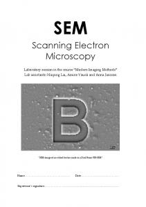
FIGS. 4,5. Surface of pratylenchu~ agilis examined with low-temperature scanning electron microscopy. 4) Anterior end. Although the nematode was embedded in water-ice, raising the temperature of the stage from - 185 to - 9 5 C etched the surface water-ice and revealed a ca. 180-p.m length of the anterior portion. T h e fine a m o r p h o u s material on the surface probably represents residual medium components. Bar = 20 p~m. 5) Anterior end and a medial segment of another specimen, also etched at - 95 C. While a specimen is plungefrozen in liquid nitrogen, differential contraction and expansion between the freezing suspension and the specimen holder produce internal prefractures, which either break (single arrows) or do not break double arrows) the e m b e d d e d nematodes. Bar = 20 v,m.
Low Temperature SEM: Wergin et al. 219
FIGS. 6,7. Frozen suspension of d a u e r larvae of Steinernema carpocapsae fractured in the cryosystem prechamber a n d lightly etched to sublime some of the surface water-ice. 6) Random longitudinal, oblique, and cross sections resulting from lack of orientation of nematodes in the suspension before freezing. Bar = 10 ~m. 7) Oblique fracture, revealing the outer cuticle and the well developed lateral field as well as the i n n e r layers of the fractured cuticle (arrow), somatic muscle (M), epidermal cord (C), and other unidentified tissues and structures. Bar = 2 txm.
220
Journal of Nematology, Volume 25, No. 2,June 1993
Fro. 8. Complementary cross fractures (a,b) through a frozen dauer larva of Steinernemacarpocapsae.T h e fracture does not follow a single flat horizontal plane; instead, many of the tissues and cell types fracture at unique, distinct planes. This fortuitous feature helps to distinguish adjacent tissues such as the cuticle, somatic muscle, and epidermal cord (arrows). Bar = 10 ~m.
Low T e m p e r a t u r e S E M : Wergin et al.
221
FIG. 9. Composite micrographs of a complementary longitudinal fracture through a frozen dauer larva of
Steinernema carpocapsae. Because the fracture tends to split membranes, the fracture plane generally follows limiting membranes over or beneath ceils, ducts, granules, and other structures. The depressions present in one image (a, arrows) correspond to bumps in the complementary image (b, arrows). Bar = 10 ~m.
222 Journal of Nematology, Volume 25, No. 2, June 1993 men orientation in TEM examinations (23), could be done before freezing to orient the specimens in a two-dimensional plane. In this study, no attempts were made to determine the ultimate resolution that could be obtained with nematodes. Relatively low magnification images (less than 4,000 x) were recorded to demonstrate the capability and flexibility of the cryotechnique. These magnifications were easily obtained with the standard cryosystem and DISCUSSION operating procedures. Highly magnified General advantages and disadvantages: The images that resolved membrane particles general advantages of low-temperature measuring 9-10 nm on fractured yeast SEM as opposed to conventional EM have cells have been obtained by substituting been discussed (3,13,14,24,25,28). Briefly, high vacuum evaporation for sputter coat(i) cryofixation is faster than chemical fix- ing (25,26). The same coating procedure, ation; (ii) samples remain largely in their possible for nematodes, may allow obserhydrated state; (iii) relatively large areas, vation of samples at 100,000x or more. Nematodes apparently are not prone to including complementary fractured faces, can be examined; (iv) the technique is non- the third disadvantage, i.e., disruption of destructive, allowing for further analysis tissues because of ice crystal formation. by other methods; (v) the procedure is Cryoprotectants can maintain viability of rapid; and (vi) frozen biological samples frozen hydrated nematode parasites of are frequently more conductive and there- plants (1,10,15-18), mammals (19), and insects (5,12). T h e free-living nematodes fore require less coating. Despite these advantages, several disad- Caenorhabditis briggsae (8) and C. elegans vantages have been r e p o r t e d for low- (33) can also be successfully frozen and temperature SEM (27). For example, (i) stored in liquid nitrogen. Therefore, unorientation and manipulation of speci- der the proper freezing conditions some mens during low temperature fixation is nematodes may exhibit little or no internal more difficult to control; (ii) until recently, damage from ice crystal formation, unlike resolution has been limited; (iii) when not many other biological organisms. Prefracproperly controlled, the formation of ice tures may still occur in the cryoprotected crystals can result in considerable disrup- preparation and probably would contribtion of tissues; (iv) samples that are frozen ute to the reduced viability that has been and viewed are difficult to save or store; reported; however, this type of freezing and, (v) contaminants, such as water vapor, damage is easily identifiable and readily may condense on the surface of the sample distinguished from damage caused by inand interfere with specimen viewing and ternal ice crystal formation. O u r previous reports have also adstructural interpretations. Fortunately, our results indicate that dressed the last two disadvantages comthese disadvantages may not pose serious monly associated with low-temperature problems for nematologists. First, the use SEM, namely, specimen storage and conof the hinged gold holders facilitates load- tamination (27,29). Frozen hydrated leaf ing of a nematode suspension. Although tissue was observed by low-temperature use of a suspension results in randomly SEM, removed from the cryostage, stored oriented specimens, embedding the un- for several weeks in liquid nitrogen, and fixed nematodes in a thin layer of water then reexamined in the SEM. No detriagar, as has been done to facilitate speci- mental changes or artifacts were observed.
tary face (Fig. 9). When structures on one face cannot be properly m a t c h e d with those on the opposing face, one must suspect the presence of an artifact. Holders used in this study permitted the specimen to be tilted so that each complementary image could be viewed in three dimensions by recording a second image containing the parallax information necessary for three-dimensional viewing (e.g., P. agilis, Fig. 10).
Low Temperature
S E M : Wergin et al.
223
FIG. 10. Complementary stereo images of an obliquely fractured frozen juvenile of Pratylenchus agilis. To attain the three-dimensional view (stereopsis), place a stereo viewer on the micrographs so that the !eft and right lenses are aligned above the left and right images, respectively. Alternatively, stereopsis can be achieved without a viewer by entraining the left and right eyes on the left and right figures, respectively. Stereopsis helps to quickly determine the true shapes and spatial relationships within an image and also is used to obtain quantitative information (stereometry) about the sizes and distances within a stereo image. Bar = 10 Ixm.
224 Journal of Nematology, Volume 25, No. 2, June 1993 Indeed, storage was associated with a minor advantage; in several cases surface debris on the specimen during the initial examination was not present during subsequent observation, apparently because of dislodgement by the gentle turbulence that had occurred in the storage container. Finally, most of the contamination on the surface of specimens consists of water-ice produced from the condensation of water vapor on the surface of the frozen specimen. This contaminant generally does not cause serious problems because it can be removed by raising the specimen temperature to - 9 5 C to sublime the ice. Advantages of complementary imaging: T h e obvious advantage of using complementary pairs is that each complementary half of the fractured specimen is available for comparison and analysis. T h e ability to view and evaluate structures in a complementary pair helps to elucidate the surface topography and the ultrastructural features of a sample. Additionally, observation of each half helps to identify surface contaminants, which are more common in low-temperature SEM than in ambientt e m p e r a t u r e SEM. These contaminants can arise d u r i n g fracturing, etching, or coating--procedures that involve moving the sample from a prechamber to and from the cold stage on the SEM and that also may require subjecting the sample to at least two changes in temperature. If the frozen sample is not properly prepared, handled, or shrouded during these procedures, t h e n water vapor, oil vapor, or other contaminants in the prechamber or the microscope can condense on the sample surface. Because many of these contaminants could easily be mistaken for structural features of the sample, viewing both complementary halves facilitates determination of whether features present on one area of a sample do not have corresponding features on the complementary area and thus are artifactual.
Advantages of stereology and stereometry: A single SEM micrograph is a two-dimensional representation o f a three-dimen-
sional specimen. Depriving the brain of the three-dimensional view can lead to erroneous conclusions about t h e relative sizes, positions, and convergence of structures within a specimen (22,30,31). Because of this factor, two micrographs that are recorded at slightly different angles and thereby contain the parallax information necessary for stereo viewing should be frequently evaluated when studying specimens. Stereometry is the mathematical process by which accurate measurements are taken from a three-dimensional image. In micrographs obtained with an SEM, stereometry is the only procedure by which an individual can m a k e t r u e m e a s u r e m e n t s o f height, determine the length of sinuous structures, construct contour maps of a surface, or mold three-dimensional models of specimens (22). T h e procedure that we have described for observing and recording low temperature stereo images of nematodes allows one to easily record the paired images that can be analyzed by stereology so that meaningful quantitative data can be obtained. In conclusion; we present results obtained with frozen hydrated samples of nematodes that were examined using lowtemperature SEM. At present, one can use this procedure to make simple observations and to identify the major tissue systems of nematodes. In addition, the technique can be used to observe complementary images of these tissues in two as well as three dimensions. Because this technique represents a new method for observing the internal features of fractured nematodes, more detailed investigations may require that low-temperature SEM studies be carried out in tandem with thin-section TEM observations. These studies will allow the cell types and organelles that are apparent but are difficult to identify in the frozen images to be compared and contrasted to similar fine-structural features that can be readily identified in the thin sections. We believe that this procedure will considerably enhance our knowledge and increase
Low T e m p e r a t u r e
SEM: Wergin et al.
225
larvae of Meloidogyne incognita and M. hapla. Nematologica 10:168-179. 17. Sayre, R. M., and S.-W. Hwang. 1975. Freezing and storing Ditylenchus dipsaci in liquid nitrogen. Journal of Nematology 7:199-202. LITERATURE CITED 18. TriantaphyUou, A. C., and E. McCabe. 1989. Efficient preservation of root-knot and cyst nema1. Asahina, E. 1959. Frost-resistance in a nematode todes in liquid nitrogen. Journal of Nematology 21: Aphelenchoides ritzema-bosi. Low Temperature Science, 423-426. 19. Van Wyk, J. A., H. M. Gerber, and W. P. Van Series B 17:51-62. 2. Bachmann, L., and Y. Talmon. 1984. Cryomi- Aardt. 1977. Cryopreservation of the infective larvae croscopy of liquid and semiliquid specimens: Direct of the common nematodes of ruminants. Ondersteimaging versus replication. Ultramicroscopy 14:211- poort Journal of Veterinary Research 44:173-194. 20. Wergin, W. P. 1982. Bibliography on the use of 218. 3. Beckett, A., and N. D. Read. 1986. Low- scanning electron microscopy (SEM) in studies of temperature scanning electron microscopy. Pp. 45- plant parasitic nematodes. Scanning Electron Micros80 in H. C. Aldrich and W. J. Todds, eds. Ultrastruc- copy 2:831-836. rural techniques for microorganisms. New York: Ple21. Wergin, W. P. 1981. Scanning electron micronum. scopic techniques and applications for use in nema4. Bird, A. F., andJ, Bird. 1991. The structure of tology. Pp. 175-204 in B. M. Zuckerman, and R. A. nematodes. San Diego: Academic Press. Rohde, eds. Plant parasitic nematodes, vol. 3. New 5. Curran, J., C. Gilbert, and K. Butler. 1992. Rou- York: Academic Press. tine cryopreservation of isolates of Steinernema and 22. Wergin, W. P. 1985. Three-dimensional imagHeterorabditis spp. Journal of Nematology 24:269- ery and quantitative analysis of SEM studies of nema270. todes. Agriculture, Ecosystems and Environment 12: 6. Echlin, P., R. Paden, B. Dronzek, and R. Wayte. 317-334. 1970. Scanning electron microscopy of labile biolog23. Wergin, W. P., and B. Y. Endo. 1976. Ultraical material maintained under controlled conditions. structure of a neurosensory organ in a root-knot Scanning Electron Microscopy 1970:49-56. nematode. Journal of Uhrastructure Research 56: 7. Gibbons, L.M. 1986. SEM guide to the mor- 258-276, phology of nematode parasites of vertebrates. Farn24. Wergin, W. P., and E. F. Erbe. 1989. Increasham Royal, UK: C.A.B. International. ing the versatility of an EMscope SP2000A Sputter 8. Haight, M., J. Frim, J. Pasternak, and H. Frey. Cryo System on a Hitachi S-570 scanning electron 1975. Freeze-thaw survival of the free-living nema- microscope. Scanning 11:293-303. tode, Caenorhabditis briggsae. Cryobiology 12:497-505. 25. Wergin, W. P., and E. F. Erbe. 1991. Increas9. Huettel, R. N., and R. V. Rebois. 1985. Cultur- ing resolution and versatility in low temperature coning plant parasitic nematodes using root explants. Pp. ventional and field emission scanning electron mi155-158 in B. M. Zuckerman, W. F. Mai, and M. B. croscopy. Scanning Microscopy 5:927-936. Harrison, eds. Plant nematology laboratory manual. 26. Wergin, W. P., and E. F. Erbe. 1991. Using Amherst: University of Massachusetts Agricultural high vacuum evaporation to obtain high resolution Experiment Station. 10. Hwang, S.-W. 1970. Freezing and storage of low temperature images of freeze-fractured memnematodes in liquid nitrogen. Nematologica 16:305- branes from yeast. Pp. 514-515 in G. W. Bailey, ed. Proceedings of the 49th annual meeting Electron Mi308. 11. Lamberti, F., and C. E. Taylor, eds. 1985. Cyst croscopy Society of America. San Francisco: San Francisco Press. nematodes. New York: Plenum Press. 27. Wergin, W. P., and E. F. Erbe. 1991. Introduc12. Popiel, I., and E.M. Vasquez. 1991. Cryopreservation of Steinernema carpocapsae and Hete- tion to the advantages and problems associated with rorhabditis bacteriophora. Journal of Nematology 23: low temperature scanning electron microscopy. Scanning 13:24-26. 432-437. 28. Wergin, W. P., and E. F. Erbe. 1992. Tech13. Read, N.D. 1991. Low-temperature scanning electron microscopy of fungi and fungus-plant inter- niques for obtaining and observing complementary actions. Pp. 17-29 in K. Mendgen and D. E. Lese- images with a low-temperature field emission SEM mann eds. Electron microscopy of plant pathogens. and subsequent comparison of the identical cells in freeze-etch replicas viewed with a TEM. Scanning 14: Berlin: Springer-Verlag. 14. Read, N. D., and C. E. Jeffree. 1991. Low- 17-30. 29. Wergin, W. P., and E.F. Erbe. 1992. Recent temperature scanning electron microscopy in biology. advancements in low temperature scanning electron Journal of Microscopy 161:59-72. 15. Riga, E., andJ. M. Webster. 1991. Cryopreser- microscopy. Supplement to Scanning 14:40-42. 30. Wergin, W. P., and E. F. Erbe. 1992. Advanvation of the pinewood nematode, Bursaphelenchus tages of complementary stereo images as viewed with spp. Journal of Nematology 23:438-440. 16. Sayre, R.M. 1964. Cold-hardiness of nema- the low temperature field emission scanning electron todes. I. Effects of rapid freezing on the eggs and microscope. Pp. 1314-1315 in G. W. Bailey, ed. Proour understanding of nematode morphology and fine structure.
226
Journal of Nematology, Volume 25, No. 2,June 1993
ceedings of the 50th annual meeting Electron Microscopy Society of America. San Francisco: San Francisco Press. 31. Wergin, W. P., and C. Pooley. 1988. Photographic and interpretive artifacts. Pp. 175-204 in R. F. E. Crang, and K. L. Klomparens, eds. Artifacts in biological electron microscopy. New York: Plenum. 32. Wergin, W. P., and A. R. Stone. 1981. Tech-
niques for preparation and examination of plant parasitic nematodes in the scanning electron microscope. Scanning Electron Microscopy 3:169-176. 33. Wood, W. B. 1988. The nematode Caenorhabditis elegans. Cold Spring Harbor, New York: Cold Spring Harbor Laboratory. 34. Zuckerman, B. M. 1980. Nematodes as biological models. New York: Academic Press.