ACTA BIOLOGICA CRACOVIENSIA Series Botanica 47/1: 173–178, 2005
USING ENZYME POLYMORPHISM TO IDENTIFY THE GAMETIC ORIGIN OF FLAX REGENERANTS ZUZANA BARTOŠOVÁ, BOHUŠ OBERT, TOMÁŠ TAKÁCˇ, ANDREJ KORMUTˇÁK, AND ANNA PRETˇOVÁ* Institute of Plant Genetics and Biotechnology, Slovak Academy of Sciences, Akademická 2, P.O. Box 39/A, 950 07 Nitra, Slovak Republic Received December 6, 2004: revision accepted April 29, 2005 Anther culture is currently the most successful method for production of doubled haploid lines in flax. Recently, ovary culture was also described as a good source of doubled haploids. In this contribution we investigated the incorporation of enzyme polymorphism of acid phosphatase and peroxidase as molecular markers for the gametic origin of flax plants derived from anther and ovary cultures.
Key words: Anther culture, ovary culture, isozymes, haploids, flax.
INTRODUCTION Flax is a very important economic fiber and oilseed plant. The use of anther and ovary cultures has advantages for flax breeding programs. Both are considered a tool for production of haploid plant material and homozygous lines supporting breeding, and more recently also for molecular marker studies. Anther culture is currently the most successful method for production of doubled haploid lines in flax (Fried et al., 1995; Bergmann and Fried, 1996; Chen et al., 1998; Pretova and Obert, 2000; Obert et al., 2004a,b), but its efficiency is still very low. Flax gynogenesis was first reported by Bartošová et al. (2003). The conditions under which the flax donor plants are cultivated seem to be one of the limiting factors for good androgenic response (Bartošová and Pret’ová, 2004). Both androgenesis and gynogenesis can be useful for breeding purposes when the gametic origin of the regenerated plants is confirmed. In both cases, regeneration can easily take place not only from the microspore or the egg cell in the ovule, but also from the surrounding diploid somatic tissue (anther wall and/or ovary tissue). For this reason, identification of the gametic origin of the regenerants is very important. For discrimination of doubled haploids originated from hybrid flax lines via anther cultures, morphological markers are the main ones used so far (Tejklova, personal communication). Chen et al. (2001) employed several *e-mail:
[email protected]
PL ISSN 0001-5296
markers (RAPD, ISSR) of dominant phenotype and codominant markers (PCR/RFLP) for identification of presumed flax doubled haploids of androgenic origin. We have chosen the polymorphism of peroxidase (EC 1.11.1.7) and acid phosphatase (EC 3.1.3.2) for our studies. Peroxidases in plants catalyze a large variety of reactions (Siegel, 1993). The involvement of peroxidases in stress-related physiological processes (Low and Merida, 1996) as well as in plant-pathogen interactions have been demonstrated (Montalbini et al., 1995; Wojtaszek, 1997). The isozyme patterns of peroxidases are intensively studied for cultivar identification in plant breeding, seed marketing and other fields of agriculture (Samec et al., 1998). Acid phosphatase is one of the critical lytic enzymes in plants, and has been reported as a marker for differentiation processes in plant development (Coppens and de Wite, 1990; Feirer and Simon, 1991). Acid phosphatase is present in tissues characterized by high metabolic activity, and indicates the need for high nutrient uptake (Raghavan, 1977). Both enzymes used in our investigations have been studied in flax during callus formation and root and shoot regeneration (McDougal et al., 1992, 1993). The objectives of our studies were (1) to identify the gametic origin of flax regenerants, and (2) to use the enzyme polymorphism of acid phosphatase and Abbreviations: RAPD – random amplified polymorphic DNA; ISSR – inter-simple sequence repeat amplification; PCR/RFLP – PCR primers produce a DNA fragment, which after digestion with an enzyme yields special RFLP (restriction fragment length polymorphism) patterns. © Polish Academy of Sciences, Cracow 2005
174
Bartošová et al.
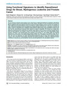
Fig. 1. Analysis of isozyme spectra in genotypes PRFGL 77 (a) and AC Emerson (b). Control samples were isozyme spectra of field plants and seedlings of the same genotypes, which were compared with the isozyme spectra of regenerants developed from microspores.
peroxidase as molecular markers for the gametic origin of flax plants derived from anther and ovary cultures.
77, obtained from the gene bank at the Research Institute of Plant Production in Piešt’any (Slovakia) and the Research Station in Morden (Canada).
MATERIALS AND METHODS
ANTHER CULTURE
Donor plant material was grown in field conditions in the experimental field at the Institute of Plant Genetics and Biotechnology, Slovak Academy of Sciences (Nitra, Slovakia), in the summer season from April to July. The cultivars used in the experiments were AC Emerson, Szegedi 30, Viking, and a new breeding line, PRFGL
Flower buds were excised before bud opening. To induce androgenesis in microspores, we collected flower buds when the microspores were in late uninucleate stage. Anther cultures were prepared as described previously (Obert et al., 2004a). The anthers of genotypes AC Emerson, PRFGL 77 and Szegedi 30 were cultivated on
175
Gametic origin of flax regenerants
Fig. 2. Zymograms of AC Emerson (a) and Viking (b). Control samples were isozyme spectra of field plants and in vitro seedlings of the same genotypes, which were compared with isozyme spectra of regenerants developed from ovules.
semiliquid IMA2.3 medium (Bartošová and Pret’ová 2004). Efficiency of induction was calculated as the ratio between the number of anthers producing calli and the overall number of anthers (150 anthers per genotype). All induced calli were transferred to MS (Murashige and Skoog, 1962) medium containing 2,4Dichlorophenoxyacetic acid (2,4-D) (1 mg/l) for 14 days, and after that to regeneration medium according to Nichterlein (2003). When the shoots developed, they were cultivated on MS medium without hormones, and after one subcultivation the isozymes in the leaves were analyzed.
OVARY CULTURE
For ovary cultures, the flower buds of genotypes AC Emerson and Viking were isolated 24–48 h before anthesis when the corolla began to overlap the green calyx slightly. The preparation of flax ovary cultures was described recently (Bartošová and Pret’ová, 2003). All induced calli were placed on MS medium with 2,4-D (1 mg/l) for 1 month and later on shoot regeneration medium with thidiazuron (TDZ) (0.5 mg/l) and benzylaminopurine (BAP) (0.5 mg/l). Regenerated shoots were excised and placed on rooting MS medium without hormones, and after one subcultivation the
176
Bartošová et al.
Fig. 3. Karyological analysis of a cell with a haploid number of chromosomes (n = 15) in mitosis (metaphase) in callus originating from ovary culture (a) and of a doubled haploid cell (2n = 30) in mitosis (anaphase) of root tip in a regenerant of gynogenic origin (b).
isozymes in the leaves were analyzed. Developed roots (1 cm long) of the regenerants were kept in ice water for 24 h and analyzed for chromosome numbers. ANALYSIS OF ISOZYME SPECTRA
When the shoots were 9 cm long, the true leaves were collected to analyze the protein spectra of selected enzymes, acid phosphatase (ACP) and peroxidase (PRX), according Krulickova et al. (2002). Ten plants of genotype PRFGL 77, 10 plants of AC Emerson derived from microspores, and 5 plants of each genotype (AC Emerson and Viking) derived from ovary cultures were analyzed. First, leaf tissues (0.1–0.5 g) were frozen in liquid N2, homogenized with extraction buffer (0.25 M TRIS; 0.05 M EDTA; 5 mM cystein-HCl; pH 6.8) at volume adjusted based on the protein content (250–500 µl), and centrifuged for 30 min at 10,500 g at 4˚C (Universal 32R, Hettich). Protein content was analyzed spectrophotometrically in supernatant according to Bradford (1976) using an ELISA reader (Elx800, Bio-Tek Instruments). Supernatant was stored at -20˚C or used directly for electrophoresis. The enzymes ACP and PRX were separated using 10% native discontinuous polyacrylamide gel (Laemmli, 1970) electrophoresis (3.5 mA per plate, 4 h, 4˚C). To detect ACP the gels were stained in 100 ml cold acetate buffer (pH 5.2) containing 0.03 g Fast Green GBC and 0.05 g 1-naphthyl phosphate for 1 h. To visualize PRX, the gels were stained in 100 ml acetate buffer (pH 4.6) supplemented with saturated benzidine R (0.2%) and 1.7 ml H2O2 (10%). Stained gels were kept in 7% acetic acid. The isozyme spectra from true leaves of 16 plants from field conditions and spectra of true leaves from 16 seedlings grown in vitro and excised after 1 subcultivation on MS medium supplemented with 2% sucrose were used as control samples. ANALYSIS OF PLOIDY LEVEL
The ploidy level of cells in calli and root tip was karyologically analyzed. The tissue of calli was fixed in
Carnoy solution (100% ethanol: glacial acetic acid, 3:1) for 7 days. Afterwards, samples were washed thoroughly with distilled water and then treated in 1 N HCl at 60˚C for 10 min. Then the macerated tissue was washed again and stained with Schiff reagent (Tomaskova, 1974) for 1.5 h. The washing step was repeated (30 min) and the calli or root tips were gently squashed with a drop of 1% acetocarmine solution. Observations employed an AXIOPLAN microscope (Opton, Germany) with CCD camera, and the images were analyzed with CHROMOVIDAS software (Germany).
RESULTS ANTHER CULTURE
The ratio of the induced anthers (anthers forming calli per 100 cultivated anthers) varied from 3.2% (PRFGL 77) to 2.6% (AC Emerson) after 4 weeks in culture. No callogenesis was observed in cultivated anthers of the Szegedi 30 genotype in this experiment. Regeneration of shoots from calli was calculated after 6 months of subculturing. The ratio of regenerating calli was 7.4% in PRFGL 77 and 27.3% in AC Emerson. OVARY CULTURE
The ratio of induced ovary segments (appearing after 1–2 months in culture) ranged from 4.5% in AC Emerson to 50.0% in Viking. Some cells in the calli were haploid, but most were diploid (Fig. 3a). Shoots appeared on the calli after 6–7 months in culture. All regenerated plantlets were doubled haploids (Fig. 3b). ISOZYME SPECTRA IN PLANTS DERIVED FROM ANTHER AND OVARY CULTURES
Analysis of the isozyme spectra in true leaves showed segregation of alleles at two loci of PRFGL 77 (PRX, Fig. 1a) and at two loci of AC Emerson (PRX, ACP; Fig. 1b), confirming the homozygosity of the regenerants
Gametic origin of flax regenerants
(*** in columns). The expected somaclonal variation appeared in 77% of PRFGL 77 and in all AC Emerson regenerants (**). Epigenetic modifications occurred in the ACP expression of PRFGL 77 and PRX of AC Emerson in seedlings (frequency 1: 31%; frequency 2: 12.5%; frequency 3: 12.5%; frequency 4: 44%; ****). Intravarietal variability (*) was also detected among field plants in ACP (25%) and PRX (50%, Fig. 1a), and only in ACP (12.5%, Fig. 1b). Compared to donor plants (2), the average number of alleles per loci increased in seedlings (ACP, PRX) and regenerants (PRX) of genotype AC Emerson (2–3), and in seedlings (ACP) and regenerants (PRX) of PRFGL 77 (2–3). Isozyme analysis in regenerants of presumed gynogenic origin (Fig. 2a,b) showed less polymorphism than in microspore-derived plants. The segregation of alleles showed a pattern indicating homozygosity of regenerants (***) at only one locus of AC Emerson (PRX) and also of Viking (PRX). Intravarietal changes in the number of isozymes occurred in 12.5% of AC Emerson field plants (Fig. 1a, *). Somaclonal variation appeared in 25% of the cases in genotype Viking (Fig. 2b, *) and in all AC Emerson (Fig. 2a, **) regenerants. New isozyme patterns were formed as epigenetic changes between 16 seedlings in Viking (Fig. 2b, **). The number of alleles per loci was higher in both regenerants and seedlings (ACP, PRX) of AC Emerson (2–3). In comparison with field plants (2), the number of alleles per loci increased (2–3) in regenerants (ACP) and seedlings (ACP, PRX) in the Viking genotype. PLOIDY LEVEL
Ploidy level analysis showed haploid as well as diploid cells in calli and in root tips of regenerated plantlets.
DISCUSSION Several protocols for flax anther culture has been published (Pretova and Obert, 2000; Chen and Dribnenki, 2002; Nichterlein, 2003; Rutkowska-Krause, 2003; Obert et al., 2004b). However, the efficiency of this biotechnological approach depends on the environmental conditions under which donor plants are grown, and also on the impact of their genetic predisposition for androgenesis and shoot regeneration in vitro. In our conditions, the donor material was grown in the field and the flower buds were collected during the summer months. Every summer season is specific, differing in the amount of precipitation and in average daily temperature. Those factors particularly influence the course of microsporogenesis, which is later displayed in the response of anthers cultivated in vitro (Bartošová and Pret’ová, 2004). The somatic tissue of the flax ovary seems to produce a special compound that supports gynogenic development from unpollinated ovules, because ovule
177
culture itself has been unsuccessful (unpubl.data). The impact of somatic tissue on induction of callogenesis in ovary culture is being studied further, as is the question of whether the ovules in cultivated ovaries are synchronous at the beginning of cultivation. Regeneration of shoots from androgenic and gynogenic calli was highly genotype-dependent, and was effected predominantly from diploid cells in calli. The diploidization very probably took place after induction of microspores in anther cultures or after induction of unpollinated ovules in ovary cultures. The origin of regenerants was analyzed using a biochemical marker system for the codominant phenotype. Exhibiting both molecular heterogeneity and variability, isozymes represent a large group of molecular markers extensively used in population genetics studies and also for analyzing ontogenic processes. The expression of isozymes can be influenced by mutations, polyploidization and chromosomal aberrations that might occur. Thus, correct analysis of isozyme banding patterns on electrophoretic gel in genetic terms requires proper determination of the factors influencing the electrophoretic phenotype (Zeidler, 2000). In our case, the isozyme spectra of regenerated plantlets originating from flax anther cultures and from ovary cultures were analyzed and compared with the spectra of field donor plants, which were stable breeding lines, and also with the spectra of seedlings. We observed several changes in banding patterns. Intravarietal differences in enzymatic activity were found between field plants of AC Emerson (ACP) and PRFGL 77 (ACP, PRX). This is related either to changes in the structural part of the locus controlling ACP or to nutrition of plants by phosphorus (Miller et al., 2001), as well as some kind of environmental stress (PRX) (Low and Merida, 1996). The zymograms of seedlings grown in vitro showed several new isozymes which probably formed as a result of in vitro culturing. Interestingly, in genotypes Viking and PRFGL 77, the intravarietal variation between the plants from the field (3 months old) was low or none, but the seedlings of these genotypes (1 month old) revealed significantly higher differences in the expression of PRX and ACP. Regenerated plantlets of both in vitro cultures varied in the number of alleles, considered somaclonal variation due to the prior callus stage. Two or three isozymes were found at a locus in the field plants and seedlings, but in regenerants only one isozyme was detected. We can assume that the donor material was heterozygous and that the plants originating from anther culture were homozygous at two loci (PRX isozymes) of PRFGL 77, at two loci (ACP and PRX) of AC Emerson and plants originating from ovary culture, and at one locus (PRX) of both the AC Emerson and Viking genotypes. The polymorphism in enzyme expression found between donor material and regenerants was low, because the starting materials were stable breeding lines.
178
Bartošová et al.
ACKNOWLEDGEMENTS This work was supported by APVT project 51–002302 and 51–0286 from the Agency for Science and Technology Support, Slovak Republic, and from the VEGA Grant Agency (2/5079/5).
REFERENCES BARTOŠOVÁ Z, and PRETˇOVÁ A. 2003. Induction of callogenesis in ovary and anther cultures of flax. In: Proceedings of the 10. scientific seminar. November 2003, Pieany (Slovakia), 2003, 25–28. BARTOSOVA Z, ROUX N, and PRETOVA A. 2003. Ovary culture in Linum usitatissimum L. Proceedings of the XI International Conference on Plant Embryology, September 2003, Brno, Czech Republic, 116. BARTOŠOVÁ Z, and PRETˇOVÁ A. 2004. Some environmental factors influence the androgenic flax response. In: Proceedings of the X Plant Physiology Days, September 2004, Bratislava (Slovakia), 17. BERGMANN R, and FRIEDT W. 1996. Haploidy and related biotechnological methods in linseed (Linum usitatissimum L.). In: Jain MS, Sopory SK, and Veilleux RE [eds.], In vitro haploids production in higher plants, vol. 1, 1–16. Kluwer Academic Publisher, Dordrecht, The Netherlands. BRADFORD MM. 1976. A rapid and sensitive method for the quantitation of microgram quantities of protein utilising the principle of protein dye binding. Analytical Biochemistry 72: 248–254. CHEN Y, and DRIBNENKI P. 2002. Effect of genotype and medium composition on flax Linum usitatissimum L. anther culture. Plant Cell Reports 21: 204–207. CHEN Y, KENASCHUK E, and DRIBNENKI P. 1998. High frequency of plant regeneration from anther culture in flax, Linum usitatissimum L. Plant Breeding 117: 463–467. CHEN Y, KENASCHUK E, and DRIBNENKI P. 2001. Inheritance of rust resistance genes and molecular markers in microsporederived populations of flax. Plant Breeding 120: 82–84. COPPENS L, and DE WITTE D. 1990. Esterase and peroxidase zymograms from barley (Hordeum vulgare L.) callus as a biochemical marker system of embryogenesis and organogenesis. Plant Science 67: 97–105. FEIRER RP, and SIMON PW. 1991. Biochemical differences between carrot inbreds differing in plant regeneration potential. Plant Cell Reports 10: 152–155. FRIEDT W, BICKERT C, and SCHAUB H. 1995. In vitro breeding of high linolenic, double haploid lines of linseed (Linum usitatissimum) via androgenesis. Plant Breeding 114: 110–118. KRULICKOVA K, POSVEC Z, and GRIGA M. 2002. Identification of flax and linseed cultivars by isozyme markers. Biologia Plantarum 45: 327–336. LAEMMLI UK. 1970. Cleavage of structural proteins during the assembly of the head of bacteriophage T4. Nature 227: 681–685.
LOW PS, and MERIDA JR. 1996. The oxidative burst in plant defense: Function and signal transduction. Physiologia Plantarum 96: 532–542. MCDOUGAL GJ, DAVIDSON D, and MILLAM S. 1992. Alterations in surface-associated peroxidases during callus development and shoot formation in explants of Linum usitatissimum. Journal of Plant Physiology 140: 195–200. MCDOUGAL GJ, MILLAM S, and DAVIDSON D. 1993. Alterations in surface-associated peroxidases during in vitro root development of explants of Linum usitatissimum. Plant Cell Tissue and Organ Culture 32: 101–107. MILLER SS, LIU J, ALLAN DL, MENZHUBER CHJ, FEDOROVA M, and VANCE CP. 2001. Molecular control of acid phosphatase secretion into the Rhizosphere of proteoid roots from phosphorus-stressed white lupin. Plant Physiology 127: 594– 606. MONTALBINI P, BUONAURIO R, and UMESH-KUMAR NN. 1995. Peroxidase activity and isoperoxidase pattern in tobacco leaves infected with tobacco necrosis virus and other viruses inducing necrotic and non necrotic alternations. Journal of Phytopathology 143: 295–301. MURASHIGE T, and SKOOG F. 1962. A revised medium for rapid growth and bio assays with tobacco tissue cultures. Physiologia Plantarum 15: 473–497. NICHTERLEIN K. 2003. Anther culture of linseed (Linum usitatissimum L.). In: Maluszynski M, Kasha KJ, Forster BP, Szarejko I [eds.], Doubled haploid production in crop plants. A manual, IAEA, 249–254. OBERT B, BARTOSOVA Z, and PRETOVA A. 2004a. Dihaploid production in flax by anther and ovary cultures. Journal of Natural Fibres 1: 1–14. OBERT B, DEDICOVA B, HRICOVA A, SAMAJ J, and PRETOVA A. 2004b. Plant regeneration from flax anther culture: genotype, pretreatment and media effect. Plant Cell Tissue and Organ Culture 79: 233–238. PRETOVA A, and OBERT B. 2000. Progress in flax androgenesis. Biotechnological Approaches for utilization of gametic cells, 165–169. COST 824, Slovenia, 2000 RAGHAVAN V. 1977. Patterns of DNA synthesis during pollen embryogenesis in henbane. Journal of Cell Biology 73: 521–526. RUTKOWSKA-KRAUSE I, MANKOWSKA G, LUKASZEWICZ M, and SZOPA J. 2003. Regeneration of flax (Linum usiatissimum L.) plants from anther culture and somatic tissues with increased resistance to Fusarium oxysporum. Plant Cell Reports 22: 110–116. SAMEC P, POSVEC Z, STEJSKAL J, NASINEC V, and GRIGA M. 1998. Cultivar identification and relationship in Pisum sativum L. based on RAPD and isozymes. Biologia Plantarum 41: 39–48. SIEGEL BZ. 1993. Plant peroxidases – an organismic perspective. Plant Growth Regulators 12: 303–312. TOMASKOVA D. 1974. Quick methods in plant cytology. University Press, Prague. WOJTASZEK P. 1997. Oxidative burst: an early plant response to pathogen infection. Biochemistry Journal 322: 681–692. ZEIDLER M. 2000. Electrophoretic analysis of plant isozymes. Acta Universitatis Palackianae Olomucensis. Facultas Rerum Naturalium, Biologica 38: 7–16.