Naylor et al. BMC Musculoskeletal Disorders 2011, 12:77 http://www.biomedcentral.com/1471-2474/12/77
RESEARCH ARTICLE
Open Access
Validity and reliability of using photography for measuring knee range of motion: a methodological study Justine M Naylor1,2*, Victoria Ko1, Sam Adie1,2, Clive Gaskin3, Richard Walker2, Ian A Harris1,2 and Rajat Mittal1,2
Abstract Background: The clinimetric properties of knee goniometry are essential to appreciate in light of its extensive use in the orthopaedic and rehabilitative communities. Intra-observer reliability is thought to be satisfactory, but the validity and inter-rater reliability of knee goniometry often demonstrate unacceptable levels of variation. This study tests the validity and reliability of measuring knee range of motion using goniometry and photographic records. Methods: Design: Methodology study assessing the validity and reliability of one method (’Marker Method’) which uses a skin marker over the greater trochanter and another method (’Line of Femur Method’) which requires estimation of the line of femur. Setting: Radiology and orthopaedic departments of two teaching hospitals. Participants: 31 volunteers (13 arthritic and 18 healthy subjects). Knee range of motion was measured radiographically and photographically using a goniometer. Three assessors were assessed for reliability and validity. Main outcomes: Agreement between methods and within raters was assessed using concordance correlation coefficient (CCCs). Agreement between raters was assessed using intra-class correlation coefficients (ICCs). 95% limits of agreement for the mean difference for all paired comparisons were computed. Results: Validity (referenced to radiographs): Each method for all 3 raters yielded very high CCCs for flexion (0.975 to 0.988), and moderate to substantial CCCs for extension angles (0.478 to 0.678). The mean differences and 95% limits of agreement were narrower for flexion than they were for extension. Intra-rater reliability: For flexion and extension, very high CCCs were attained for all 3 raters for both methods with slightly greater CCCs seen for flexion (CCCs varied from 0.981 to 0.998). Inter-rater reliability: For both methods, very high ICCs (min to max: 0.891 to 0.995) were obtained for flexion and extension. Slightly higher coefficients were obtained for flexion compared to extension, and with the Marker compared to the Line of Femur Method. For intra- and inter-rater reliability, the mean differences (within 2 degrees) and 95% limits of agreement (within 5 degrees) were generally clinically acceptable for both methods. Conclusion: Photography potentially offers a superior method of measurement over standard goniometry as visualising the centre of the knee is simplified in a two-dimensional plane and the permanent record provides greater assessor transparency as well as opportunity to confer. The Marker and Line of Femur Methods have moderate to substantial validity, but the inter- and intra-rater repeatability for trained observers are excellent with both methods yielding small mean differences with narrow limits of agreement. The Line of Femur Method offers the added advantage that it does not rely on inter-clinician consistency in identifying the greater trochanter.
* Correspondence:
[email protected] 1 South West Sydney Clinical School, University of New South Wales, Liverpool, NSW, Australia Full list of author information is available at the end of the article © 2011 Naylor et al; licensee BioMed Central Ltd. This is an Open Access article distributed under the terms of the Creative Commons Attribution License (http://creativecommons.org/licenses/by/2.0), which permits unrestricted use, distribution, and reproduction in any medium, provided the original work is properly cited.
Naylor et al. BMC Musculoskeletal Disorders 2011, 12:77 http://www.biomedcentral.com/1471-2474/12/77
Background Goniometry for measuring knee range of motion (ROM) is well entrenched in the orthopaedic and rehabilitative communities. It is a measure of some importance as severe restriction in range has ramifications for gait [1-3], function [3-5], and the need for manipulation [6,7]. Further, knee flexion and extension ROMs are incorporated into orthopaedic knee scoring tools to assess disease severity [6], are frequently used to track recovery after various knee surgeries [7-10], and are also used as clinical indicators by which to monitor and benchmark physiotherapy or rehabilitative performance [11]. In light of its extensive use, the clinimetric properties of knee goniometry (namely validity, reliability and responsiveness) are critical to appreciate. The clinimetric properties of goniometry have been reviewed [12,13]. In general, intra-observer reliability of goniometry has been shown to be acceptable [14-19] and this is usually (though not always [18]) provided the observer is practised in the procedure. However, the validity and inter-rater reliability of knee goniometry has not been consistently shown to be impressive nor acceptable [14-24]. Differences between studies in how the latter clinimetric properties were analysed, the goniometric tools used, and even subject body habitus, are likely contributors to the varying results between studies (Table 1). Whilst most studies evaluating the validity of clinical knee goniometry via comparisons with measures obtained from radiographic images (the ‘proxy criterion’ or ‘best available’ estimate) have demonstrated a significant correlation using intra-class or Pearson product moment correlations [14,15,17,21,22], the strength of the relationship has varied considerably. Further, studies which provide a clinically relevant contextual estimate of validity [16,23] indicate that the variation between clinical goniometry and the proxy criterion could be as large as 20 degrees [23]. That the radiographic and the clinical measures inevitably use different landmarks in part explains why variation between the two methods is considerable. Rater error, therefore, is not the only source of variation and thus the level of acceptable variation when assessing validity will necessarily be greater than what would be acceptable for inter-rater variability. Inter-rater goniometric reliability likewise suffers from clinical and statistical uncertainty (Table 1). Inter-rater reliability consistently underperforms intra-rater reliability [14,15,18-20], with inter-rater differences (up to 18 degrees [24]) often exceeding what would be considered a clinically relevant difference. Greater inter-rater reliability for flexion more so than extension angles is typically shown [14,15,18,19,21]. The position of measurement (supine or sitting [24]) and professional background of the assessor [23] have been shown to be confounders. Consequently, the responsiveness of the
Page 2 of 10
measure (the ability of the tool to detect a real change) is greatly undermined when different measurers (observers) are involved. In other words, the capacity to detect small changes with standard goniometry when multiple clinicians are involved is highly questionable. Agreement between observers for knee goniometry relies largely on consistency in the identification of the bony landmarks on the proximal femur (greater trochanter, GT) and the distal tibia (lateral malleolus, LM), and visualising the sagittal axis of movement for the knee joint. Previous investigators cite the visual estimation of the axis as the ‘Achilles heel’ of knee goniometry [14,15,24]. Our own anecdotal evidence illuminates the identification of the GT as a primary source of interobserver error. Figures 1a and 1b illustrate the inconsistency in GT marker placement between experienced orthopaedic physiotherapists. Differing degrees of adiposity overlying the GT often contribute to the difficulty clinicians have in locating the GT in some individuals (Figures 2a and 2b). In light of the inter-rater reliability concerns in particular, we proposed the use of photographic records for tracking knee ROM. Karkouti and Marks [25] pursued this methodology for measurement of active knee extension and flexion (up to 30 degrees) in standing. They observed fair (ICC 0.40 to 0.59) to very high (ICC 0.8 to 1.0) intra-tester reliability. We propose that the permanent record of knee ROM afforded by a photographic trail provides greater assessor transparency as well as opportunity to confer about measured ROM at a later time if required. We also propose that photography potentially offers a superior method of measurement over standard goniometry as visualising the centre of the knee is simplified in a two-dimensional plane. In this study, we aimed to describe the validity and reliability of two measurement approaches for obtaining knee ROM, both of which used photographic records. One method required raters to measure knee range off a photograph using the skin markers placed over the GT and LM by a clinician as reference points (’Marker Method’). As the identification of GT could itself be a source of error, the second method bypassed the GT marker, requiring the raters to estimate the line of femur instead (’Line of Femur Method’). Both methods required the raters to estimate the centre of the knee joint.
Methods Design and Participants
This methodology study was nested within a large randomised controlled trial investigating the effectiveness of rehabilitation strategies after total knee replacement (TKR) (Australian New Zealand Clinical Trials Registry, ACTRN12609000476235). A subset of patients presenting
Naylor et al. BMC Musculoskeletal Disorders 2011, 12:77 http://www.biomedcentral.com/1471-2474/12/77
Page 3 of 10
Table 1 Overview of validity and inter-rater reliability for knee goniometry Study
Validity
Lavernia et al [23]
Mean difference between radiologic and goniometric 1-way ANOVA method determined a significant measurement up to 13 degrees with wide (up to 20 difference between raters for visual and measured degrees) 95% limits of agreement range of motion
Lenssen et al [24]
Inter-rater
Mean difference (95% limits of agreement) between raters differed by position tested and angle: 1.4 degrees (-16.2 to 19 degrees) (flexion/supine); 2.7 (-6.7 to 12.1) (flexion, sitting); 2.2 (-6.2 to 10.6) (extension, sitting)
Goniometric tool and test position Standard goniometer; supine. Long-arm goniometer; sitting and supine.
Edwards et al [16]
22% of the goniometric measurements were different Correlation coefficient, 0.91 (type not specified). by 5 degrees or more
Standard goniometer; supine.
Brosseau et al [14]
r = 0.975-0.987 (flexion), r = 0.390-0.514 (extension).
ICC, 0.959-0.982 (flexion); ICC 0.856-0.926 (extension). Both devices performed similarly.
Parallelogram and Universal goniometer; supine.
Brosseau et al [15]
r = 0.73-0.78 (flexion), r = 0.33-0.48 (extension).
ICC, 0.82-0.88 (flexion); ICC 0.43-0.52 (extension). Both devices performed similarly, but the parallelogram provided more advantages to the clinician.
Parallelogram and Universal goniometer; supine.
ICC 0.90 (flexion); ICC, 0.86 (extension)
Standard goniometer; positioned varied.
ICC 0.99
Standard goniometry; sidelying.
ICC 0.84-0.93 (flexion); ICC 0.59-0.80 (extension). All devices performed similarly.
3 different standard goniometers; position not specified. Extended goniometer; supine position.
Watkins et al [19] Gogia et al [22]
ICC 0.98-0.99.
Rothstein et al [18]
Lawrence, cited by Johnson [17]
r = 0.94 (extension and flexion angles combined).
Cleffken et al [20]
Intra-observer error +/- 2.3 degrees for end flexion (aggregate standard deviation). Inter-observer error +/- 2.6 degrees end extension and +/-4.2 degrees end flexion Passive end of range flexion - Smallest detectable difference 0 ± 6.4 degrees
Electronic digital inclinometer; supine
Pearson product moment correlation (r), intra-class correlation (ICC).
to the pre-operative education programme prior to their TKR and who consented to participate in the RCT, were invited to partake in this smaller methodology trial. To get a scatter in age and body size, healthy volunteers (onsite healthcare workers) were also invited to participate through word-of-mouth. Pregnancy and inability to speak English were the only exclusion criteria applied. Written informed consent was obtained from all participants and the study was approved as a sub-study within the larger RCT by the Executive of the Human Research Ethics Committee of Sydney South West Area Health Service. Protocol Protocol for measuring the participant
On the study day, each participant was positioned (horizontally) supine on a radiographic table in the radiology department. An investigator (JN) arbitrarily positioned the participant’s knee (left or right) in either a flexed (between approximately 20-130 degrees) or extended position (between full extension and approximately 20 degrees of flexion). The investigator ensured that the selected position was pain free for the subject, and this
position was maintained with use of sandbags and high density foam supports if required. Care was taken using visual inspection to ensure the leg was neither abducted nor adducted, and in neutral rotation. A second investigator [Clinician 1, a physiotherapist of 12 years experience, VK] located and placed adhesive skin markers over the posterior lip of the GT and the middle of the LM. This same investigator took a photograph of the participant’s knee whilst in this position using a digital camera (Canon PowerShot A470, 7.1 megapixels, 3.4× optical zoom). The clinician stood between 1 and 1.2 m from the table to get the entire lower limb in full view, and held the camera lens level with and parallel to the participant’s knee. The participant maintained the position whilst the markers were removed and a third investigator [Clinician 2, an orthopaedic registrar of 2 years experience, SA] entered the room to repeat the process undertaken by first clinician. Clinician 1 and 2 alternated the sequence for who placed the markers first. Upon completion of the second photograph, a lateral knee radiograph was taken of the patient in the same position. To minimise the burden on the radiology
Naylor et al. BMC Musculoskeletal Disorders 2011, 12:77 http://www.biomedcentral.com/1471-2474/12/77
Page 4 of 10
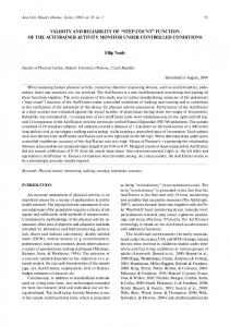
A
A
B
B
Figure 1 Inconsistency in GT marker placement between experienced orthopaedic physiotherapists. (Note the position of the marker (black circle) in relation to the scar (black straight line)).
services, the assessments were spread over two radiology departments utilising similar protocols. The x-ray beam (63 kV, 6.3 mAs) was perpendicular to the plane of the leg and at the level of the knee joint. The radiographs were taken using a 35 × 43 cm plate placed 100 cm from the x-ray beam. The whole body effective dose was