Imashimizu et al. Genome Biology (2015) 16:98 DOI 10.1186/s13059-015-0666-5
RESEARCH
Open Access
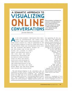
Visualizing translocation dynamics and nascent transcript errors in paused RNA polymerases in vivo Masahiko Imashimizu1, Hiroki Takahashi2, Taku Oshima3, Carl McIntosh1, Mikhail Bubunenko1, Donald L. Court1 and Mikhail Kashlev1*
Abstract Background: Transcription elongation is frequently interrupted by pausing signals in DNA, with downstream effects on gene expression. Transcription errors also induce prolonged pausing, which can lead to a destabilized genome by interfering with DNA replication. Mechanisms of pausing associated with translocation blocks and misincorporation have been characterized in vitro, but not in vivo. Results: We investigate the pausing pattern of RNA polymerase (RNAP) in Escherichia coli by a novel approach, combining native elongating transcript sequencing (NET-seq) with RNase footprinting of the transcripts (RNET-seq). We reveal that the G-dC base pair at the 5′ end of the RNA-DNA hybrid interferes with RNAP translocation. The distance between the 5′ G-dC base pair and the 3′ end of RNA fluctuates over a three-nucleotide width. Thus, the G-dC base pair can induce pausing in post-translocated, pre-translocated, and backtracked states of RNAP. Additionally, a CpG sequence of the template DNA strand spanning the active site of RNAP inhibits elongation and induces G-to-A errors, which leads to backtracking of RNAP. Gre factors efficiently proofread the errors and rescue the backtracked complexes. We also find that pausing events are enriched in the 5′ untranslated region and antisense transcription of mRNA genes and are reduced in rRNA genes. Conclusions: In E. coli, robust transcriptional pausing involves RNAP interaction with G-dC at the upstream end of the RNA-DNA hybrid, which interferes with translocation. CpG DNA sequences induce transcriptional pausing and G-to-A errors.
Background RNA polymerase (RNAP) transcribes DNA of different structural and chemical sequences. Interaction of RNAP with some of these sequences results in transcriptional pausing, which occurs on average every 100 bp of transcribed DNA in vitro [1]. Regulation of elongation via pausing has a variety of physiological consequences [1]. In prokaryotes, the RNAP pausing/anti-pausing system that utilizes RfaH protein controls expression of genes involved in DNA transfer and virulence [2, 3]. Many regulatory events derived from pausing appear to be localized in promoter-proximal regions in eukaryotes or the 5′ untranslated region (UTR) of mRNA genes in prokaryotes * Correspondence:
[email protected] 1 Center for Cancer Research, National Cancer Institute, Frederick, MD 21702, USA Full list of author information is available at the end of the article
[2, 4–6]. For example, eukaryotic RNAPII tends to pause in a region located ≤100 bp downstream of a transcription start site, and is controlled by accessory protein factors such as NELF/DSIF [4, 7]. These paused polymerases allow a rapid transcription response to environmental stimuli and are used during development in higher eukaryotes [4, 6]. The RNAPII pausing at promoter-proximal regions in eukaryotes also plays a critical role in protecting these regions from adopting repressive chromatin structures, thereby maintaining an open promoter complex for highly expressed genes [8, 9]. In prokaryotes, pausing plays a key role in transcription attenuation and termination and in synchronization of transcription and translation [1, 3, 10]. An elongation complex (EC) consists of RNAP bound to double-stranded DNA and the RNA-DNA hybrid with the 3′ end of the RNA positioned in the active
© 2015 Imashimizu et al. This is an Open Access article distributed under the terms of the Creative Commons Attribution License (http://creativecommons.org/licenses/by/4.0), which permits unrestricted use, distribution, and reproduction in any medium, provided the original work is properly credited. The Creative Commons Public Domain Dedication waiver (http:// creativecommons.org/publicdomain/zero/1.0/) applies to the data made available in this article, unless otherwise stated.
Imashimizu et al. Genome Biology (2015) 16:98
center of the enzyme [11]. The hybrid length fluctuates between 9-bp and 10-bp length depending on the translocation state of RNAP. After phosphodiester bond formation, the movement of the RNA-DNA hybrid back along the catalytic cleft vacates the active center, enables binding of the next NTP and reduces the length of the RNA-DNA hybrid from 10 to 9 bp in a process called translocation [1]. Translocation is a smooth process except in cases where certain DNA sequences impose an intrinsic translocation barrier [1, 12]. This block of translocation as well as the inhibition of the bond formation after translocation causes RNAP pausing [1]. Protein factors exist that strengthen or weaken pausing by targeting translocation, such as the archaeal/eukaryotic Spt5 and bacterial NusG/NusA [3, 13, 14] as well as the Nun/N transcription termination/antitermination proteins of lambdoid phages [1, 15]. Pausing of EC within the post-translocated or pretranslocated state is enhanced when an RNA hairpin is formed immediately upstream of the hybrid [16, 17]. Some pausing signals in Escherichia coli, such as ops sequence, involve backtracking of RNAP along DNA [18]. Backtracking stabilizes pausing [12, 19] and leads to extrusion of one or more nucleotides of the 3′ RNA end beyond the active center [20]. A stably backtracked EC forms a roadblock to DNA replication [21], which can be highly toxic to the cell [22–24]. A direct assessment of transcription fidelity by RNA-seq in vivo and in vitro showed that an error at the 3′ end of a nascent RNA causes long transcription pausing by inducing RNAP backtracking [25]. It was also shown that transcription errors cause some heritable phenotypic changes [26, 27], which have been thought to affect aging [28] and carcinogenesis [29, 30]. Bacterial GreA and GreB or eukaryotic TFIIS proteins induce endonucleolytic RNA cleavage of any extruded 3′ RNA, with or without errors, thereby allowing renewed transcription in the backtracked EC [31, 32], which ensures better fidelity and removes the DNA replication barrier [22–25]. Extensive biochemical and single-molecule experiments have identified the steps involved in pausing in vitro [1]: Pausing can be caused by (i) a misalignment of incoming NTP and complementary template DNA base within the active site of the post-translocated RNAP [33], and (ii) an intrinsic barrier caused by DNA sequence during forward translocation from the pretranslocated state [13, 34]. This latter type of pausing can be stabilized by backtracking [12]. However, little is known about how broadly these mechanisms for pausing identified in vitro are involved in transcription regulation in vivo. In the present work, we employed native elongating transcript sequencing (NET-seq) [35] to identify RNAP pause sites and error hotspots in the E. coli chromosome by making an assumption that transcription errors
Page 2 of 17
contribute to pausing in vivo. After paused RNAP complexes are isolated from the genome, RNases are used to trim excess RNA from the 5′ ends leaving only the nascent RNA that is protected by RNAP. Thus, RNET-seq stands for RNase footprinting followed by NET-seq. A previous in vitro study showed that an RNAP forming an EC protects different lengths of the 3′-proximal transcript from trimming by RNases A and T1 depending on the EC translocation state [36]. Post-translocated, pretranslocated, and backtracked complexes protect 14nucleotide (nt), 15-nt and >15-nt segments of the RNA, respectively [36]. Importantly, because the very 3′ end of the RNA is extruded to a narrow pore from the active center of the enzyme during backtracking, the extruded RNA remains inaccessible to RNases increasing in length as backtracking increases [36]. Thus, paused RNAP in either the pre- and post-translocated states as well as at different backtracked distances were monitored over the entire genome. The unique properties of our RNET-seq approach provided an opportunity to dissect the core mechanisms of different types of pausing in living cells.
Results Gre factors reduce pausing in the 5′ UTR genome-wide
We employed RNET-seq on the wild-type (WT) E. coli strain and an isogenic strain deficient in genes for GreA and GreB (ΔgreAB). Gre factors and their eukaryotic analog TFIIS rescue backtracked complexes of RNAP [1]. Briefly, the cells were rapidly lysed via spheroplasting, and the transcribing RNAPs were released from the genomic DNA by digestion with DNase I (Fig. 1A). Any ribosomes involved in co-transcriptional translation were separated from RNAP by digestion with RNase A. During the cell lysis heparin was present to inhibit nonspecific binding of RNAPs to DNA and RNA [37]. All RNAPs, including those associated with the fragmented double-stranded DNAs and their 5′-truncated nascent RNAs, were immobilized on Ni2+-NTA beads through the hexa-histidine-tagged β’ subunit [38] and then extensively washed with a high-salt buffer (see “Materials and methods”). The purification was done in the native conditions not involving DNA-protein crosslinking. The 5′ ends of the transcripts in ECs were trimmed with RNase T1/V1 (V1 digests double-stranded RNAs in nascent transcripts, which are resistant to T1) to leave a minimal length of RNA protected by RNAP (Fig. 1A). The RNases were subsequently removed by further washing of the beads. Next, elution with imidazole generated ECs carrying ~6- to 30-nt long transcripts (Fig. 1B; Fig. S1A in Additional file 1). The predominant RNA length distribution was consistent with nucleotide lengths of nascent RNA protected by RNAP from in vitro digestion by different RNases in active and backtracked ECs (Fig. 1B; ≥14 nt) [36]. The imidazole eluate also contained shorter